characterization of pantoea ananatis isolated from garlic
TRANSCRIPT

Research Article
Characterization of Pantoea ananatis Isolated from Garlic and Shallot
Karakterisasi Pantoea ananatis yang Diisolasi dari Bawang Putih dan Bawang Merah
Nanik Nurjanah1), Tri Joko 2), & Siti Subandiyah2)*1)Agricultural Quarantine Agency Class 1 Ambon
Jln. Rurehe, Waihaong, Ambon, Maluku 971112) Department of Crop Protection, Faculty of Agriculture, Universitas Gadjah Mada
Jln. Flora 1, Bulaksumur, Sleman, Yogyakarta 55281
*Corresponding author. E-mail: [email protected]
ABSTRACTThe new disease on garlic (Allium sativum) and shallot (A. cepa L. aggregatum group) have been found in several
production centers of garlic and shallot in Tawangmangu and Temanggung, Central Java. The infected plants showedsymptoms of leaf blight accompanied by chlorosis. The objective of this study was to determine the pathogen thatcauses leaf blight and chlorosis based on the phenotypic characterization and gyrB gene sequences analysis. Theresearch started from the isolation of pathogen, physiological and biochemical test, DNA extraction, and sequenceanalysis of gyrB using gyrB 01-F and gyrB 02-R primer. The results showed that the isolated bacterial pathogen havea yellow pigment, slimy colonies with regular borders, convex, gram-negative, non-spore, facultative anaerobic, motile,catalase production, indole production, and acid production from D-glucose, D-mannitol, sucrose, and lactose. Fromthe pathogenicity test, it was found that the bacteria produced the typical symptom of leaf blight. Characterization ofpathogens based on gyrB gene sequence revealed that the pathogen was placed in the group of Pantoea ananatis.
Keywords: Alium cepa, Alium sativum, gyrB, leaf blight, Pantoea ananatis
INTISARIPenyakit baru pada bawang putih (Allium sativum) dan bawang merah (A. cepa L. aggregatum group) telah
ditemukan di beberapa sentra produksi bawang putih dan bawang merah di Tawangmangu dan Temanggung, JawaTengah. Tanaman yang terinfeksi menunjukkan gejala hawar daun disertai klorosis. Tujuan penelitian untuk mengetahuikarakter patogen berdasarkan fenotipik dan sekuen gen gyrB. Penelitian dimulai dengan isolasi bagian tanaman yangsakit, uji fisiologi dan biokimia, ekstraksi DNA dengan metode CTAB/NaCl dan amplifikasi gen gyrB menggunakanprimer gyrB 01-F and gyrB 02-R. Hasil uji menunjukkan koloni berlendir, cembung, pigmen berwarna kuning, gramnegative, tidak berspora, aerob fakultatif, motil, produksi katalase, indol, membentuk asam dari D-glukosa, D-monnitol,sukrosa dan laktosa, dan patogenesitas positif. Karakterisasi patogen berdasarkan sekuen gen gyrB, menunjukkanpatogen hawar daun berkerabat dekat dengan Pantoea ananatis.
Kata kunci: Alium cepa, Alium sativum, gyrB, leaf blight, Pantoea ananatis
Jurnal Perlindungan Tanaman Indonesia, Vol. 21, No. 2, 2017: 120–126DOI: 10.22146/jpti.27407
INTRODUCTIONPantoea ananatis is an important bacterial plant
pathogen in the world and has a very wide host range.P. ananatis can survive in a various ecosystem as thesaprofit, endophytes, epiphytes and pathogens. P.ananatis is reported to cause center rot disease ofonion, and accounted for 100% loss in some fields(Gitaitis & Gay, 1997). While maize white spot (MWS)caused by this pathogen could cause up to 60%yield loss (Miller et al., 2016).
In Indonesia, P. ananatis has never been reportedto infect plants until recently, although the tropical
climate conditions in Indonesia are also suitable for thedevelopment of P. ananatis. The risks and possibilityof the pathogen to spread in Indonesia is highlyconsidered by the import commodities from countriesthat have reported the presence of these bacteria.According to Carr et al. (2010), the development ofcenter rot symptoms in onion bulbs during storageposes a significant problem. The onion is often storedfor months prior to grading and marketing will resultin a more severe infection. However, onion bulbsthat are infected solely by P. ananatis remain firm andexhibit subtle or no external symptoms, they can be
Submitted August 11, 2017; accepted November 9, 2017

difficult to detect on grading lines. The onion bulbscan be a source of inoculum for other plants, althoughthe onion is not cultivated in Indonesia.
The metabolic pattern of P. ananatis from cropdebris is similar to those recovered directly from lesionsor from healthy leaves, suggesting that these bacteriain crop debris could act similarly to epiphytic isolates,being a source of inoculum for further infections. P.ananatis can survive as epiphytes in the leavesof healthy maize plants, non-host plants and in cropdebris, and possibly multiply there (Sauer et al.,2015). The bacteria infect host through flowers,mechanical injury, wound insect bite and frictioninjuries plants with the current crop of strong winds(Azad et al., 2000). Tobacco thrips of Frankliniellafusca are a vector of P. ananatis caused center rot inonion (Gitaitis et al., 2003).
The gyrB gene codes for the b-subunit of DNAgyrase, a type II DNA topoisomerase, which introducesnegative supercoils into closed circular DNA molecules.One of the reasons why the gyrB gene is selectedfor phylogenetic studies is that, as horizontal genetransfer (HGT) occurs infrequently in informationalgenes that are involved in transcription and translation,it is assumed not to undergo HGT (Harayama & Kasai,2006). GyrB gene sequences have been widely usedfor the identification of bacterial species. GyrB genesequence is more suitable for determining geneticrelationships and the identification of bacterial thanthe 16S rRNA (Parkinson et al., 2009; Takeda et al.,2010).
The objectives of this study was to characterizebacterial pathogen isolated from onion and garlic basedon phenotypic properties and gyrB gene sequencesanalyses.
MATERIALS AND METHODS
Survey and sampling were carried out based onpurposive random sampling method (Sumardiyonoet al., 2011; Windari et al., 2015; Ismiyatuningsih etal., 2016). Symptomatic tissue showing leaf blightand chlorosis diseases were collected from severalfields of garlic and shallot production area in CentralJava.
Isolation of Bacterial PathogenBacteria were isolated from the affected tissue
according to Joko et al. (2011a) with slight modification.A one gram of the sample was crushed in a smalltube 500 μl sterilized ddH2O. One loopful of the
suspension was streak onto YP agar (yeast extract 5g, peptone 10 g, agar 15 g, water 1000 ml, pH 6.8)plate medium and it was incubated for 2 days at 28oC(Wibowo et al., 2010; Wardhika et al., 2014). Yellowcolonies were formed on YP agar plate, and singlecolonies were subcultured onto YP agar slants.
Physiological and Biochemical CharacterizationThe bacterial isolates were then characterized as
follows (Lelliot & Stead, 1987; Joko et al., 2000;Schaad et al., 2001):
Gram reaction with 3% KOH is to differentiatebacteria based on the structure of the cell wall.Gram-negative bacterial will become gummy uponmixing with a loop, while gram-positive bacterialwill not.
Catalase test is to detect the presence of catalaseenzyme in bacteria that is able to hydrolyze hydrogenperoxide (H2O2) into water and oxygen. If the bacteriumhas a catalase enzyme, it will form gas bubbles.
Anaerobic growth test aims to determine whetherthe bacteria can grow in aerobic or anaerobic conditionsin media containing bromotimol blue covered withsterile oil paraffin (anaerobic) and without paraffin(aerobic). A color change from blue to yellow in bothtubes is recorded as positive for anaerobic growth/fermentation.
Indole test uses Kovac’s method, that is reagentswhich contains hydrochloric acid and p-dimethylaminobenzaldehyde in amyl alcohol. The Bacteriathat produce the enzyme tryptophanase can convertthe amino acid tryptophan to by-products that includeindole. The indole which is produced was detectedby adding Kovac’s reagent which produced cherryred (Aneja, 2003).
Oxidase test is to detect the presence of pathogenicbacteria cytochromeoxidase. The bacteria are streakedonto filter paper that has been containing tetramethyl-p-phenylenediamine dihydrochloride 1%, The strainwas rated oxidase-positive if a purple color developswithin 10s, delayed positive if coloration developswithin 10-60s and negative if no color developsafter 60 seconds.
Nitrate reduction test is to determine the abilityof bacteria to reduce nitrate to nitrite compounds.The bacteria were grown in NA medium containing0.1% KNO3, then it is incubated at room temperature(24h and 48h). Each tube was added with 1 mlreagent A (1 g α-naphthyl amine in 200ml of aceticacid 30%) and 1 ml reagent B (0.5 g of sulfanilic
Nurjanah et al.: Characterization of Pantoea ananatis Isolated from Garlic and Shallot 121

acid in 150ml of 30% acetic acid). The color changeoccurs in the culture of bacteria, nitrate is reduced toform a perfect nitrogen gas (N2) or ammonium gas(NH3), a positive reaction happens when the redcolor is formed about 30 minutes and medium cracks.
Arginine dihydrolase test is to detect the conditionof the growth of anaerobic bacteria in Thornleymedia (arginine medium with phenol red dye), andcovered with liquid paraffin to create anaerobicconditions and incubated at 28oC for 4 days. Thepositive reaction was occurred when there was achange in the medium from pink to red indicatesarginine compound is hydrolyzed to urea and ornithine.
Gelatin hydrolysis test is to detect the presenceof proteolytic enzymes. The bacteria are grown ingelatin medium and incubated for 7-14 days at roomtemperature. Before being observed, the tube wascooled at 4°C for 30 min (until control is gelled)every day to check for gelatin liquefaction. The nutrientgelatine medium inoculated with a gelatine negativebacterium will remain solid after the cold treatment(Leboffe & Pierce, 2010).
H2S production. The bacteria are grown in SIMmedium, that is medium containing peptone andsodium thiosulfate as a sulfur source. The presence ofH2S is indicated by a formation of a black precipitateat the stabbing side.
The presence of urease is detectable when theorganisms are grown in a urea broth mediumcontaining the pH indicator phenol red. An increasein alkalinity is indicated by a magenta red color (pHapprox. 9.0) was evidence of urease activity.
Hypersensitivity reaction (HR) on tobacco leaves.The suspension of P. ananatis were grown for 48 hin YP broth (yeast peptone medium at pH 6.8) byshaking at 120 rpm, then diluted to approximately108 cfu/ml (Kido et al., 2010) and injected in themesophyll which is located between the bones oftobacco leaves.
Pathogenicity test using 108−109cfu/ml concentration,the suspension was injected and sprayed under theleaf epidermis and sterile dH2O as negative control.
DNA ExtractionBacterial genomic DNA was extracted using mini
preparation DNA isolation technique with slightmodification (Joko et al., 2007a; 2007b; Danaatmadjaet al., 2009).As much of 1.5 ml of cell culture wascentrifuged at 5,000 g for 2 min. The pellet of DNAwas diluted with 540 µl of TE buffer (0.1 M Tris-
HCl, 0.1 M EDTA pH 8), added with 30 µl 10% SDSand then incubated at 37oC for 60 min. Afterwards,the pellet was added with 100 µl of 5 M NaCl and80 µl of CTAB/NaCl and then incubated at 65oC for10 min prior to addition of 750 µl of chloroformisoamyl alcohol (24:1) and centrifuged at 12,000 gfor 5 min. The upper layer was transferred into 1.5 mlEppendorf tube, added with 600 µl of phenol/chloroform isoamylalcohol (25:24:1) and thencentrifuged at 12,000 g for 5 min. The supernatant wastransferred again into new 1.5 ml Eppendorf tube.As much of 0.6 times volume of isopropanol wasadded and centrifuged at 12,000 g for 5 min. Thepellet was rinsed with ethanol 70%, air-dried andthen diluted with 20 µl of TE buffer.
gyrB Gen Sequence AnalysisThe amplification of gyrB gene of P. ananatis was
done using primer set of gyrB 01-F (5’-TAARTTY G AY G AYA A C T C Y TAYA A A G T- 3 ’ )(R=A/G;Y=C/T); and gyrB 02-R (5’-CMCCYTCCACCARGTAMAGTTC-3’) (M=A/C) (Pérez-y-Terrón et al., 2009). PCR Master mix consisted of30 μl Taq ready mix PCR kit (KAPA), 6 μl forwardprimer, 6 μl reverse primer, 12 μl nuclease free water.The DNA sample is then added 1 μl according tothe number of bacterial isolates. DNA amplificationwas done in a Bio-Rad thermocycler as follows:predenaturation at 95°C for 5 min, denaturation at95°C for the 30 s, annealing at 55°C for 1 min, extensionat 72°C for 1 min, and a final extension at 72°C for10 min. The cycle is repeated 35 times (Joko et al.,2011b). Samples were run on a 2% agarose gelstained with ethidium bromide and visualised undera UV light for the presence of amplified products(Joko et al., 2012).
DNA Sequencing was carried out by submittingthe PCR products to 1st BASE company. Nucleotidesequence was edited using Genetyx program 7th version(Genetyx, Japan) (Mahfut et al., 2016a; 2016b). Thesequences of the isolates were matched to referencestrains from Gene Bank Database based on 16S rRNAgene fragments to figure out their similarity. Nucleotidesequence was analyzed using Basic Local AlignmentSearch Tool (BLAST) at www.ncbi.nlm.nih.gov.Some reference strains with similarity close to 100%were determined (Suharti et al., 2017; Widyaningsih etal., 2017). The phylogenetic tree was constructed usingbootstrap method with 1,000 times replication; whileMultiple Sequence Alignment was analyzed usingMEGA 7 program (Joko et al., 2014; Dwimartina et
Jurnal Perlindungan Tanaman Indonesia Vol. 21 No. 2122
al., 2017). An unrooted phylogram was obtained bythe neighbor joining (NJ) method. The stability ofthe tree was assessed by 1000 bootstrap replicationswith the neighbor-joining method and Jukes-Cantordistance analysis. An interior branch test was done(heuristic option, 1000 replications) to check the treetopology for robustness. Some reference strains withsimilarity close to 100% were determined.Additionally, the Poisson correction was applied NJfor distance estimation, and the complete deletion optionwas used in handling gaps or missing data obtainedfrom alignments (Kumar et al., 2016).
RESULTS AND DISCUSSION
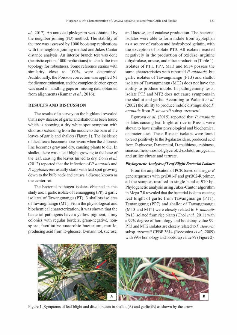
The results of a survey on the highland revealedthat a new disease of garlic and shallot has been foundwhich is showing a dry white spot symptom withchlorosis extending from the middle to the base of theleaves of garlic and shallots (Figure 1). The incidenceof the disease becomes more severe when the chlorosisline becomes gray and dry, causing plants to die. Inshallot, there was a leaf blight growing to the base ofthe leaf, causing the leaves turned to dry. Conn et al.(2012) reported that the infection of P. ananatis andP. agglomerans usually starts with leaf spot growingdown to the bulb neck and causes a disease known asthe center rot.
The bacterial pathogen isolates obtained in thisstudy are: 1 garlic isolate of Temanggung (PP), 2 garlicisolates of Tawangmangu (PT), 3 shallots isolatesof Tawangmangu (MT). From the physiological andbiochemical characterization, it was shown that thebacterial pathogens have a yellow pigment, slimycolonies with regular borders, gram-negative, non-spore, facultative anaerobic bacterium, motile,producing acid from D-glucose, D-mannitol, sucrose,
and lactose, and catalase production. The bacterialisolates were able to form indole from tryptophanas a source of carbon and hydrolyzed gelatin, withthe exception of isolate PT3. All isolates reactednegatively in the production of oxidase, argininedihydrolase, urease, and nitrate reduction (Table 1).Isolates of PT1, PP7, MT3 and MT4 possess thesame characteristics with reported P. ananatis, butgarlic isolates of Tawangmangu (PT3) and shallotisolates of Tawangmangu (MT2) does not have theability to produce indole. In pathogenicity tests,isolate PT3 and MT2 does not cause symptoms inthe shallot and garlic. According to Walcott et al.(2002) the ability to produce indole distinguished P.ananatis from P. stewartii subsp. stewartii.
Egorova et al. (2015) reported that P. ananatisisolates causing leaf blight of rice in Russia wereshown to have similar physiological and biochemicalcharacteristics. These Russian isolates were foundto react positively to the β-galactosidase, produced acidfrom D-glucose, D-mannitol, D-melibiose, arabinose,sucrose, meso-inositol, glycerol, d-sorbitol, amygdalin,and utilize citrate and tartrate.
Phylogenetic Analysis of Leaf Blight Bacterial IsolatesFrom the amplification of PCR based on the gyr B
gene sequences with gyrB01-F and gyrB02-R primer,all the samples resulted in single band at 970 bp.Phylogenetic analysis using Jukes-Cantor algorithmin Mega 7.0 revealed that the bacterial isolates causingleaf blight of garlic from Tawangmangu (PT1),Temanggung (PP7) and shallot of Tawangmangu(MT3 and MT4) were closely related to P. ananatisPA13 isolated from rice plants (Choi et al., 2011) witha 99% degree of homology and bootstrap value 99.PT3 and MT2 isolates are closely related to P. stewartiisubsp. stewartii CFBP 3614 (Rezzonico et al., 2009)with 99% homology and bootstrap value 89 (Figure 2).
Figure 1. Symptoms of leaf blight and discoloration in shallot (A) and garlic (B) as shown by the arrow
A B
Nurjanah et al.: Characterization of Pantoea ananatis Isolated from Garlic and Shallot 123

AssaysIsolates
PT1 PT3 PP7 MT2 MT3 MT4Gram assay - - - - - -OF-test O O O O O OCatalase + + + + + +Oxidase - - - - - -Urease - - - - - -Arginine dihydrolase - - - - - -Levan formation + + + + + +H2S production - - - - - -Nitrate reduction - - - - - -Indole production + - + - + +Gelatin liquefaction - - + + + +Motility + + + + + +Acid production or growth onAyers media containing:Glucose + + + + + +Lactose + + + + + +Sucrose + + + + + +Mannitol + + + + + +Maltose - - - - - -
Hypersensitive reaction + + + - + +Pathogenicity + - + - + +
Description: PP (garlic isolate of Temanggung), PT (garlic isolate of Tawangmangu), MT (shallot isolate of Tawangmangu).+ : indicated positive reaction; - : indicated no reaction; O : indicated oxidative
Table 1. Physiological and biochemical analyses of bacterial isolates from garlic and shallot
Figure 2. Phylogenetic tree showing the relationship of the leaf blight bacterial isolates and the closely related strainsavailable in the GenBank; on the basis of the alignment of gyrB gene sequences, a phylogenetic tree wasconstructed using the neighbor-joining method; the stability of the tree was assessed by 1000 bootstrapreplications with the neighbor-joining method and Jukes-Cantor distance analysis; the sequence ofXanthomonas axonopodis pv. alli strain LMG 580 was used as an outgroup
Jurnal Perlindungan Tanaman Indonesia Vol. 21 No. 2124

CONCLUSION
The use of other phylogenetic markers beside16S rRNA is necessary to achieve unambiguousidentification at the species level or below. In thepresent study we describe a method for identifyingPantoea ananatis using partial sequence of gyrB asa phylogenetic marker. Since gyrB is a universallydistributed gene in all prokaryotes, we are currentlyextending our work to similar studies of other bacterialfamilies with the aim of establishing the use of gyrBas a universal phylogenetic marker.
ACKNOWLEDGEMENT
The authors with to thank Australian Center forInternational Agricultural Research (ACIAR) forsupporting this work trough research grant (ACIARHORT 2009/056) to SS.
LITERATURE CITED
Aneja, K.R. 2003. Experiments in Microbiology,Plant Pathology and Biotechnology. Fourth Edition.New Age International Publishers, New Delhi.607 p.
Azad, H.R., G.J. Holmes, & D.A. Cooksey. 2000.A New Leaf Blotch Disease of Sudan GrassCaused by Pantoea ananas and Pantoea stewartii.Plant Disease 84: 973–979.
Carr, E.A., J.M. Bonasera, A.M. Zaid, J.W. Lorbeer,& S.V. Beer. 2010. First Report of Bulb Diseaseof Onion Caused by Pantoea ananatis in NewYork. Plant Disease 94: 916.
Choi, O., J.Y. Lim, Y.S. Seo, I. Hwang, & J. Kim.2012. Complete Genome Sequence of the RicePathogen Pantoea ananatis Strain PA13. Journalof Bacteriology 194: 531.
Conn, K., J. Lutton, & S. Rosenberger. 2012. OnionDisease Guide. Seminis Vegetable Seeds, California,USA. 72 p.
Danaatmadja, Y., S. Subandiyah, T. Joko, & C.U.Sari. 2009. Isolasi dan Karakterisasi Ralstoniasyzygii. Jurnal Perlindungan Tanaman Indonesia15: 7−12.
Dwimartina, F., T. Arwiyanto, & T. Joko. 2017.Potential of Endophytic and Rhizobacteria as anEffective Biocontrol for Ralstonia syzygii subsp.syzygii. Asian Journal of Plant Pathology 11:191–198.
Egorova, M., E. Mazurin, & A.N. Ignatov. 2015.First Report of Pantoea ananatis Causing GrainDiscolouration and Leaf Blight of Rice in Russia.New Disease Reports 32: 21.
Gitaitis, R.D. & J.D. Gay. 1997. First Report of LeafBlight, Seed Stalk Rot, and Bulb Decay of Onionby P. ananatis in Georgia. Plant Disease 81:1096.
Gitaitis, R.D., R.R. Walcott, M.L. Wells, J.C. DiazPerez, & F.H. Sanders. 2003. Transmission of P.ananatis, Causal Agent of Center Rot of Onion,by Tobacco Thrips, Frankliniella fusca. PlantDisease 87: 675–678.
Harayama, S. & H. Kasai. 2006. Bacterial PhylogenyReconstruction from Molecular Sequences. In E.Stackebrandt (ed.) Molecular Identification,Systematics, and Population Structure of Prokaryotes,Chap. 5. Springer, Berlin.
Ismiyatuningsih, T. Joko, & S. Hartono. 2016. Surveyand Detection of Pectobacterium atrosepticumin Major Potato-Growing Areas in Central JavaProvince, Indonesia. Ilmu Pertanian 1: 1−6.
Joko, T., S. Subandiyah, & S. Somowiyarjo. 2000. TheRole of Extracellular Protein on the Pathogenicityof Xanthomonas campestris pv. citri. JurnalPerlindungan Tanaman Indonesia 6: 32−38.
Joko, T., H. Hirata, & S. Tsuyumu. 2007a. SugarTransporter (MfsX) of Major Facilitator Super-family is Required for Flagella-Mediated Patho-genesis in Dickeya dadantii 3937. Journal ofGeneral Plant Patholology 73: 266−273.
Joko, T., H. Hirata, & S. Tsuyumu, 2007b. A SugarTransporter (MfsX) is also Required by Dickeyadadantii 3937 for in Planta Fitness. Journal ofGeneral Plant Patholology 73: 274−80.
Joko, T., D. Kiswanti, S. Subandiyah, & Hanudin.2011a. Occurence of Bacterial Soft Rot ofPhalaenopsis Orchids in Yogyakarta and WestJava, Indonesia, p. 255–265. In Y. Koentjoro(ed.), Proceeding of Internasional Seminar on“Natural Resources, Climate Change, and FoodSecurity in Developing Countries”, 27−28 June2011. Surabaya, Indonesia.
Joko, T., N. Kusumandari, & S. Hartono. 2011b.Optimasi Metode PCR untuk Deteksi Pectobacteriumcarotovorum, Penyebab Penyakit Busuk LunakAnggrek. Jurnal Perlindungan Tanaman Indonesia17: 54–59.
Joko, T., M.P. Koentjoro, S. Somowiyarjo, M.S.Rohman, A. Liana, & N. Ogawa. 2012. Response ofRhizobacterial Communities in Watermelon toInfection with Cucumber Green Mottle MosaicVirus as Revealed by Cultivation-DependentRISA. Archives of Phytopathology and PlantProtection 45: 1810−1818.
Joko, T., A. Subandi, N. Kusumandari, A. Wibowo,& A. Priyatmojo. 2014. Activities of Plant CellWall-Degrading Enzymes by Bacterial Soft Rot
Nurjanah et al.: Characterization of Pantoea ananatis Isolated from Garlic and Shallot 125
of Orchid. Archives of Phytopathology and PlantProtection 47: 1239−1250.
Kido, K., M. Hasegawa, H. Matsumoto, M., Kobayashi,& Y. Takikawa. 2010. Pantoea ananatis Strainsare Differentiated Into Three Groups Based onReactions of Tobacco and Welsh Onion and onGenetic Characteristics. Journal of GeneralPlant Pathology 76: 208–218.
Kumar, S., G. Stecher, & K. Tamura. 2016. MEGA7:Molecular Evolutionary Genetics Analysis Version7.0 for Bigger Datasets. Molecular Biology andEvolution 33: 1870–1874.
Leboffe, M.J. & B.E. Pierce. 2010. MicrobiologyLaboratory Theory and Application, 3rd ed.Morton Publishing Company, Englewood, Co.656 p.
Lelliot, R.A. & D.E. Stead. 1987. Methods in PlantPathology. Vol. 2. Methods for the Diagnosis ofBacterial Diseases of Plants. British Society forPlant Pathology by Blackwell ScientificPublication, Oxford. 216 p.
Mahfut, T. Joko, B.S. Daryono, & S. Somowiyarjo.2016a. Survei Odontoglossum Ringspot Virus(ORSV) yang menginfeksi anggrek alam tropisdi Indonesia. Jurnal Perlindungan TanamanIndonesia 17: 54–59.
Mahfut, T. Joko, & B.S. Daryono. 2016b. MolecularCharacterization of Odontoglossum RingspotVirus (ORSV) in Java and Bali, Indonesia. AsianJournal of Plant Pathology 10: 9−14.
Miller, A.M., J.E.F. Figueiredo, C.L. Chaves, E.A.Ruas, M.I. Balbi-Peña, N.B. Colauto, & L.D.Paccola-Meirelles. 2016. Genomic Variability ofP. ananatis in Maize white Spot Lesions Assessedby AFLP Markers. Genetics and MolecularResearch 15: 1–13.
Parkinson, N., C. Cowie, J. Heeney, & D. Stead. 2009.Phylogenetic Structure of Xanthomonas Determinedby Comparison of gyrB Sequences. InternationalJournal of Systematic and Evolutionary Microbiology59: 264– 274.
Pérez-y-Terrón, R., M.C. Villegas, A. Cuellar, J.Muñoz-Rojas, M. Castañeda-Lucio, I. Hernández-Lucas, R. Bustillos-Cristales, C.L. Bautista-Sosa,J.A. Munive, R. Caicedo-Rivas, & L.E. Fuentes-Ramírez. 2009. Detection of P. ananatis, CausalAgent of Leaf Spot Disease of Maize, in Mexico.Australasian Plant Disease Notes 4: 96–99.
Rezzonico, F., T.H. Smits, E. Montesinos, J.E. Frey,& B. Duffy. 2009. Genotypic Comparison ofPantoea agglomerans Plant and Clinical Strains.BMC Microbiology 9: 204.
Sauer, A.V., K.R. Rocha, R.M. Gonçalves, & W.F.Meirelles. 2015. Survival of Pantoea ananatis,Causal Agent of Maize White Spot Disease inCrop Debris. Agronomy Science and Biotechnology1: 21–24.
Schaad, N.W., J.B. Jones, & W. Chun. 2001.Laboratory Guide for Identification of PlantPathogenic Bacteria. Third Edition. Minnesota:The American Phytopathological Society. 373 p.
Suharti, T., T. Joko, & T. Arwiyanto. 2017. DeteksiBakteri Patogen Terbawa Benih Akor (Acaciaauriculiformis A. Cunn. ex Benth.). Jurnal Hamadan Penyakit Tumbuhan Tropika 17: 19–36.
Sumardiyono, C., T. Joko, Y. Kristiawati, & Y.D.Chinta. 2011. Diagnosis dan PengendalianPenyakit Antraknosa pada Pakis dengan Fungisida.Jurnal Hama dan Penyakit Tumbuhan Tropika11: 194–200.
Takeda, K., Y.Q. Kang, K. Yazawa, T. Gonoi, & Y.Mikami. 2010. Phylogenetic Studies of NocardiaSpecies Based on gyrB Gene Analyses. Journalof Medical Microbiology 59: 165–171.
Walcott, R.R, R.D. Gitaitis, A.C. Castro, F.H. Sanders,& J.C. Diaz-Perez. 2002. Natural Infestation ofOnion Seed by P. ananatis, Causal Agent ofCenter Rot. Plant Disease 86: 106–111.
Wardhika, C.M., Suryanti, & T. Joko. 2014. EksplorasiBakteri Agens Pengendali Hayati Fusariumsolani dan Meloidogyne incognita pada Lada.Jurnal Perlindungan Tanaman Indonesia 18:90−95.
Wibowo, A., T. Joko, S. Subandiyah, I. Mariska, Y.Supriyati, Y. Suryadi, & I. Roostika. 2010.Peningkatan Ketahanan Tanaman Pisang KepokKuning Terhadap Penyakit Darah MelaluiVariasi Somaklonal dan Simbiosis Endofitik.Jurnal Perlindungan Tanaman Indonesia 16:15–21.
Widyaningsih, S., S.N.H. Utami, T. Joko, & S.Subandiyah. 2017. Development of Disease andGrowth on Six Scion/Rootstock Combinations ofCitrus Seedlings under Huanglongbing Pressure.Journal of Agricultural Science 9: 229−238.
Windari, U., T. Joko, & S. Subandiyah. 2015.Deteksi Penyakit Bacterial Fruit Blotch padaMelon Menggunakan Elisa. Jurnal PerlindunganTanaman Indonesia 19: 1–5.
Jurnal Perlindungan Tanaman Indonesia Vol. 21 No. 2126