“avaliaÇÃo de teste de flexÃo para cerÂmicas...
TRANSCRIPT

FACULDADE DE ODONTOLOGIA DE PIRACICABA
JOÃO PAULO LYRA E SILVA
“AVALIAÇÃO DE TESTE DE FLEXÃO PARA CERÂMICAS ODONTOLÓGICAS”
“ASSESSMENT OF TEST PARAMETERS OF DENTAL CERAMICS”
PIRACICABA
2015

ii

iii
UNIVERSIDADE ESTADUAL DE CAMPINAS
FACULDADE DE ODONTOLOGIA DE PIRACICABA
JOÃO PAULO LYRA E SILVA
“AVALIAÇÃO DE TESTE DE FLEXÃO PARA CERÂMICA ODONTOLÓGICAS”
“ASSESSMENT OF TEST PARAMETERS OF DENTAL CERAMICS”
Tese de Doutorado apresentada ao Programa de Pós-Graduação em Materiais Dentários da Faculdade de Odontologia de Piracicaba da Universidade Estadual de Campinas para obtenção do título de Doutor em Materiais Dentários.
Doctorate thesis presented to the Dental Materials Post graduation Program of Piracicaba Dental School of the State University of Campinas to obtain the Ph.D.
grade in Dental Materials.
Orientador: Prof. Dr. Lourenço Correr Sobrinho
ESTE EXEMPLAR CORRESPONDE À VERSÃO
FINAL DA TESE PELO ALUNO JOÃO PAULO
LYRA E SILVA, E ORIENTADA PELO PROF. DR.
LOURENÇO CORRER SOBRINHO.
__________________________
Assinatura do Orientador
PIRACICABA
2015
iv
FICHA CATALOGRÁFICA
Agência de fomento: Não se aplicaNº processo: Não se aplica
Ficha catalográficaUniversidade Estadual de Campinas
Biblioteca da Faculdade de Odontologia de PiracicabaMarilene Girello - CRB 8/6159
Lyra e Silva, João Paulo, 1981- L995a LyrAvaliação de teste de flexão para cerâmicas odontológicas / João Paulo Lyra e
Silva. – Piracicaba, SP : [s.n.], 2015.
LyrOrientador: Lourenço Correr Sobrinho. LyrTese (doutorado) – Universidade Estadual de Campinas, Faculdade de
Odontologia de Piracicaba.
Lyr1. Cerâmicas odontológicas. 2. Análise de elementos finitos. I. Correr Sobrinho,
Lourenço,1960-. II. Universidade Estadual de Campinas. Faculdade deOdontologia de Piracicaba. III. Título.
Informações para Biblioteca Digital
Título em outro idioma: Assessment of test parameters of dental ceramicsPalavras-chave em inglês:Dental ceramicsFinite element analysisÁrea de concentração: Materiais DentáriosTitulação: Doutor em Materiais DentáriosBanca examinadora:Lourenço Correr Sobrinho [Orientador]Veridiana Resende Novais SimamotoSimonides ConsaniRafael Pino VittiGilberto Antônio BorgesData de defesa: 06-07-2015Programa de Pós-Graduação: Materiais Dentários
Powered by TCPDF (www.tcpdf.org)

v

vi

vii
RESUMO
Os objetivos neste estudo foram (1) investigar a influência de diferentes métodos
de ensaio de resistência à flexão de cerâmicas odontológicas de acordo com a
norma ISO 6872; (2) avaliar o efeito do proporcionamento do teste de resistência à
flexão biaxial para cerâmicas odontológicas por Análise de Elementos Finitos.
Estudo 1 - dez discos em cera (Ø 12 mm x espessura de 1.2 mm) e barras em
resina acrílica (25 mm de comprimento x 5 mm de espessura e 2 mm de altura)
foram usinadas usando o sistema CAD/CAM. Em seguida, os padrões em cera e
resina usinados foram incluídos em revestimento para técnica de cera perdida e
pastilhas de cerâmica à base de disilicato de lítio foram injetadas. Para a análise
de elementos finitos, três modelos tridimensionais foram gerados usando
elementos hexaédricos e a análise foi realizada de acordo com os testes. Estudo 2
- três modelos tridimensionais de elementos finitos para teste de resistência à
flexão biaxial foram gerados usando elementos hexaédricos simulando as
condições da ISO 6872 e em proporções dimensionais de 75% e 50%. Todas as
pontas aplicadoras foram consideradas superfícies de contato e uma carga de 120
N foi aplicada. Os materiais foram considerados como homogêneos, lineares,
elásticos e isotrópicos. Os discos cerâmicos assumiram as propriedades
mecânicas do dissilicato de lítio (96 GPa e 0,23 coeficiente de Poisson). Fricção
entre os sistemas de carga e os cilindros foi desconsiderada. Restrições foram
aplicadas nas extremidades do disco cerâmico, para evitar o deslocamento do
espécime. Análise estática estrutural de contato foi considerada linear (MSC Marc
2010, MSC Software Corporation) e os resultados foram analisados usando von
viii
Mises (VM) e Máximo Principal (MPS). No estudo 1, diferenças significantes
foram observadas para os valores de resistência à flexão obtidos com diferentes
métodos, com maiores valores para resistência à flexão biaxial que para a flexão
de três pontos. Maiores concentrações de tensão foram encontradas nas amostras
nas áreas correspondentes ao contato da ponta aplicadora e suportes para ambos
os métodos. Os resultados no estudo 2 mostram que maior concentração de
tensão foi observado no ponto de carregamento e nas áreas de apoio, para todos
os modelos do teste. O mesmo padrão de distribuição de tensão foi produzido
para as diferentes proporções de piston on three ball. Já no estudo 1, pode-se
concluir que: (I) para os valores de resistência à flexão, diferenças estatísticas
foram verificadas para o mesmo material, quando foram utilizados diferentes
métodos de ensaio (resistência à flexão uniaxial e biaxial). Para o estudo 2, de
acordo com as análises de Von Mises e Resistência Máxima Principal, reduzindo a
proporção da ISO 6872 houve aumento de concentração de tensão, no entanto, a
distribuição de tensões nos discos de teste foi influenciada pelos diferentes design
do teste.
Palavras-chave: Análise por elementos finitos; Cerâmicas odontológicas;
Resistência Flexural, Flexão Biaxial
ix
ABSTRACT
The aims of these studies were: (1) to assess the influence of different test
methods for assessing the flexural strength of dental ceramics according to the
ISO 6872 standard; (2) to evaluate effect of proportioning biaxial flexural strength
test on ceramic dental by Finite Element Analysis. Study 1 - ten waxed discs (12
mm in diameter and 1.2 mm thickness) and acrylic resin beams (25 mm length x 5
mm thickness and 2 mm height) were milled using CAD/CAM system. Then, the
patterns were invested and lithium disilicate-based ceramic ingots were pressed.
For FEA, three-dimensional (3D) models were generated and meshed using eight-
node hexahedral elements and analysis was performed according to the tests.
Study 2 - three models of 3D finite element of the biaxial flexure tests were
generated and meshed using eight-node hexahedral elements simulating
conditions of ISO 6872 parameters and its proportion of 75% and 50%. All loading
systems were considered contacting surfaces and a 10 N load was applied. The
materials were assumed as homogeneous, linear-elastic and isotropic. Ceramic
disc assumed the mechanical property of lithium disilicate (96 GPa and 0.23
Poisson’s ratio). Friction between the loading systems and the cylinder was
considered negligible. Constraints were applied at the edges of the ceramic disc, to
avoid the dislodgement of the specimen. Static structural analysis considering non-
linear contact was performed (MSC Marc 2010, MSC Software Corporation) and
the results were analyzed using von Mises (VM), and Maximum Principal (MPS). In
study 1, significant differences were observed for the flexural strength values
x
obtained with the different testing methods, with higher values for the biaxial
flexural test than for the three point bending. Higher stress concentration was
found in the specimens at the contact areas corresponding to the loading point and
supports for the both test methods. The results of study 2 showed that more stress
concentration was revealed at load point and support areas for all test designs.
The same pattern of stress concentration was produced for the different
proportions of piston on three ball test. For study 1 it was possible to conclude that:
(I) for flexural strength values, statistical differences were verified for the same
material when different testing methods (uniaxial and biaxial flexural strength) were
performed. For study 2, according to the von Misses and Maximum principal stress
analysis reducing the proportion of ISO 6872 the stress concentration increases,
however, the stress concentration at the testing discs was influenced by the
different test design.
Key Word: Finite element analysis; Dental ceramics; Flexural strength, Biaxial
flexural strength

xi
SUMÁRIO
DEDICATÓRIA.................................................................................................xiii
AGRADECIMENTOS......................................................................................xvii
INTRODUÇÃO .................................................................................................... 1
CAPÍTULO 1- Influence of ISO 6872 testing methods for assessing the
flexural strength of dental ceramics ................................................................ 5
CAPÍTULO II - PROPORTIONING THE BIAXIAL FLEXURAL TEST FOR
DENTAL CERAMICS EVALUATION: A FINITE ELEMENT ANALYSIS ......... 22
CONSIDERAÇÕES ........................................................................................... 34
CONCLUSÃO .................................................................................................... 36
REFERÊNCIAS ................................................................................................. 37

xii

xiii
DEDICATÓRIA
A Deus,
Sou eternamente grato a Deus pelo dom da vida e pela possibilidade de utilizá-la
para o bem. Obrigado Pai, por sua inestimável bondade e por permitir mais esta
conquista. Agradeço pelo acolhimento nos momentos difíceis, pela ajuda na
superação dos obstáculos e provações.
Aos meus pais, Francisco e Maria de Jesus,
Exemplos de superação e conquista, vejo a figura de vocês. Duas pessoas
íntegras que conseguiram criar os filhos com princípios, dando a maior riqueza
que uma pessoa pode receber em sua vida: a educação. Deus me abençoou com
uma enorme fortuna dando-me uma família tão unida como a nossa. Obrigado Pai
e Mãe por seu amor e carinho, pela disponibilidade, por suas orações diárias,
pelas preocupações e pelo grande esforço em minha formação. Pai, Mãe, esta
vitória pertence a vocês. Obrigado por tudo!
Aos meus irmãos, Francisco (in memoriam), Paulo, Carlos e Adriana,
Agradeço pelo enorme carinho e pela compreensão que sempre tiveram e por
participarem efetivamente da minha formação. Sou muito orgulhoso por ter vocês
como irmãos, pois são exemplos de vida para mim! Cada palavra deste trabalho
tem a participação de vocês.
xiv
Às minhas cunhadas Mirani, Luciana e Janice e ao meu cunhado Vanderlei,
Pelo amor, carinho, amizade, em fim tudo que já fizeram por mim e por serem tão
importantes na minha vida.
À Sofia,
Mesmo distante, todas as vitórias da minha vida, serão dedicadas a você, você é
muito especial para mim e sempre ocupará um lugar no meu coração. O Papai te
ama.
À Sabrina,
Por estar ao meu lado, me dando força, me ensinando a ser um ser humano
melhor, apesar do pouco tempo já faz parte da minha vida e ocupa um grande
espaço no meu coração. Obrigado por tudo meu amor!

xv
AGRADECIMENTOS ESPECIAIS
Ao Prof. Lourenço Correr Sobrinho,
agradeço pela fantástica oportunidade de compartilhar conhecimentos e de
receber opiniões sempre tão relevantes. Muito obrigado pela amizade, confiança,
por sua disponibilidade e pela ajuda crucial na realização deste estudo.
Ao Prof. Carlos José Soares,
tenho imensa gratidão por seus inestimáveis auxílios em todos os momentos de
minha formação profissional e por sua amizade. Muito obrigado por tudo!
Ao Prof. Alfredo Júlio Fernandes Neto,
muito obrigado pela amizade e por todos os ótimos conselhos. Mais ainda, por
mostrar a responsabilidade que os Cirurgiões-dentistas têm com a sociedade
como profissionais da área de saúde e o potencial dos mesmos para propiciar
mais conforto à vida das pessoas.
Ao Prof. Adérito Soares da Mota,
agradeço muito por todos os conselhos e lições. Muito obrigado pelos vários
ensinamentos sobre a arte da Odontologia. Espero poder aprender com você̂
muito mais como pessoa e profissional nos próximos anos. Que Deus abençoe o
seu caminho e continue te iluminando!

xvi
aos Professores, Mário Alexandre Coelho Sinhoreti, Simonides Consani,
Mario Fernando de Goes, Marcelo Giannini, Regina Maria Puppin Rontani,
Américo Bortolazzo Correr, Ana Rosa Costa Correr e demais funcionários do
programa de Pós-Graduação em Materiais Dentários, na pessoa de Marcos
Blanco e Selma Segalla. Muito obrigado pela boa convivência, por seus grandes
ensinamentos e por me darem a oportunidade de aprender tanto com vocês.
Levarei esse conhecimento com o nome da FOP-UNICAMP com muito orgulho!
Aos amigos Lucas Dantas e Luís Raposo, pela amizade e por toda ajuda ao
longo do doutorado, sem vocês este trabalho não seria possível! Por me ajudar
nos momentos mais difíceis, tenho vocês como irmãos.
A todos os colegas que participaram indiretamente dessa jornada, em
especial Rafael Pacheco, Thiago Preto, Eveline Soares, Fabian Murilo, Gabriel
Abuna,
Aos demais integrantes da República, Anderson (Dinho) e Bruno Barreto,
pela paciência, amizade, e pelas conversas, sou muito grato por tudo!
xvii
AGRADECIMENTOS
À Universidade Estadual de Campinas – UNICAMP,
Pela oportunidade de uma formação concreta fundamentada em uma estrutura
sólida de ótima qualidade, na pessoa do Reitor José Tadeu Jorge
À Faculdade de Odontologia de Piracicaba – FOP-UNICAMP,
Pela formação tão completa que tive nesta Instituição. Sou grato por todos os
grandes mestres que tive e por todos os ensinamentos que recebi. Levarei o nome
desta escola com muito orgulho. Agradeço ao diretor Guilherme Elias Pessanha
Henriques e diretor associado Francisco Heiter Neto.
À Faculdade de Odontologia da Universidade Federal de Uberlândia,
Pela grande oportunidade de associação e troca de conhecimentos e também pela
ótima receptividade.
À CAPES,
pela concessão de bolsa, a qual teve extrema importância no desenvolvimento
deste trabalho.
AO CNPq,
Pelo apoio (Grant 303928/2009–3), para realização desse trabalho.

xviii
xix
“Se eu enxerguei mais longe é porque estava
sobre os ombros de gigantes”
Albert Einstein

xx

1
INTRODUÇÃO
Na Odontologia contemporânea, existe uma procura cada vez mais
acentuada por procedimentos estéticos devido à inserção da população em uma
sociedade na qual a aparência tem importância significativa na aceitação e
autoestima (Resende 2003).
As cerâmicas odontológicas, com uma série de características intrínsecas
desejáveis, como biocompatibilidade, alta resistência à compressão e abrasão,
estabilidade de cor, radiopacidade, estabilidade química, coeficiente de expansão
térmica próximo ao da estrutura dentária e excelente potencial para simular a
aparência dos dentes naturais, apresentam-se como um dos principais materiais
na ciência e arte da reconstrução dentária (Lehner 1998, Resende 2003, Reskalla
2005).
A confecção de restaurações em cerâmica livre de metal tornou-se possível
graças ao surgimento da odontologia adesiva e de cerâmicas reforçadas. Esses
sistemas baseiam-se no desenvolvimento de materiais de infraestrutura, em
substituição às ligas metálicas que, associados às cerâmicas de cobertura, podem
proporcionar excelente resultado estético sem comprometer o desempenho
mecânico indispensável à longevidade clínica da restauração (Anusavice 2005,
Reis 2006).
Desde a introdução até os dias atuais, os materiais cerâmicos têm evoluído
quanto à composição e diferentes métodos de processamento, podendo ser
2
classificadas em: vítreas (feldspáticas, leucita e dissilicato de lítio,), à base de
alumina (óxido de alumina) e à base de zircônia (policristais de zircônia
estabilizados por óxido) (Conrad et al., 2007).
No presente estudo foi utilizada a cerâmica à base de dissilicato de lítio IPS
Empress 2 (Ivoclar Vivadent), a qual é aquecida e injetada no interior de um bloco
de revestimento. Os cristais de dissilicato de lítio são densamente dispostos e
unidos à matriz vítrea. Em 2009, foi introduzido no mercado um novo sistema com
aumento na quantidade de dissilicato de lítio, denominado IPS e.max Press
(Ivoclar vivadent). Este sistema surgiu com o intuito de estender a indicação para
prótese parcial fixa de três elementos, até o segundo pré-molar, sendo também
indicado para confecção de coroas unitárias anteriores e posteriores, inlays,
onlays e facetas laminadas. A resistência à flexão varia entre 300 – 400 MPa
(Giordano 2000, Cattel 2001, Itinoche 2002).
Métodos para determinar a resistência à flexão de materiais cerâmicos são
uniaxial (por exemplo, três ou quatro pontos para flexão de barras) ou testes de
flexão biaxial (por exemplo,piston on ring, piston on three ball, ball on ring e ring on
ring). Ensaios de resistência à flexão biaxial têm várias vantagens sobre os testes
uniaxiais porque estados de tensão multiaxial são produzidos e falhas de borda
são eliminados (Thompson 2004).
Os testes de flexão uniaxial de três ou quatro pontos têm sido utilizados há
muito tempo para determinar a resistência mecânica das cerâmicas dentais.
Entretanto, na maioria das aplicações protéticas ocorrem situações de cargas
biaxiais (Huang &Hsue 2011).

3
Já, os testes de flexão biaxial são utilizados extensivamente para
determinar a resistência à flexão biaxial de materiais cerâmicos. Em função das
coroas totalmente cerâmicas serem normalmente confeccionadas como
laminados, existe a necessidade de formular equações que correlacionem a
resistência à flexão biaxial das cerâmicas odontológicas multilaminadas à carga de
fratura de discos multilaminados submetidos aos ensaios de flexão (Thompson,
2004).
Como resultado, os testes de flexão biaxial se tornam cada vez mais
popular como um meio de medir a resistência coesiva das cerâmicas
odontológicas (Cattell, 1999). Nestes testes, um disco com espessura fina é
suportado por um anel (ou três esferas), ao redor da sua borda e carregado
através de um anel menor coaxial, um pistão, ou uma esfera em sua região
central. O disco é submetido a um momento biaxial em sua região central e as
tensões são biaxiais nesta região (Huang & Hsue 2011).
Entretanto, as informações do comportamento estrutural interno dos
espécimes durante a aplicação de carga não são mostradas nos ensaios
mecânicos destrutivos, visto que estas cargas geram tensões que resultam em
deformações estruturais, podendo acentuar de acordo com a geometria e
propriedades mecânicas, ultrapassando o limite plástico do material (Soares,
2006; Soares et al., 2008). Desse modo, a associação dos ensaios destrutivos
com metodologias não-destrutivas e teóricas como o método de elementos finitos
(MEF), é necessária para análise da interferência de pequenos fatores no ensaio
mecânico.

4
Os objetivos do presente estudo in vitro e in sítico, composto por dois
artigos científicos, foram:
1. Avaliar o efeito do tipo de ensaio mecânico na resistência à flexão de uma
cerâmica odontológica (Capítulo 1);
2. Avaliar o efeito do dimensionamento do teste de resistência à flexão biaxial
no teste de cerâmicas odontológicas (Capítulo 2).
5
CAPÍTULO 1- Influence of ISO 6872 testing methods for assessing the
flexural strength of dental ceramics
ABSTRACT
The aim of this study was to assess the influence of different test methods for
assessing the flexural strength of dental ceramics according to the ISO 6872
standard. Stress distribution was evaluated on the biaxial flexural and three-point
bending testing schemes by Finite Element Analysis (FEA) and the laboratory tests
were also carried out. Using CAD/CAM system, ten wax disc (Ø:12 mm; thickness:
1.2 mm) and beam (length 25 mm; thickness, 5 mm, height 2 mm) patterns were
milled. Then, the patterns were invested and lithium dissilicate-based ceramic
ingots were pressed. For FEA, three-dimensional (3D) models were generated and
meshed using eight-node hexahedral elements and analysis was performed
according to the tests. Results were analyzed using von Mises stress and
Maximum Principal Stress. Significant differences were observed for the flexural
strength values obtained with the different testing methods, with higher values for
the biaxial flexural test than for the three point bending. Higher stress concentration
was found in the specimens at the contact areas corresponding to the loading point
and supports for the both test methods. it was possible to conclude that: for flexural
strength values, statistical differences were verified for the same material when
different testing methods (uniaxial and biaxial flexural strength) were performed.

6
Key words: biaxial flexural strength, dental ceramics, finite element analysis,
flexural strength, ISO standard, three point bending.
1. Introduction
One restorative material that has developed the most in the last 25 years
are dental ceramics, with a significant progress in mechanical properties, allowing
restorations with excellent masticatory resistance and esthetics properties. Those
changes have resulted in increased demand for ceramic restorations in dentistry
(Rizkalla & Jones, 2004). Laboratory mechanical tests are very important for
improving dental ceramics, namely as, flexural strength tests, including uni and
biaxial set ups.
For uniaxial three-point bending test, particular concerns involve wedging
stresses at contact points and counter moments produced by friction at the
loading-point-specimen interface (Quinn & Morrell 1991). As an alternative to
uniaxial test, biaxial flexural test has been used to determine fracture strength of
dental ceramics (Thompson 2004), since in most prosthetic applications, biaxial
loads situations occur (Huang & Hsue 2011). Biaxial flexural strength tests have
several advantages over uniaxial tests because multiaxial stress states are
produced and boundary faults are eliminated. Thus, biaxial flexural strength tests
are becoming increasingly popular for assessing the strength of dental ceramics
(Cattell 1999). For these tests, a thin disk is supported by three balls near its
7
periphery and charged through a piston at its central region. The disc is subjected
to a biaxial moment at its central region and stresses are biaxial at this region
(Huang & Hsue 2011).
However, information regarding internal structural behavior of the specimen
during the load application is not available in destructive mechanical tests. This
occur since these loads produce stresses that results in structural distortion, and it
may increase according to specimen geometry and mechanical properties,
exceeding the plastic regimen to structure failure. In this case, destructive
mechanical tests associated to non-destructive methods as finite element analysis
(FEA) are necessary to analyze the influence of mechanical tests on the results
(Soares, 2006; Soares et al., 2008).
Therefore, the aim of this study was to assess the influence of different test
methods for assessing the flexural strength of dental ceramics according to ISO
6872 standard. Stress distribution was evaluated on the biaxial flexural and three
point bending schemes by FEA and laboratory tests were performed as well. Then,
two work hypotheses were proposed: 1) the flexural strength for a lithium
dissilicate-based dental ceramic would not be influenced by the flexural strength
test method (uniaxial and biaxial); 2) the stress distribution for the specimens
would not differ between the both tests.
8
2. Materials & methods
2.1. Specimen preparation
A dental CAD/CAM system (VIPI MINI; Vipi Produtos Odontológicos,
Pirassununga, SP, Brazil), was used for milling ten wax disc (Ø:12 mm; thickness:
1.2 mm) and beam (length: 25 mm; thickness: 5 mm; height: 2 mm) patterns
according to ISO 6872 standard. Wax patterns (discs and beams) were then
invested in phosphate-based material (IPS PressVest Speed, Ivoclar Vivadent),
and wax was eliminated in an automatic furnace (Vulcan A- 550, Degussa-Ney,
Yucaipa, CA, USA) at 850ºC for one hour. Lithium dissilicate ceramic ingots (IPS
e.max Press, Ivoclar-Vivadent, Schaan, Liechtenstein) were pressed into molds in
an automatic press furnace (EP 600, Ivoclar-Vivadent). After cooling, specimens
were divested and then finished with wet silicon papers up to 1200-grit and
polished in a polishing machine (Struers DP 10, Panambra, São Paulo, SP, Brazil)
with diamond paste (10 µm). The dimensions of each specimen were verified with
a digital caliper (Mitutoyo Corp, Kawasaki, Japan) with ±0.05 mm tolerance.
2.2. Biaxial Flexural testing
The flexural strength was measured using biaxial flexural strength test
scheme, according to ISO 6872 standard for dental ceramics. To support the test
specimen, three hardened steel balls 3.2 mm in diameter, were positioned 120°
apart on a support circle with 10 mm diameter (Fig. 1). Disc shaped specimens
were positioned concentrically on the supports and loading was applied at their

9
center with a flat piston 1.2 mm in diameter. Test was carried out in an universal
testing machine (Instron 4411, Instron Corp.) at a cross-head speed of 0.5 mm/min
until fracture.
Biaxial flexural strength (MPa) was calculated using the following equation,
according to ISO 6872 guidelines:
𝑺 = −𝟎,𝟐𝟑𝟖 𝟕𝑷 𝑿− 𝒀 /𝒅𝟐 (1)
where, S is the maximum centre tensile stress (MPa), P is the total load causing
fracture (N), X = 𝟏+ 𝒗 𝑰𝒏 (𝒓𝟐/𝒓𝟑)𝟐 + (𝟏− 𝒗)/𝟐 ((𝒓𝟐/𝒓𝟑)𝟐 , Y = 𝟏+ 𝒗 𝟏+
𝑰𝒏(𝒓𝟐/𝒓𝟑)𝟐 + (𝟏− 𝒗)(𝒓𝟐/𝒓𝟑)𝟐 . In which, 𝒗 is the Poisson’s ratio (assumed as
0.25); 𝒓𝟏 is the radius of support circle (mm); 𝒓𝟐 is the radius of loaded area (mm);
r3𝒓𝟐 is the radius of specimen (mm); and d is specimen thickness at fracture origin
(mm).
2.3 . Uniaxial Flexural testing (Three-point bending)
For three-point bending test, ceramic beams were placed flat on a jig with
rounded supporting rods 2.0 mm in diameter, placed 20 mm apart. Specimens
were loaded at the center with a rounded 2.0 mm rod at a crosshead speed of 0.5
mm/min until fracture, using an universal testing machine (Instron 4411, Instron
Corp., Canton, MA, USA), according to testing scheme proposed by ISO 6872.
For flexural strength calculation (σ), the following equation was used: σ=
10
3Wl/2bd2, where W is fracture load (N); l is span length between bearers (mm) and
loading points (here a = L/2); b is specimen width (mm); and d is specimen
thickness (mm).
2.4 . Statistical analysis
After checking for normality and homogeneity, the mean flexural strength
values for the both groups were compared using one-way analysis of variance
(ANOVA) followed by Tukey post hoc test at a 95% confidence level.
2.5 . Finite Element Analysis
Three-dimensional (3D) models simulating the experimental groups were
generated and meshed using eight-node hexahedral elements (MSC Mentat 2010,
MSC Software Corporation, Santa Ana, CA, USA) (Fig. 1A e 1B). The ceramic
specimens were considered homogeneous, linear-elastic and isotropic. Mechanical
properties for lithium dissilicate-based ceramic were obtained by literature review
and defined considering the elastic modulus as 120 GPa (E) and Poisson’s ratio
0.25. Loading/supporting assemblies were generated following the laboratory tests,
according to ISO 6872 standard. Contacts were simulated assuming a 0.3
coefficient of friction (Novais et al., 2011), and same theoretical loading was
applied perpendicularly to beams. Static structural analysis was performed
considering non-linear contacts and constraints at X and Z axes. Qualitative and
nodal quantitative results were analyzed using von Mises and Maximum Principal
Stress.

11
Fig. 1. Numerical models of: (A) Biaxial flexural testing scheme (piston on three
ball); and (B) Uniaxial flexural testing scheme (three point bending) (*according to
ISO 6872 standard).
3 Results
3.1. Flexural Strength
Mean and standard deviations of flexural strength data for the two
experimental groups are shown in Table I. Significant differences were observed
for the flexural strength values obtained with the different testing methods, with
higher values for the biaxial flexural test than for the three point bending.
Table I – Mean flexural strength (MPa) and standard deviation (±) for the biaxial
flexural (piston on three ball) and uniaxial flexural (three point bending) testing
A B
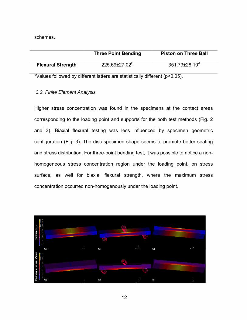
12
schemes.
Three Point Bending Piston on Three Ball
Flexural Strength 225.69±27.02B 351.73±28.10A
*Values followed by different latters are statistically different (p<0.05).
3.2. Finite Element Analysis
Higher stress concentration was found in the specimens at the contact areas
corresponding to the loading point and supports for the both test methods (Fig. 2
and 3). Biaxial flexural testing was less influenced by specimen geometric
configuration (Fig. 3). The disc specimen shape seems to promote better seating
and stress distribution. For three-point bending test, it was possible to notice a non-
homogeneous stress concentration region under the loading point, on stress
surface, as well for biaxial flexural strength, where the maximum stress
concentration occurred non-homogenously under the loading point.
13
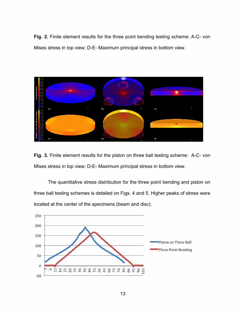
Fig. 2. Finite element results for the three point bending testing scheme: A-C- von
Mises stress in top view; D-E- Maximum principal stress in bottom view.
Fig. 3. Finite element results for the piston on three ball testing scheme: A-C- von
Mises stress in top view; D-E- Maximum principal stress in bottom view.
The quantitative stress distribution for the three point bending and piston on
three ball testing schemes is detailed on Figs. 4 and 5. Higher peaks of stress were
located at the center of the specimens (beam and disc).

14
Fig. 4. Graph plot of Maximum Principal Stress for three point bending and piston
on three ball testing schemes (*Values obtained from the central nodes at the
bottom of specimens).
Fig. 5. Graph plot of von Mises stress for three point bending and piston on three
ball testing schemes (*Values obtained from the central nodes at the bottom of
specimens).
4 Discussion
Flexural strength is defined as a material property of resisting stresses that
cause bending, without fracture (Dauvillier et al., 2000; Walker et al. 2006). This
property combines compression and tensile forces and can be measured by
uniaxial or biaxial tests (Suansuwan & Swain, 2001; Yilmaz et al., 2007; Pick et al.
2010).
15
Since this property is related to the elastic behavior, it is also composition-
dependent, being inherent to each material, and should not vary when the same
material is evaluated by different methods. However, this study showed significant
difference for the flexural strength values of the same ceramic material when
different testing methods (uniaxial and biaxial flexural schemes) were used. Thus,
the first hypothesis was rejected, since the flexural strength of the lithium
dissilicate-based ceramic was influenced by the test method.
Several factors can influence results in a bending test, as follows: structural
parameters, such as inclusion of voids and cracks (Huang & Hsue 2011), flaws on
surface or within volume, specimens dimension, gradients, and state voltage (Ban
& Anusavice, 1990). Furthermore, the effect of geometry (Alshehri 2011), shape
and size of specimen and test conditions (Jin et al., 2004) can strongly influence
the results of stress tests. Surface defects were identified as fracture initiation sites
in 86.6% to 96.6% of specimens (Rodrigues et al., 2008). The influence of many of
these factors can be eliminated or reduced by following the ISO 6872 (2008) for
three-point bending and biaxial flexural test in dental ceramics. This standard
describes exactly how specimens should be made, followed by accurate
measurement procedures, including necessary testing equipment (often illustrated
with diagrams), control of environmental parameters, sampling and data
processing (Della Bona et al., 2011).
Due to the difficulties in obtaining specimens without imperfections and to
perfectly perform any laboratory testing method, the use of standardizations should
lead to better control and less influence of technique for in vitro studies.

16
Finite element analysis showed differences for tensile stress distribution
between testing methods. As the stress distribution in the specimens was affected
by the testing scheme, the second hypothesis was also rejected. For the three-
point bending test, high tensile stress concentration was observed in large areas in
the opposite region to the point load, which is compatible with the characteristics of
this mechanical test. For the biaxial flexural test, tensile stresses were distributed
along the area between support points, with highest concentration peaks in the
opposite area to the load point, with no stress concentration on the edges, as
reported in other studies (Abu-Hassan et al, 1998; Fischer et al, 2008). Due to
these characteristics, biaxial flexural strength may produce less data variation
(Wen et al., 1999).
For three-point bending test specimens, the majority of fractures occurred in
the specimen center, at the area below loading, which is consistent with reports
from other studies (Fischer et al., 2008). For this test, all specimens showed
failures with two fracture fragments. However, for biaxial flexural strength test
specimens, due to stress distribution, fractures occurred in different directions,
resulting in two or three disc fragments. Thus, according to finite element analysis,
all failures occurred in regions where high stress concentrations were observed,
which shows a relation between the results obtained in laboratory testing with finite
elements analysis.
5 Conclusion

17
Within the limitations of this study, it was possible to conclude that: (I)
significant differences were found in the flexural strength for the same ceramic
material with the both testing methods evaluated; (II) all laboratory failures
occurred in regions in which high stress concentration was observed in the finite
element analysis.
References
Abu-Hassan MI, Abu-Hammad OA, Harrison A. Strains and tensile stress
distribution in loaded disc-shaped ceramic specimens. An FEA study. J Oral
Rehabil. 1998 Jul;25(7):490-5.
Alshehri SA. An investigation into the role of core porcelain thickness and
lamination in determining the flexural strength of In-Ceram dental materials. J
Prosthodont. 2011 Jun;20(4):261-6.
Ban S, Anusavice KJ. Influence of test method on failure stress of brittle dental
materials. J Dent Res. 1990 Dec;69(12):1791-9.
Cattell MJ, Knowler JC, Clarke RL, Lynch E. The biaxial flexural strength of two
pressable ceramic systems.J Dent 1999;27:183–96.
18
Dauvillier BS, Feilzer AJ, De Gee AJ, Davidson CL. Visco-elastic parameters of
dental restorative materials during setting. J Dent Res. 2000 Mar;79(3):818-23.
Della Bona A, Wozniak WT, Watts DC. International dental standards--order out of
chaos? Dent Mater. 2011 Jul;27(7):619-21.
Fischer J, Stawarczyk B, Hämmerle CH. Flexural strength of veneering ceramics
for zirconia. J Dent. 2008 May;36(5):316-21.
Huang CW, Hsueh CH. Piston-on-three-ball versus piston-on-ring in evaluating the
biaxial strength of dental ceramics. Dent Mater. 2011 Jun;27(6):e117-23. Epub
2011 Apr 2.
ISO 6872. Dentistry-dental ceramics. International Organization for
Standardization; 2008.
Jin J, Takahashi H, Iwasaki N. Effect of test method on flexural strength of recent
dental ceramics. Dent Mater J. 2004 Dec;23(4):490-6.
Pick B, Meira JB, Driemeier L, Braga RR. A critical view on biaxial and short- beam
uniaxial flexural strength tests applied to resin composites using Weibull,
fractographic and finite element analyses. Dent Mater. 2010 Jan;26(1):83-90.
19
Quinn GD, Morrell R. Design-data for engineering ceramics—a review of the
flexure test. J Am Ceram Soc 1991;74:2037–66.
Rizkalla AS, Jones DW. Mechanical properties of commercial high strength
ceramic core materials. Dent Mater 2004;20:207-12.
Rodrigues SA Jr, Ferracane JL, Della Bona A. Flexural strength and Weibull
analysis of a microhybrid and a nanofill composite evaluated by 3- and 4-point
bending tests. Dent Mater. 2008 Mar;24(3):426-31.
Soares CJ, Martins LR, Fonseca RB, Correr-Sobrinho L, Fernandes Neto AJ.
Influence of cavity preparation design on fracture resistance of posterior Leucite-
reinforced ceramic restorations. J Prosthet. Dent.2006;95(6):421-9.
Soares CJ, Soares PV, Santos-Filho PC, Armstrong SR. Micro tensile specimens
attachment and shape-finite element analysis. J Dent Res. 2008 Jan;87(1):89-93.
Soares PV, Santos-Filho PC, Martins LR, Soares CJ. Influence of restorative
technique on the biomechanical behavior of endodontically treated maxillary
premolars. Part I: fracture resistance and fracture mode. J Prosthet
Dent.2008;99(1):30-7.
20
Scherrer SS, Wiskott AH, Coto-Hunziker V, Belser UC. Monotonic flexure and
fatigue strength of composites for provisional and definitive restorations. J Prosthet
Dent 2003;89:579–88.
Scherrer SS, Quinn JB, Quinn GD, Kelly JR. Failure analysis of ceramic clinical
cases using qualitative fractography. Int J Prosthodont 2006;19:185–92.
Suansuwan N, Swain MV. Determination of elastic properties of metal alloys and
dental porcelains. J Oral Rehabil. 2001 Feb;28(2):133-9.
Thompson GA. Determining the slow crack growth parameter and Weibull two-
parameter estimates of bilaminate disks by constant displacement-rate flexural
testing.Dent Mater. 2004 Jan;20(1):51-62.
Walker MP, Haj-Ali R, Wang Y, Hunziker D, Williams KB. Influence of
environmental conditions on dental composite flexural properties. Dent Mater. 2006
Nov;22(11):1002-7.
Wen MY, Mueller HJ, Chai J, Wozniak WT. Comparative mechanical property
characterization of 3 all-ceramic core materials. Int J Prosthodont. 1999 Nov-
Dec;12(6):534-41.

21
Yilmaz H, Aydin C, Gul BE. Flexural strength and fracture toughness of dental core
ceramics. J Prosthet Dent. 2007 Aug;98(2):120-8.

22
CAPÍTULO II - PROPORTIONING THE BIAXIAL FLEXURAL TEST FOR
DENTAL CERAMICS EVALUATION: A FINITE ELEMENT ANALYSIS
SUMMARY
Numeric simulations using Finite Elements Analysis (FEA) have been widely
used in the biomedical industry and specifically in dental field in the last years.
Thus, the aim of this study was to evaluate the proportioning of the biaxial flexural
test for dental ceramic evaluation by finite element analysis. Three-dimensional
(3D) finite element models were generated and meshed using eight-node
hexahedral elements simulating the biaxial bending test according to ISO 6872
parameters for dimensions (100%) and the proportioning of the original test set up
to 75% and 50%. The loading piston and supporting spheres were considered
contacting surfaces and a 120 N load was applied. Ceramic discs assumed lithium-
disilicate mechanical properties (120 GPa and 0.23 Poisson’s ratio) and were
consideres homogeneous, linear-elastic and isotropic. Friction between contact
bodies and cylinder was considered negligible. Constraints were applied at the
edges of the ceramic disc. Static structural analysis considering non-linear contacts
was performed and results were analyzed using von Mises (VM), and Maximum
Principal stress (MPS). FEA showed higher stress concentration at the load point
and support contacting areas to the disc for all test dimensions. Similar stress
distribution pattern was verified for the different proportions used for the piston on
three ball test. According to von Misses and Maximum principal stress analysis,
reducing the dimensions of the biaxial flexural test proposed by ISO 6872 caused

23
increased stress concentration; however, stress distribution pattern at testing discs
was not influenced by the proportioned test designs. Thus, the dimensions of the
biaxial bending test set up can be reduced by proportioning without affecting
flexural strength results.
INTRODUCTION
Dental ceramics are brittle materials, thus, sensitive to tensile stresses.
Different test methods have been proposed to evaluate the mechanical properties
of ceramics. Test designs for evaluating the mechanical properties of monolithic
specimens and bond strength of bilayer specimens can be based on uniaxial
flexural tests, such as three-point bending (non-uniform central stress field) and
four-point bending (uniform central stress field), and on biaxial flexural tests
(reduced edge failures as compared with the two previous test designs) (1).
Three-point bending test is largely dependent on the surface roughness of
specimen. Therefore, strength measurement of brittle materials using biaxial
flexural conditions rather than uniaxial flexural is often considered more reliable,
because maximum tensile stresses occur within central loading area, and
unfinished edge failures are eliminated. This allows slightly uneven specimens to
be tested and results are not affected by surface conditions. Thus, biaxial flexural
test should produce less variation in data for strength determination on brittle
materials (2).

24
Due to the costs involved for in vivo studies, numeric simulations and in vitro
testing approaches are frequently used by scientists and manufacturers of
biomedical devices. Study design analysis is first accomplished on computer
and/or in simulated oral environment conditions previously to be tested in clinical
experiments. When best design or material has been refined, the actual
experiment may be conducted. Modeling and simulation step saves time and costs,
reducing risks from conducting the study, or clinical trial, in vivo (3).
Virtual prototyping (numeric simulation) using Finite Elements Analysis (FEA)
has been widely applied in biomedical industry for development, design,
engineering, testing, certificating and production of several materials. The use of
FEA in dental research has been significantly refined during last decade (4). Thus,
the aim of this study was to evaluate the proportioning of the biaxial flexural test for
dental ceramic evaluation by finite element analysis. The hypothesis to be tested
would be that the different dimensions of the biaxial flexural test would not alter the
stress distribution in the disc specimens.
MATERIALS AND METHODS
Strain and stresses under a given loading can be calculated by finite element
(FEA) analysis on the basis of specimen geometry, boundary conditions, and
material properties. Numeric methods such as FEA showed good correlations with
experimental methods. Three-dimensional (3D) finite element models were
25
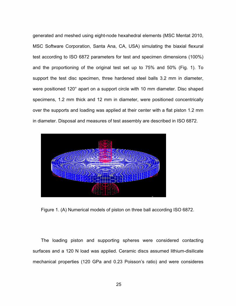
generated and meshed using eight-node hexahedral elements (MSC Mentat 2010,
MSC Software Corporation, Santa Ana, CA, USA) simulating the biaxial flexural
test according to ISO 6872 parameters for test and specimen dimensions (100%)
and the proportioning of the original test set up to 75% and 50% (Fig. 1). To
support the test disc specimen, three hardened steel balls 3.2 mm in diameter,
were positioned 120° apart on a support circle with 10 mm diameter. Disc shaped
specimens, 1.2 mm thick and 12 mm in diameter, were positioned concentrically
over the supports and loading was applied at their center with a flat piston 1.2 mm
in diameter. Disposal and measures of test assembly are described in ISO 6872.
Figure 1. (A) Numerical models of piston on three ball according ISO 6872.
The loading piston and supporting spheres were considered contacting
surfaces and a 120 N load was applied. Ceramic discs assumed lithium-disilicate
mechanical properties (120 GPa and 0.23 Poisson’s ratio) and were consideres

26
homogeneous, linear-elastic and isotropic (5). Friction between contact bodies and
cylinder was considered negligible. Constraints were applied at the edges of the
ceramic disc. Static structural analysis considering non-linear contacts was
performed (MSC Marc 2010, MSC Software Corporation). Qualitative and nodal
quantitative results were analyzed using using von Mises (VM) and Maximum
Principal stress (MPS).
RESULTS
FEA showed higher stress concentration at load point and support contacting
areas to the disc for all test dimensions. Similar stress distribution pattern was
verified for the different proportions used for the piston on three ball test scheme.
According to von Misses and Maximum principal stress analysis, reducing the
dimensions of the biaxial flexural test proposed by ISO 6872 caused increased
stress concentration; however, stress distribution pattern at testing discs was not
influenced by the proportioned test designs (Figs. 2-4).
Graph plots were obtained from the stress verified in the nodes located at the
bottom of the finite element models considering the biaxial flexural test dimensions
of ISO 6872 in 100%, 75% e 50%). Von Mises stress values are detailed in Figure
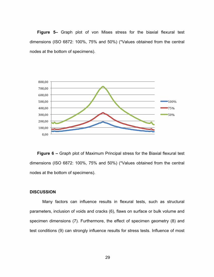
5. Models proportioned to 75% and 50% of the original ISO 6872 dimensions
exhibited higher peaks of stress near the point load. ISO 6872 model (100%)
showed lower stress concentration on the disc than the other models, with lower
stresses at loading point. Despite the different stress peaks, the stress distribution

27
pattern was similar for the three models. The detailed Maximum Principal Stress
showed compression stress at the loaded surface and tensile stress at opposite
area for all biaxial flexural test dimensions (Fig. 6).
Figure 2. Finite element results for piston on three-ball test according to ISO
6872 original dimensions (100%): VM- von Mises stress in top view (a-c); MPS -
Maximum principal stress in bottom view (d-f).
Figure 3. Finite element results for piston on three-ball test proportioned to
28
75% of ISO 6872 original dimensions: VM- von Mises stress in top view (a-c); MPS
- Maximum principal stress in bottom view (d-f).
Figure 4. Finite element results for piston on three-ball test proportioned to
50% of ISO 6872 original dimensions: VM- von Mises stress in top view (a-c); MPS
- Maximum principal stress in bottom view (d-f).
29
Figure 5– Graph plot of von Mises stress for the biaxial flexural test
dimensions (ISO 6872: 100%, 75% and 50%) (*Values obtained from the central
nodes at the bottom of specimens).
Figure 6 – Graph plot of Maximum Principal stress for the Biaxial flexural test
dimensions (ISO 6872: 100%, 75% and 50%) (*Values obtained from the central
nodes at the bottom of specimens).
DISCUSSION
Many factors can influence results in flexural tests, such as structural
parameters, inclusion of voids and cracks (6), flaws on surface or bulk volume and
specimen dimensions (7). Furthermore, the effect of specimen geometry (8) and
test conditions (9) can strongly influence results for stress tests. Influence of most

30
of these factors can be reduced, or eliminated, by following ISO 6872 standards
(10).
For biaxial flexural test it is necessary to be careful during specimen
preparation in order to not introduce defects that are not present in regular clinical
situations (11). Thus, those types of tests are extremely dependent on surface
polishing and specimen edges (12). According to Yilmaz, (13) it is impossible to
eliminate all defects during specimen fabrication, which may cause several
variations in flexural strength values.
Intending to minimize interferences from internal flaws inherent from the
processing of brittle materials, especially ceramics, the present study proposed to
reduce the dimensions of ISO 6872 biaxial flexural load/support devices and
specimens by proportioning them. This fact would increase the accuracy of test
results, since this approach can reduce intrinsic flaws in the specimens.
Additionally, this reduction would decrease the costs to produce ceramic
specimens. By proportioning ISO 6872 original dimensions (100%) to 75% and
50% there was no difference among groups regarding stress direction and
distribution. Specimen proportioning did not influence the test also, resulting in a
biaxial bending moment, with higher stress accumulation in specimen center
because of the same load applied for all models (120 N). The proportioned
specimens showed compressive stresses in the region contacting with the loading
point and tensile stress in the area opposed to the load application, as it would be
expect for biaxial flexural test, not influencing the test (Figs 2, 3 and 4 d-e).
31
However, model proportioning changed stress concentration Thus, the
hypotheses was rejected. Stress values at the testing discs were influenced by the
different test dimensions. This could be explained by the fact that the dimensions
were proportioned, but, the loading was not altered among the models. In order to
make load proportioning, laboratory tests should generate data to support FEA.
Obtaining adequate mechanical properties that accurately characterize
materials behavior is essential for appropriate knowledge of dental materials. This
fact evidences the importance in providing suitable properties, which will be
decisive to results accuracy achieved with FEA method. Therefore, interaction
between laboratory and computational methodologies seems very important for
understanding biomechanical behavior of dental structures and restorative
materials (14).
The research design of this in silico study shows some intrinsic limitations,
such as using numeric analysis, only. However, our findings are in accordance to
previous investigations for biaxial flexural test. Future studies with experimental
assessments which may overcome these limitations would be of benefit.
Within the limitations of this study, finite element analysis showed that the
dimensions of the biaxial bending test set up can be reduced following
proportioning without affecting flexural strength results.
32
REFERENCES
1. Lin WS, Ercoli C, Feng C, Morton D. The effect of core material, veneering
porcelain, and fabrication technique on the biaxial flexural strength and weibull
analysis of selected dental ceramics. J Prosthodont. 2012 Jul;21(5):353-62.
2. Huang CW, Hsueh CH. Piston-on-three-ball versus piston-on-ring in evaluating
the biaxial strength of dental ceramics. Dent Mater. 2011 Jun;27(6):e117-23. Epub
2011 Apr 2.
3. Magne P. Virtual prototyping of adhesively restored, endodontically treated
molars. J Prosthet Dent. 2010 Jun; 103(6):343-51.
4. Ausiello P, Apicella A, Davidson CL, Rengo S. 3D-finite element analyses of
cusp movements in a human upper premolar, restored with adhesive resin-based
composites. J Biomech 2001;34:1269-77.
5. DeHoff PH, Barrett AA, Lee RB, Anusavice KJ. Thermal compatibility of dental
ceramic systems using cylindrical and spherical geometries. Dent Mater 2008;
24:744-52.
6. Huang CW, Hsueh CH. Piston-on-three-ball versus piston-on-ring in evaluating
the biaxial strength of dental ceramics. Dent Mater. 2011 Jun;27(6):e117-23. Epub
2011 Apr 2.

33
7. Ban S, Anusavice KJ. Influence of test method on failure stress of brittle dental
materials. J Dent Res. 1990 Dec;69(12):1791-9.
8. Alshehri SA. An investigation into the role of core porcelain thickness and
lamination in determining the flexural strength of In-Ceram dental materials. J
Prosthodont. 2011 Jun;20(4):261-6.
9. Jin J, Takahashi H, Iwasaki N. Effect of test method on flexural strength of
recent dental ceramics. Dent Mater J. 2004 Dec;23(4):490-6.
10. ISO 6872. Dentistry-dental ceramics. International Organization for
Standardization; 2008.
11. Kelly JR. Perspectives on strength. Dent Mater 1995;11:103-10.
12. Zeng K, Oden A, Rowcliffe D: Flexure tests on dental ceramics. Int J
Prosthodont 1996;9:434-9.
13. Yilmaz H, Aydin C, Gul BE. Flexural strength and fracture toughness of dental
core ceramics. J Prosthet Dent. 2007 Aug;98(2):120-8.

34
14. Soares CJ, Soares PV, Santos-Filho PC, Armstrong SR. Micro tensile
specimens attachment and shape-finite element analysis. J Dent Res. 2008
Jan;87(1):89-93.
CONSIDERAÇÕES
Ensaios mecânicos têm sido aplicados na avaliação do comportamento
físico-mecânico dos materiais odontológicos, tais como as cerâmicas
odontológicas. Esses testes são essenciais para o estudo, desenvolvimento e
implementação dos materiais. Os testes mais comumente utilizados na
odontologia objetivam na verificar das propriedades mecânicas ou a
qualidade/resistência de união na avaliação dos materiais. Entretanto, muitos dos
testes utilizados para caracterização dos materiais restauradores não são
realizados nos padrões necessários, levando a resultados ambíguos para
materiais similares. A falta de parametrização dos ensaios mecânicos aplicados na
avaliação dos materiais odontológicos tem sido fator de frequentes investigações
como forma de se obter resultados laboratoriais mais consistentes. Possibilitando
assim, predizer o desempenho dos mesmos nas aplicações clínicas com maior
confiabilidade.
O presente estudo demonstrou que os testes de flexão empregados na
verificação de cerâmicas odontológicas têm suas configurações modificadas de tal
forma que resultados bastante divergentes podem ser obtidos para um mesmo

35
material. Algumas aplicações dos ensaios mecânicos podem mesmo exceder a
indicação destes, fazendo com que resultados pouco confiáveis sejam obtidos.
Assim, é indicado maior padronização dos ensaios mecânicos utilizados no teste
de materiais odontológicos, devendo estes, serem executados de acordo com as
normas adequadas de forma que os mesmos ofereçam resultados mais confiáveis
e que possuam maior validade clínica. A aplicação de novas metodologias é
encorajada no sentido de romper paradigmas que possam existir sobre a
caracterização dos materiais odontológicos.

36
CONCLUSÃO
Dentro das limitações do presente estudo in vitro e in silico, foi possível
concluir no capítulo 1: (I) foram encontradas diferenças significativas na
resistência à flexão para o mesmo material cerâmico com ambos os métodos de
ensaio avaliados; (II) todas as falhas laboratoriais ocorreram em regiões onde a
concentração tensão elevado foi observado na análise de elementos finitos. Já no
capítulo 2 pode se concluir: a análise por elementos finitos mostrou que o
dimensionamento do teste de flexão biaxial, configurado pode ser reduzido
proporcionalmente sem afetar os resultados de resistência à flexão.

37
REFERÊNCIAS
ANUSAVICE, K. J. Materiais dentários de Phillips. 11 ed. Rio de Janeiro:
Guanabara Koogan. 2005. 412p.
BOTTINO, M. A. ; QUINTAS, A. F.; MIYASHITA, E.; GIANNINI,V. Estética em
reabilitação oral metal-free. São Paulo: ArtesMédicas, 2001.
CATTELL MJ, KNOWLER JC, CLARKE RL, LYNCH E. The biaxial flexural
strength of two pressable ceramic systems.J Dent 1999;27:183–96.
CONRAD HJ, et al. Current ceramic materials and systems with clinical
recommendations: a systematic review. J ProsthetDent. 2007; 98(5): 389-404.
COSTA, J. L. V.; et al. O estágio atual das cerâmicas odontológicas. PCL, 2006, 8
(40): 193-198.
CRAIG, R. G.; POWERS, J.M. Materiais Dentários Restauradores. 11 ed. São
Paulo: Santos , 2004.
DIB, L. L.; SADDY, M. S. (Coord).Atualização na clínica odontológica: estética
e prótese. vol.3. São Paulo: ArtesMédicas, 2006.
38
GIORDANO R. A comparation of all-ceramic restorative systems.Part 1.Gen Dent.
1999;47(6):566-70.
GIORDANO, R. A. A coparison of all-ceramic restorative system, Part 2.Gen Dent,
Chicago, Jan-Feb 2000. 48(1): 38-45.
GOMES E. A. et al. Cerâmicas odontológicas: o estado atual. Cerâmica 54
(2008) 319-325
HUANG CW, HSUEH CH. Piston-on-three-ball versus piston-on-ring in evaluating
the biaxial strength of dental ceramics.Dent Mater. 2011 Jun;27(6):e117-23. Epub
2011 Apr 2.
ITINOCHE, M.K. Estudo da ciclagem mecânica na resistência à flexão de
cerâmicas. (Tese de Doutorado). São José dos Campos:Faculdade de
Odontologia de São José dos Campos da UNESP;2002.
LEHNER, C. et al. Six-year clinical results of leucite reinforced glass ceramic inlays
and onlays. Acta Med. Dent. Helv. 2000. 3: 137-46.
MEZZOMO, E.; SUZUKI, R. M. et al. Reabilitação Oral Contemporânea. São
Paulo: Santos, 2006.

39
PARREIRA, G. G.; SANTOS, L. M. Cerâmicas Odontológicas-conceitos e
técnicas. São Paulo: Santos, 2005.
REIS, R.S.; CASEMIRO,L.; SILVA,F. R. Sistema Cercon: Sistema de zircônia
frezada por computador para prótese “metal-free”. In:LAURIA DIB,L.; SADDY,M.S.
(Coord.) Atualização Clínica em Odontologia. 1 ed. São Paulo: Artes Médicas,
2006.
RESENDE, C. A. Tipos de cerâmicas odontológicas. In: Cerâmicas
odontológicas [monografia]. Piracicaba: Faculdade de Odontologia de Piracicaba
Unicamp; 2003..
RESKALLA, H.N.F.; CHAVES FILHO, H.D.M.; SALGADO, I.O.; CHAVES NETTO,
H.D.M.; PANZERI, H. Estudo sobre resistência da união de porcelana em ligas de
Ni-Cr com e sem Berílio e ligas experimentais com titânio. J BrasClinOdontolInt,
2005; 9 (50/51): 234 – 43.
SILVA, J.S.A. et al. Cerâmicas Odontológicas. In: LIMA, F. Prótese dentária:
Fundamentos e técnicas reabilitação oral para todos. Florianópolis/SC: Ed.
Ponto, 2010, p. 253-276
SOARES CJ, MARTINS LR, FONSECA RB, CORRER-SOBRINHO L,
FERNANDES NETO AJ. Influence of cavity preparation design on fracture
40
resistance of posterior Leucite-reinforced ceramic restorations. J
ProsthetDent.2006;95(6):421-9.
SOARES PV, SANTOS-FILHO PC, MARTINS LR, SOARES CJ. Influence of
restorative technique on the biomechanical behavior of endodontically treated
maxillary premolars. Part I: fracture resistance and fracture mode. J Prosthet
Dent.2008;99(1):30-7.
SOARES PV, SANTOS-FILHO PC, QUEIROZ EC, ARAÚJO TC, CAMPOS RE,
ARAÚJO CA,SOARES CJ. Fracture resistance and stress distribution in
endodontically treated maxillary premolars restored with composite resin. J
Prosthodont. 2008 Feb;17(2):114-9.(a)
SOARES CJ, SOARES PV, SANTOS-FILHO PC, Armstrong SR.
Microtensilespecimes attachment and shape-finite element analysis. J Dent Res.
2008 Jan;87(1):89-93. (b).
SOARES CJ, SANTANA FR, CASTRO CG, SANTOS-FILHO PC, SOARES PV,
QianF,Armstrong SR. Finite element analysis and bond strength of a glass post
tointraradicular dentin: comparison between microtensile and push-out tests. Dent
Mater. 2008 Oct;24(10):1405-11. (c)
41
SOBRINHO, L.C.; BORGES, G.A.;SINHORETI,M.A.C.;CONSANI,S. Materiais
cerâmicos Cap.6. In: MIYASHITA,E.; FONSECA,A.S (Coord.). Odontologia
Estética: o estado da arte. São Paulo: ArtesMédicas, 2004.
THOMPSON GA. Determining the slow crack growth parameter and Weibull two-
parameter estimates of bilaminate disks by constant displacement-rate flexural
testing.Dent Mater. 2004 Jan;20(1):51-62.