atividade antimicobacteriana, toxicidade aguda...
TRANSCRIPT
1
UNIVERSIDADE FEDERAL DA GRANDE DOURADOS
ATIVIDADE ANTIMICOBACTERIANA, TOXICIDADE
AGUDA, GENOTOXICIDADE E MUTAGENICIDADE DA
FLAVONA (2-FENIL-4H-1-BENZOPIRANO-4-ONA) EM
CAMUNDONGOS
VANESSA VILAMAIOR DE SOUZA
DOURADOS MS
2014

2
VANESSA VILAMAIOR DE SOUZA
ATIVIDADE ANTIMICOBACTERIANA, TOXICIDADE
AGUDA, GENOTOXICIDADE E MUTAGENICIDADE DA
FLAVONA (2-FENIL-4H-1-BENZOPIRANO-4-ONA) EM
CAMUNDONGOS
Dissertação apresentada à Universidade
Federal da Grande Dourados – Faculdade
de Ciências da Saúde, para obtenção do
Título de Mestre em Ciências da Saúde.
Orientador: Dr. Julio Henrique Rosa Croda
DOURADOS MS
2014
3
Agradeço
A Deus, ter me dado forças, luz e sabedoria.
Aos meus pais Elis Regina Sousa e Antonio Alves pelos momentos de plenitude e apoio
familiar incondicionais. A Vocês, minha eterna gratidão.
Aos meus irmãos Alexsandra e Geandro pelo incentivo e pela presença sempre constante
em minha vida. Gostaria de agradecer especialmente minha irmã por toda a ajuda,
incentivo, dedicação e paciência.
A professora Drª Candida Kassuya por toda ajuda e paciência desde o início do mestrado.
Sou imensamente grata por tudo.
Aos professores doutores: Candida Kassuya, Fábio Negrão, Anelise Samara e Alexéia
Barufatti, pela composição da banca de defesa.
Ao professor Dr. Julio Henrique pela orientação e ajuda.
Ao professor Dr. Rodrigo Juliano pela disposição em ajudar, por abrir as portas de seu
laboratório.
Aos meus companheiros nessa caminhada que me auxiliaram quando necessário Flávio
Henrique Cláudia Berno e Roberto. A vocês, muito obrigada pelo acolhimento, ajuda e
paciência.
Aos meus amigos e colegas de mestrado Wesley Barbieri, Karine Sartori, Ana Claudia
Piccinelli e Giseli Traesel obrigada pelo incentivo, pelos momentos de descontração e
ajuda.
Aos técnicos do LPCS e da FCS que me ajudaram com experimentos e reagentes Flora
Martinez, Mariana Tatara, Lujan Sanabria Débora Brait, Priscilla Ely
ii

4
Dedico aos meus pais Elis Regina e Antônio
Aos meus irmãos Alexsandra e Geandro
A minha sobrinha Isabella
iii

5
Sumário
1 INTRODUÇÃO ............................................................................................................. 1
2 REVISÃO DE LITERATURA .................................................................................... 3
2.1 Tuberculose: Doença e epidemiologia .................................................................. 3
2.2 Tratamento ............................................................................................................. 4
2.3 Resistência micobacteriana ................................................................................... 6
2.4 Novas alternativas terapêuticas para o tratamento da tuberculose ..................... 7
2.5 Produtos naturais com atividade antimicobacteriana .......................................... 8
2.5.1 Flavonóides ...................................................................................................... 8
2.5.2 Flavona ........................................................................................................... 10
2.5.2.1 Propriedades químicas.....................................................................10
2.5.2.2 Atividades biológicas........................................................................11
2.6 Testes in vitro ......................................................................................................... 12
2.7 Toxicologia de produtos naturais ........................................................................ 13
2.8 Considerações sobre a genotoxicidade ................................................................. 14
3 OBJETIVOS ............................................................................................................... 16
3.1 Objetivo Geral....................................................................................................16
3.2 Objetivos Específicos.........................................................................................16
4 REFERÊNCIAS BIBLIOGRÁFICAS ...................................................................... 17
5 ANEXO I ..................................................................................................................... 23
5.1 Artigo Científico .................................................................................................... 23
ANEXO II ...........................................................................................................................44
ANEXO III .........................................................................................................................63
iv
6
Resumo
Flavonóides são compostos polifenólicos encontrados em algumas plantas medicinais,
estudos científicos demonstram que, alguns flavonóides tem atividades anti-inflamatória,
anticarcinogênica e antioxidante. A flavona (2-fenil-4H-1-benzopirano-4-ona) é um
flavonóide com atividade de estimulação cardíaca, antibacteriana, antialérgica e de inibição
ou estimulação de enzimas. Dessa forma, esse trabalho avaliou os efeitos
antimicobacterianos da flavona in vitro sobre a cepa H37Rv ATCC 27294. Foi realizado
também os testes de toxicidade aguda, genotoxicidade e mutagenicidade com a flavona in
vivo. A concentração inibitória mínima (CIM) da flavona foi mensurada nas concentrações
de 0.98 a 250 µg/mL. Modelos experimentais de toxicidade aguda, genotoxicidade e
mutagenicidade foram desenvolvidos em camundongos fêmea por via oral, sendo a flavona
administrada por gavagem no ensaio de toxicidade aguda nas doses de 175, 560, 1792 e 2000
mg/Kg-1 e em 175, 560, 1792 mg/Kg1 nos ensaios de genotoxicidade e mutagenicidade.
Foram observados sinais hipocráticos bem como, foram efetuadas análises bioquímicas,
hematológicas, genotóxicas (ensaio cometa, micronúcleo) e mutagênicas (fagocitose). A
flavona apresentou atividade antimicobacteriana de CIM = 31,25 µg/mL. Nos dados obtidos
verificou-se aumento no recrutamento de macrófagos para o tecido esplênico houve uma
média de 55% de células fagocitadas, quando comparado ao controle negativo com 37% de
células fagocitadas, estes dados demonstram que a flavona poderia desempenhar atividade
imunoestimulatória. Nos testes in vivo, não foram observados sinais de toxicidade e nem
alterações nos parâmetros bioquímicos, hematológicos, genotóxicos e mutagênicos. A
análise macroscópica de órgãos como: rins, fígado e pulmões não apresentou nenhuma
diferença significativa entre os grupos. Desta forma, o presente estudo demonstra a flavona
apresenta atividade antimicobacteriana in vitro (eficácia contra Mycobacterium
tuberculosis). Além disso, a flavona não é tóxica nos modelos realizados nesse trabalho.
Esses resultados juntos levam a demonstração da eficácia da flavona contra micobactérias e
a segurança de um composto presente na natureza.
Palavras-chave: Atividade antimicobacteriana, flavona, toxicidade, mutagenicidade
ii

7
Abstract
Flavonoids are polyphenolic compounds found in medicinal plants. Scientific studies show
that some flavonoids have anti-inflammatory, anticarcinogenic and antioxidant activities.
The flavone (2-phenyl-4H-1-benzopyran-4-ona) is a flavonoid with cardiac, antibacterial
and antiallergic stimulating activity as well as inhibition or stimulation of enzymes. Thus,
this study evaluated the antimycobacterial effects of flavone in vitro on the strain H37Rv
ATCC 27294. Tests of acute toxicity, genotoxicity and mutagenicity with flavone in vivo
were also performed. The minimum inhibitory concentration (MIC) of flavone was
measured at concentrations of 0.98 to 250 µg/mL. Experimental models of acute toxicity,
genotoxicity and mutagenicity were developed in female mice and they were orally
administered with a flavone by gavage in acute toxicity test at doses of 175, 560, 1792 and
2000 mg/ Kg-1 and 175, 560, 1792 mg/ Kg-1 in the tests of genotoxicity and mutagenicity.
Hippocratic signals were observed as well as biochemistry, hematology, genotoxic (comet
assay, micronucleus) and mutagenic (phagocytosis) analysis were performed. The flavone
showed antimicrobial activity of MIC = 31.25 µg/mL. In the data there was an increase in
the recruitment of macrophages into the splenic tissue was an average 55% phagocytosis of
cells compared to the negative control in 37% phagocytosed cells, these data demonstrate
that the flavone could play immunostimulatory activity. In the in vivo tests, no signs of
toxicity and no changes in biochemical, hematological, genotoxic and mutagenic parameters
were observed. The macroscopic analysis of organs such as kidneys, liver and lungs showed
no significant difference between groups. Thus, this study demonstrates for the flavone has
antimicrobial activity in vitro (efficacy against Mycobacterium tuberculosis). In addition,
the flavone is not toxic models performed in this work. These results together lead to
demonstration of efficacy of flavone against mycobacteria and the safety of a compound
present in nature.
Key words: Antimycobacterial activity, tuberculosis, flavone, toxicity, mutagenicity.
iii

8
Listas de siglas e abreviaturas
ANVISA - Agência Nacional de Vigilância Sanitária
AIDS - Síndrome de Imunodeficiência Adquirida
CEUA - Comitê de Ética em Uso de Animais
CIM - Concentração Inibitória Mínima
COBEA - Colégio Brasileiro em Experimentação Animal
E - Etambutol
Et - Etionamida
HIV - Vírus da Imunodeficiência Adquirida
I - Isoniazida
OMS - Organização Mundial da Saúde
R - Rifampicina
REDOX - Redução-Oxidação
REMA – Resazurin Reduction Microtiter Assay
S - Estreptomicina
SUS - Sistema Único de Saúde
TB - Tuberculose
MDR - Cepas Multidroga Resistentes
MTT – Dimethyl Thiazolyl Diphenyl Tetrazolium Salt
XDR - Cepas Extremamente Resistentes
UFC - Unidade Formadora de Colônia
Z - Pirazinamida
V
9
1 INTRODUÇÃO
A tuberculose (TB) é uma doença crônica infecciosa transmissível, causada pela bactéria
de morfologia bacilar, Mycobacterium tuberculosis também conhecido como Bacilo de
Koch. Vários aspectos devem ser considerados para a manutenção da TB em nosso meio
destacando-se, a posição socioeconômica desfavorável, comorbidade de tuberculose
HIV/AIDS, etilismo, diabetes, tabagismo, dependência química, dificuldade de acesso aos
serviços de saúde, o envelhecimento da população, a urbanização desordenada e processos
migratórios contribuíram de maneira significativa para que a doença ainda hoje não tenha
sido erradicada [1,2].
A forma pulmonar da TB é a mais comum, o indivíduo pode adquiri-la a medida que
entra em contato com o bacilo em suspensão emitido por um paciente portador. Cerca de um
terço da população mundial está infectada com o M. tuberculosis, e a chance de desenvolver
a forma ativa da doença é de 10% durante a vida [3].
A TB está concentrada em um grupo de 22 países, no ano de 2012 foi registrado um
total de 8,6 milhões de pessoas que desenvolveram a doença, deste montante, 1,3 milhão
evoluiu para óbito [4]. No mesmo período, no Brasil, foram notificados 71.230 casos, a
taxa de incidência foi de, 36.7/100.000 habitantes, colocando o Brasil na 17ª posição entre
os países com alta carga da doença [5,6].
O tratamento da TB é prolongado, dura no mínimo seis meses. O uso inadequado e o
abando do tratamento podem resultar no surgimento de cepas monorresistentes,
caracterizadas pela resistência do bacilo a pelo menos um medicamento utilizado para o
tratamento e cepas multidroga resistentes (MDR) que são resistentes às drogas isoniazida e
rifampicina. A resistência é agravada quando as cepas se tornam resistentes a pelo menos
rifampicina e isoniazida, uma quinolona (ofloxacina, levofloxacina, moxifloxacina) e um
medicamento injetável de segunda linha (amicacina, canamicina, capreomicina , são então
consideradas cepas extremamente resistentes (XDR) [7,8].
A elevação das taxas de incidência da TB e o surgimento de cepas resistentes ocasionou
a necessidade da realização de novos ensaios de triagem, bem como, a síntese de novos
fármacos e testes de susceptibilidade. Dessa forma, a indústria farmacêutica procura a
implementação de terapias medicamentosas que sejam eficientes tanto, na redução do
período de tratamento quanto na eliminação de infecções persistentes. [9]
2
Dentro dos compostos utilizados como base para as novas formulações, as pesquisas
apontam para a utilização de plantas medicinais, as quais, são fonte de compostos como os
metabólitos secundários (fontes potencias de drogas) contidos em extratos e óleos essenciais
de importância terapêutica [10].
O conhecimento popular é responsável por propagar dentro das comunidades as
propriedades terapêuticas das plantas, e é a partir das indicações populares que vários
estudos químicos e farmacológicos têm início [11] Nos últimos anos, a pesquisa sobre
plantas medicinais tem atraído à atenção em todo o mundo por serem fonte de substâncias
químicas que são potenciais substratos para síntese de novas drogas [12].
Assim, se faz necessária à busca por drogas eficazes no tratamento de diversas doenças
sendo necessários estudos acerca da segurança e eficácia de seu uso. A partir da pesquisa
realizada pelo nosso grupo de pesquisa, o qual extraímos fase metanólica das folhas de
Annona sylvatica (CIM = 184,33 µg/mL). A fração FAE (Fração Acetado de etila), resultante
do fracionamento, teve CIM = 115,2 µg/mL. Detectou-se ainda a CIM do composto isolado
luteolina (236,8 µg/mL) a qual pertence a classe das flavonas, em sua estrutra ocorre a
ligação de hidroxilas nas posições C3, C4, C5 e C7
O presente estudo teve como finalidade verificar a atividade antimicobacteriana in vitro
de flavona sem nenhum radical de hidroxila (2-fenil-4H-1-benzopirano-4-ona) e análise
toxicológica in vivo através dos testes de toxicidade aguda, genotoxicidade e
mutagenicidade.
3
2. REVISÃO DE LITERATURA
2.1 Tuberculose: Doença e epidemiologia
A infecção tuberculosa é endêmica, causada pelo Mycobacterium tuberculosis ou
bacilo de Koch pode afetar praticamente todos os órgãos, mas tem especial predisposição
pelos pulmões, o que provavelmente se deva, a alta oxigenação do órgão e de o bacilo ser
aeróbio estrito. A evolução ocorre em ciclos lentos, podendo apresentar as mais diversas
complicações [13, 14, 15].
A partir do momento que o bacilo adentra ao organismo, rapidamente acomete o
pulmão que forma como resposta tubérculos e corpos cavernosos. Quando o indivíduo
infectado tosse, espirra ou fala, expectora milhares de gotículas (1 a 10 µm de diâmetro) no
ar, cada uma contém em média um quarto de bacilos, que podem se manter suspensos no ar
por várias horas, podendo assim, serem aspirados e contaminarem outras pessoas. As vias
aéreas são a principal forma de propagação da doença [16, 17]. Outro meio de contágio é, a
inoculação direta que ocorre através da pele lesionada, é mais comum em patologistas ou
outros profissionais de laboratórios que manipulam tecidos infectados [18].
Após atingir os alvéolos pulmonares, os bacilos sofrem a ação do sistema
imunológico. A resposta imunológica inata desencadea uma resposta inflamatória que
envolve os macrófagos alveolares locais e o recrutamento de neutrófilos e monócitos,
aumento progressivo de linfócitos T e B, muitas vezes a infecção poderá ser eliminda por
esse mecanismo [19].
Os macrófagos, neutrófilos e monócitos circundam os macrófagos infectados,
células dentríticas e fibroblastos, formando o característico granuloma tuberculoso. O
granuloma é o principal mecanismo que limita a disseminação da micobactéria, criando
assim a interação da resposta entre os linfócitos T e os macrófagos ativados pelo infeferon-
, os quais impedem a multiplicação do Mycobacterium tuberculosis, assim a doença pode
apresentar longos períodos de latência, e reativar quando ocorrer algum desequilíbrio da
reposta do sistema imunológico frente ao bacilo [20].
O risco de doença pulmonar ativa é baixo após uma exposição ao organismo, mas
aumenta sob condições de estresse ou em um ambiente confinado no qual ocorrem
4
exposições repetidas [21, 22]. Segundo estimativas, 1/3 da população mundial está infectada
pelo M. tuberculosis, desse total 5 a 10% irá desenvolver a doença. Pacientes co-infectados
TB/HIV existe um incremento de 10% ao ano de desenvolvimento de doença ativa. Nesses
casos está recomendado o tratamento da tuberculose latente [24].
Os fatores que contribuem para o desenvolvimento da doença podem estar associados
ao ambiente, ao hospedeiro (idade, sexo, estado nutricional, imunológico e doenças
intercorrentes) e a linhagem do M. tuberculosis [25, 26].
Ainda podem predispor o desenvolvimento da doença fatores como: o abuso de
drogas injetáveis, a infecção recente nos últimos 2 anos, a silicose, o diabetes mellitus, a
gastrectomia, o uso prolongado com corticosteroide, a doença renal em estágio avançado, as
síndromes de mal absorção crônicas, ou baixo peso corporal (10% ou mais de peso abaixo
do ideal) e o etilismo [27,28].
Estima-se que, no ano de 2012, ocorreram 8,6 milhões de casos incidentes, dos quais
400.000 seriam pacientes coinfectados com HIV e 1,3 milhão de óbitos entre pacientes não
portadores de HIV [29, 30]. No mesmo período, no Brasil, foram notificadas 71.230 pessoas,
a taxa de incidência foi de aproximadamente de 36.7/ 100.000 habitantes e uma taxa de cura
de 69,2% e a de abandono11,9% [31, 32].
Os gastos financeiros são relevantes, visto que, a tuberculose dentre as doenças
infecciosas, é a nona causa de internação e ocupa o vigésimo sétimo lugar em gastos com
internação no Sistema Único de Saúde (SUS), e é a quarta causa de mortalidade no Brasil
[5]. Estima-se que entre 2013 – 2015 serão necessários US$ 8 bilhões por ano nos países de
renda média e baixa para o tratamento e controle da tuberculose, desse montante US$ 5
bilhões serão destinados para o tratamento da tuberculose comum, US$ 2 bilhões para o
tratamento de MDR-TB e XDR-TB e US$ 1 bilhão para o tratamento de pacientes com
HIV/TB.
2.2 Tratamento
A descoberta de novas drogas para o tratamento da tuberculose se iniciou no final
dos anos de 1940, nesse período houve a descoberta da estreptomicina (monoterapia). Com
o surgimento de cepas resistentes fez-se necessário a descoberta de fármacos, como ácido
para-aminossalicílico (PAS), isoniazida, pirazinamida, Etionamida, etambutol e
capreomicina [33].

5
Atualmente a terapia é baseada em: Rifampicina (R), Isoniazida (I), Pirazinamida
(Z), Etambutol (E), Estreptomicina (S) e Etionamida (Et) - via oral, cujos os mecanismos
estão descritos no quadro 1. A forma de tratamento é baseada em casos novos, falência
terapêutica e recidiva: Esquema I (Básico), 2RHZ / 4RH, recomendado para todas as formas
de tuberculose pulmonar e extrapulmonar. Esquema II, 2 RHZ/7RH, recomendado para a
forma meningo encefálica da tuberculose; Esquema III (reforçado), 2RHZE/4RHE,
recomendado nos casos de recidiva após cura ou retorno após abandono; esquema IV,
3SZEEt/9EEt, recomendado nos casos de falência de tratamento. A duração do tratamento é
caracterizada no esquema I (6 meses), esquema II (9 meses), esquema III (6 meses) e
esquema IV (12 meses) [28].
Quando ocorre falência no esquema terapêutico básico, é proposto um esquema
constituído por estreptomicina, etambutol, terizidona, pirazinamida e uma quinolona
(levofloxacina ou ofloxacina). Na impossibilidade de se utilizar a estreptomicina, esta é
substituída por amicacina. Em 2009 o etambutol foi incluído no esquema de tratamento, mais
especificamente, na fase intensiva (engloba os dois primeiros meses) com o objetivo de
diminuir a transmissibilidade e evitar o uso da rifampicina neste momento, para assim
minimizar a resistência [34,35,36].
Quadro 1 . Mecanismo de ação dos principais antimicobacterianos [37]
Medicamento Mecanismo de ação
Rifampicina Liga de forma irreversível ao RNA-polimerase DNA-
dependente, impedindo a produção de RNA e a síntese de
proteínas
Isoniazida Quelação de íons cobre essenciais para a célula bacteriana;
interfere também na enzima micolase-sintetase, importante
na sínte de ácido micólico
Etambutol Inibição da síntese de ácido nucleicos da célula bacteriana
Pirazinamida Provavelmente semelhante a isoniazida
Estreptomicina Se liga de forma irreversível ao ribossomo bacteriano,
produzindo bloqueio ou alterações profundas na síntese de
proteínas
Etionamida Age na enzima nicotinamida adenina-dinucleotídeo
6
2.3 Resistência micobacteriana
Uma preocupação recente, é a resistência dos bacilos frente ao tratamento, a
resistência pode ser classificada como: monorresistente - o bacilo se torna resistente a pelo
menos um medicamento utilizado para o tratamento ou polirresistente - o bacilo se torna
resistente a mais de um medicamento, mas não à combinação de isoniazida e rifampicina.
As cepas MDR apresentam resistência a rifampicina, isoniazida, enquanto que, as
cepas XDR apresentam resistência a pelo menos rifampicina e isoniazida, uma quinolona
(ofloxacina, levofloxacina, moxifloxacina) e um medicamento injetável de segunda linha
(amicacina, canamicina, capreomicina). [7,35].
As formas XDR-TB e MDR- TB não respondem ao tratamento preconizado de seis
meses com drogas de primeira linha, podendo levar dois anos ou mais para tratar com drogas
menos eficazes, mais tóxicas e com custo mais elevado. Para o tratamento da forma MDR -
falência do esquema básico e resistência à rifampicina e isoniazida, a terapia é composta por:
Estreptomicina (S), etambutol (E), ofloxacina (O), pirazinamida (Z) e terizidona (T) [30].
A resistência do patógeno causador da tuberculose aos medicamentos utilizados no
tratamento, constitui uma barreira para a obtenção do sucesso do esquema terapêutico
adotado. Pode ocorrer por conta de um contato prévio do M. tuberculosis com o
medicamento ou a falha do uso da medicação (principal fator para a formação da resistência).
O contato inicial bacilo-fármaco ocorre por conta da administração de esquemas terapêuticos
inadequados, irregularidade na administração do medicamento e o controle insatisfatório do
indivíduo durante o tratamento [38,39].
Aproximadamente 3,7% dos pacientes portadores de tuberculose, possuem a forma
resistente da doença. Em 2012, cerca de 450 mil pessoas teriam sido diagnosticadas com
MDR-TB, o que corresponde a um aumento de 42% em relação ao ano anterior. A maioria
dos casos esteve concentrado na China, Índia e Rússia. Embora os casos de XDR sejam mais
raros, estudos apontam que aproximadamente 9,6% de MDR possuem características de
XDR-TB [30].
Em 2013, no Brasil foram notificados 148 casos novos de monorresistência, 50 de
polirresistência, 525 de multirresistência e 21 de resistência extensiva [40]. O aumento das
taxas de incidência de MDR-TB e XDR-TB tem impulsionado a busca de novas alternativas
farmacológicas efetivas que possam reduzir o período de tratamento, bem como minimizar
as reações adversas causadas pela terapia.

7
2.4 Novas alternativas terapêuticas para o tratamento da tuberculose
Planta medicinal é qualquer espécie de planta que contenha substâncias que podem
ser utilizadas para fins terapêuticos ou ainda possua princípios ativos que sirvam como
precursores da síntese de novos medicamentos [41]. As plantas e, por conseguinte os
produtos naturais são utilizados para fins medicinais. Atualmente os fitoterápicos são
responsáveis por cerca de 40% dos fármacos disponíveis no mercado, sendo 70%
antimicrobianos e antitumorais [42,43].
A busca de novos fármacos menos tóxicos e mais ativos para o tratamento da
tuberculose tem estimulado os pesquisadores a investigar novos compostos para o tratamento
desta doença [44]. As propriedades terapêuticas dos produtos naturais vem sendo alvo de
estudo há décadas, mas principalmente desde a descoberta e industrialização da aspirina e
penicilina. Outros exemplos clássicos de plantas medicinais utilizadas como fármacos são:
o ácido acetilsalicílico (aspirina) proveniente da planta Salix alba L. analgésico, antitérmico,
anti-inflamatório e antiagregante plaquetário, a vincristina e a vimblastina da planta
Catharanthus roseus utilizadas no tratamento de alguns tipos de câncer e ainda a digoxina e
a digitoxina, potentes glicosídeos cardiotônicos extraídos de Digitalis purpúrea L. e a D.
lanata, respectivamente [45].
As plantas produzem metabólitos secundários que possuem características químicas
variadas e são encontrados em grupos - famílias ou gêneros - de plantas, os produtos do
metabolismo secundário constituem os chamados “produtos naturais”. Adicionalmente são
utilizados em escala industrial para produção de inseticidas, corantes, flavorizantes e
medicamentos [46].
A diversidade química das plantas permite o isolamento de metabólitos
farmacologicamente significativos, através de vias metabólicas secundárias elas produzem
alcalóides, flavonóides, isoflavonóides, cumarinas, taninos, glicosídeos, poliacetilenos,
terpenos e óleos, que, por vezes, são específicos a determinadas famílias, gêneros ou
espécies. Estes complexos químicos podem estar presentes em diferentes partes da planta,
por isso, é importante pesquisar a planta em sua totalidade, a fim de, identificar quais locais
possuem maiores concentrações de ativos [47,48].

8
2.5 Plantas e produtos naturais com atividade antimicobacteriana
As propriedades antimicobacterianas de plantas medicinais estão sendo cada vez
mais analisadas em diferentes partes do mundo. Estudos anteriores demonstram sua
atividade contra M. tuberculosis, como Clavija procera B. Stahl, S.
aintabensis e T. sibthorpii, que apresentaram atividade contra estirpes resistentes [49, 50].
Assim como outras plantas também aprsentaram atividade antimicobacteriana tais como:
Faurea saligna Harv, Parinari curatellifolia Planch ex Benth [52] Abelmoschus esculentus
Moench [51]. Aristolochia taliscana Gancho [53]; Securidaca longepedunculata Fres .;
Maerua edulis (Gilg & Gilg-Ben.) DeWolf [54] Acorus cálamo L. var. americanus [53].
A maioria dos medicamentos sintéticos que estão disponibilizados no mercado
possuem origem de produtos naturais [55]. Estima-se que 50% dos medicamentos utilizados
para o tratamento de infecções sejam oriundos de produtos naturais ou semi- sintéticos,
sendo 19,4% dos produtos naturais utilizados para síntese de medicamentos sintéticos [56].
Algumas flavonas já foram isoladas e testadas frente cepas virulentas de M.
tuberculosis, apresentando uma CIM considerável como: 3'-O-dimetoxi-5, 6,4 '-
trihiidroxiflavona (MIC 200 mcg / mL) cirsimaritin (MIC de 50 ng / mL), eupatilin (MIC de
50 ng / mL), salvigenin (MIC 100 mcg / ml), [9], 7 - metoxiflavona (MIC 12,5-50 ug / ml)
e 5,4 '-di-hidroxi-7-metoxiflavona (MIC 25-50 ug / mL) [57].
O nosso grupo de pesquisa da rede de pesquisa Pró Centro–Oeste verificou a
atividade do extrato bruto e frações e compostos da Annona sylvatica. A fração hexânica
apresentou atividade, assim foi possível isolar o composto ativo, a luteolina com uma MIC
de 236,89 mcg/mL [58].
A apigenina (4’,5,7-triidroxiflavona) foi testada a fim de inibir a hialuronidase,
utilizado como única fonte de carbono para a estirpe de M. tuberculosis H37Rv. Através
desse estudo, verificou-se que a apigenina inibiu a atividade da hialuronidase, havendo assim
a inibição da micobactéria [59]. Provavelmente, o mecanismo de ação da flavona ocorra
devido a essa inibição, uma vez que o efeito possa ser atribuído à ausência do substituinte
(por exemplo, metoxilo e hidroxilo) no anel A e B, fazendo com que não haja bloqueio
estérico efeito entre o grupo carbonilo na posição C-4 da flavona, fornecendo também uma
maior interação eletrostática, hidrofóbicas, ligações de hidrogênio com os receptores.
9
Fazendo com que haja ação proteínas e consequentemente inibindo algumas enzimas
bacterianas e interferindo nas suas vias de síntese [60].
2.5.1 Flavonóides
Entre a classe de compostos secundários presentes em plantas medicinais com
propriedades farmacológicas merece destaque os flavonóides (polifenóis), que são
compostos formados por um esqueleto de difenil propano (C6C3C6) e dois anéis benzênicos
ligados a um anel pirano, de forma livre (aglicona) ou ligados a açúcares (glicosídeos) [61]
(Figura. 1) A classe é dividida em mais de 10 diferentes subclasses dentre elas os flavonóis,
flavonas, isoflavonas, antocianinas e flavononas [62].
Os flavonóides estão distribuídos no reino vegetal e podem estar presentes em todas
as partes das plantas, desde as raízes até as flores e frutos. Distinguem-se entre si pela
coloração, vários representantes possuem a cor amarela, e agem na atração de insetos para a
polinização de plantas [63].
Para que as diferentes classes de flavonóides sejam formadas, ocorrem modificações
estruturais, como: adição ou redução, metilação de grupos hidroxila ou do núcleo dos
flavonóides, hidroxilação, dimerização (biflavonóides), glicosilação de grupos hidroxila (O-
glicosídeos) ou em algum núcleo carbônico (C-glicosídeos) [64].
Os flavonóides têm atividade antioxidante, devido à capacidade de estabilizar
radicais livres e espécies reativas de oxigênio, essas propriedades se devem aos grupos
hidroxilas ligadas à estrutura do anel aromático. A ação antioxidante pode ser potencializada
com a adição de grupos hidroxilas, caso ocorra ligação a glicosídeos pode ocorrer à redução
da atividade antioxidante. Estes fatores contribuem para a deslocalização de elétrons nos
núcleos aromáticos, concedendo a estabilidade do radical que passa a não ter energia
suficiente para reagir [65].
Os flavonóides possuem atividade anti- hemolítica, anticarcinogênica, peroxidação
lipídica, formação de radicais superóxido e apoptose celular [66,67,68]. Além disso, têm
propriedades de modulação do reparo do DNA, antialérgicas, antimicrobianas,
vasoprotetoras, antitumorais e anti-inflamatórias [69,70,71,72,73].
10
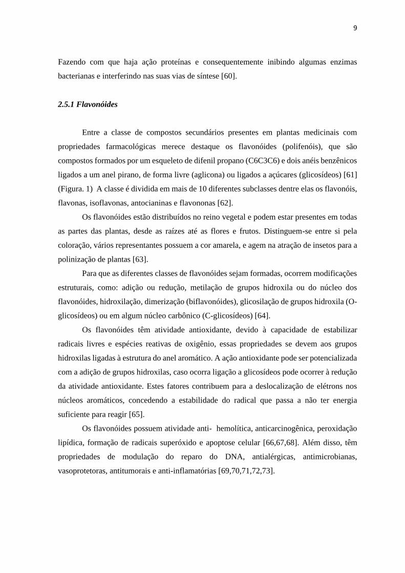
Figura 1. Estrutura química dos flavonóides [64].
2.5.2 Flavona
2.5.2.1 Propriedades químicas
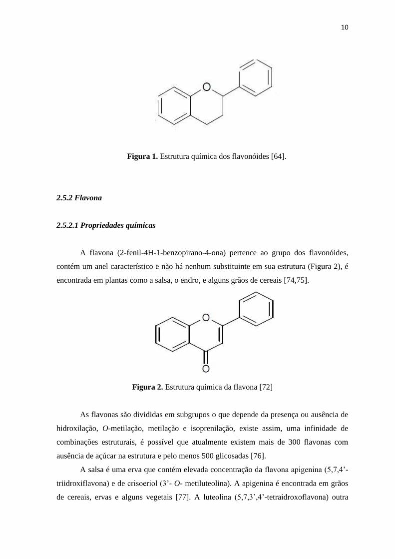
A flavona (2-fenil-4H-1-benzopirano-4-ona) pertence ao grupo dos flavonóides,
contém um anel característico e não há nenhum substituinte em sua estrutura (Figura 2), é
encontrada em plantas como a salsa, o endro, e alguns grãos de cereais [74,75].
Figura 2. Estrutura química da flavona [72]
As flavonas são divididas em subgrupos o que depende da presença ou ausência de
hidroxilação, O-metilação, metilação e isoprenilação, existe assim, uma infinidade de
combinações estruturais, é possível que atualmente existem mais de 300 flavonas com
ausência de açúcar na estrutura e pelo menos 500 glicosadas [76].
A salsa é uma erva que contém elevada concentração da flavona apigenina (5,7,4’-
triidroxiflavona) e de crisoeriol (3’- O- metiluteolina). A apigenina é encontrada em grãos
de cereais, ervas e alguns vegetais [77]. A luteolina (5,7,3’,4’-tetraidroxoflavona) outra
11
flavona encontrada em abundância está presente no brócolis, cenoura e cebola. A pimenta
vermelha e o aipo são as maiores fontes tanto de apigenina quanto de luteolina [78,79]..
Algumas flavonas metoxiladas conferem sabor amargo a frutas cítricas, como a
nobiletina (5,6,7,8,3’,4’- hematoxiflavona), sinesetina (5,6,7,3’,4’- pentametoxiflavona),
tangeritina (5,6,7,8,4’- pentametoxiflavona) [80].
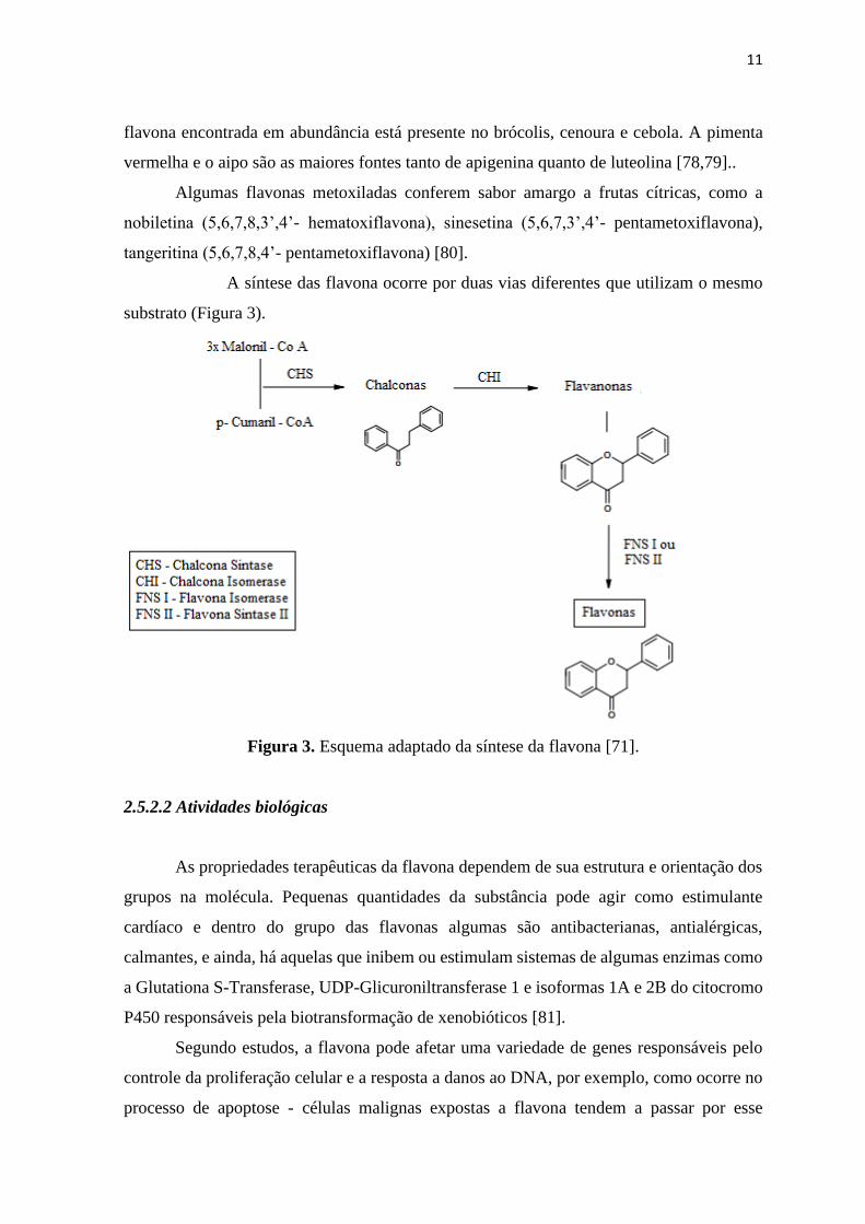
A síntese das flavona ocorre por duas vias diferentes que utilizam o mesmo
substrato (Figura 3).
Figura 3. Esquema adaptado da síntese da flavona [71].
2.5.2.2 Atividades biológicas
As propriedades terapêuticas da flavona dependem de sua estrutura e orientação dos
grupos na molécula. Pequenas quantidades da substância pode agir como estimulante
cardíaco e dentro do grupo das flavonas algumas são antibacterianas, antialérgicas,
calmantes, e ainda, há aquelas que inibem ou estimulam sistemas de algumas enzimas como
a Glutationa S-Transferase, UDP-Glicuroniltransferase 1 e isoformas 1A e 2B do citocromo
P450 responsáveis pela biotransformação de xenobióticos [81].
Segundo estudos, a flavona pode afetar uma variedade de genes responsáveis pelo
controle da proliferação celular e a resposta a danos ao DNA, por exemplo, como ocorre no
processo de apoptose - células malignas expostas a flavona tendem a passar por esse
12
processo. Células normais quando lesionadas podem recuperar sua capacidade proliferativa
quando expostas a esse metabólito [82]
O potencial terapêutico da flavona faz desta, um possível alvo para aplicação na área
farmacêutica. 65 Estudos epidemiológicos sugerem que, uma alta ingestão de flavona pode
estar associada a uma redução de alguns tipos de câncer como, por exemplo, câncer do
pulmão e do cólon, além da inflamação crônica e da osteoporose [83,84,85].
Células neurais com estresse oxidativo tiveram maior sobrevida quando expostas ao
extrato de Scutellaria baicalensis (solidéu-de-baicak) que apresenta em sua composição
quatro flavonas: wogonia (5,7-driidroxi-8-metoxiflavona), baicaleína (5,6,7-
triidroxiflavona), skullpflavona I (5,2’-diidroxi-7,8-dimetoxiflavona), skullpflavona II (5,6’-
diidroxi-6,7,8,2’-tetrametoxiflavona), nestes estudos as flavonas teriam apresentado o efeito
de sequestro direto de espécies reativas de oxigênio [86].
A epigenina, luteolina, nobiletina e a tangeritina, tem atividades antimutagênica e
inibem o crescimento de algumas células cancerosas (carcinoma de células escamosas) [78].
As flavonas epigenina e a luteolina atuam inibindo a atividade da proteína de
resistência a multidroga em 47 e 53% respectivamente, a epigenina na dose de 80 µl mol. L-
1 inibem alguns tipos de câncer como o de cólon, pele, tireóide e células de leucemia e ainda
pode prevenir e tratar a doença proliferativa prostática. A epigenina inibe a topoisomerase I
e a luteolina promove apoptose em células malignas a medida que causa danos ao DNA
[78,79,80,81].
2.6 Testes in vitro
Novas metodologias que visam a triagem da viabilidade celular tem sido
implementadas, as metodologias mais modernas são REMA (Resazurin Reduction
Microtiter Assay) e MTT (dimethyl thiazolyl diphenyl tetrazolium salt) dentre outros,
diferem-se da turbidimetria e da contagem de UFC (Unidade formadora de colônia) por
serem mais rápidas, terem um custo menor e um alto rendimento [87].
Para o ensaio do REMA é utilizada como substância reveladora a Resazurina, que
tem potencial REDOX (Óxido-Redução), com mudança colorimétrica é um indicador de
fluorescência que indica o metabolismo celular. A análise de proliferação de células serve
para avaliar a viabilidade celular e auxilia na descoberta de drogas [88].
13
O corante resazurina é um indicador de viabilidade celular que utiliza o poder redutor
natural de células vivas para converter resazurina para a molécula fluorescente, resorufina. A
resazurina é um composto não-tóxico, permeável que é de cor azul e não fluorescente. Ao
entrar em contatos com as células é reduzida a resorufina, que produz fluorescência
vermelho brilhante. As células viáveis convertem a resazurina em resorufina, gerando,
assim, uma medida quantitativa da viabilidade e citotoxicidade [86]. Assim foi possível
determinar a concentração inibitória mínima (CIM) da flavona frente ao M. tuberculosis
H37Rv ATCC 27294.
O teste MTT é utilizado para micobactérias e avalia a ação antimicobacteriana de
compostos isolados frente a uma cepa padronizada, e determina a quantidade de cepas
viáveis. Nesta técnica o azul de tetrazólio liga-se a enzimas desidrogenases mitocondriais
formando um substrato cromogênico o qual é um indicador de oxi-redução [87].
2.7 Toxicologia de produtos naturais
As plantas medicinais são utilizadas para o tratamento de enfermidades mesmo
havendo fármacos sintéticos utilizados para essas mesmas doenças. Mas, assim como ocorre
com as drogas sintéticas é importante cautela na utilização destas plantas, uma vez que,
podem ser dotadas de substâncias tóxicas que podem levar a morte [26].
Alguns dos efeitos tóxicos de substâncias presentes em plantas são: i) hepatotóxicos
como apiol, safrol, lignanas e alcaloides pirrolizidínicos; ii) a ação tóxica renal que pode ser
provocada por terpenos e saponinas e ii) algumas dermatites ocasionadas por espécies com
abundância de lactonas sesquiterpênicas. Um exemplo é o confrei (Symphytum officinale L.)
tradicionalmente utilizado pela população com cicatrizante por conta da alantoína presente,
contudo, a plantas também apresenta alcaloides pirrolizidínicos que além de hepatotóxicos
são comprovadamente carcinogênicos [48].
Os óleos essenciais obtidos das plantas medicinais também podem ter propriedades
tóxicas, alguns provenientes de frutos cítricos têm alto índice de defurano cumarinas em sua
constituição e como consequência possuem atividade fotossensibilizante, os óleos da canela,
funcho e alho tem um nível alto de cinamaldeído e podem causar reação alérgica de contato,
o óleo da noz-moscada pode ocasionar alucinações o que se deve provavelmente pela
presença da miristicina e da elemicina [88].

14
No Brasil os estudos de toxicidade pré-clínica para fitoterápicos são normatizados
pela Resolução N° 90/04 da Agência Nacional de Vigilância Sanitária que é baseada nas
normas preconizadas pela OMS que por sua vez recomenda que sejam realizados estudos de
toxicidade aguda e de genotoxicidade quando houver uso contínuo e prolongado do
medicamento em humanos [20, 89,90].
Testes in vivo são importantes para que seja feita a observação dos efeitos dos
extratos das plantas nos modelos animais utilizados. Destaca-se, o modelo utilizado de
toxicidade aguda. Os testes de toxicidade aguda visam estabelecer um estudo que determina
qual é a espécie mais sensível e o índice de letalidade, os extratos, compostos ou frações são
administrados em uma ou várias doses em um período de 24 horas [91] A maioria dos estudos
pré-clínicos de produtos sintéticos ou naturais envolve a utilização de parâmetros
bioquímicos, hematológicos e anatomopatológicos [92].
2.8 Genotoxicidade e mutagenicidade
Os agentes genotóxicos apresentam a capacidade de interagir com o DNA, formando
alterações oxidativas ou rompendo a fita de DNA, o que compromete sua replicação e a
transmissão genética. Normalmente a lesão é reparada pelo próprio organismo ou as células
são eliminadas, quando a infecção é persistente, pode ocorre mutação (alterações
hereditárias), que pode se perpetuar nas células filhas durante o processo de replicação,
nesses casos o agente causador é denominado mutagênico [93].
Os ensaios de genotoxicidade desempenham um papel significativo na síntese de
novos fármacos, devem ser efetuados nas fases iniciais, a fim de presumir uma possível
atividade genotóxica e/ou carcinogênica e para subsidiar na obtenção de novas estruturas
químicas com menor atividade tóxica [94,95]. Os ensaios de genotoxicidade in vivo
detectam a genotoxicidade e a potencial carcinogenicidade de agentes químicos ou físicos.
Esses danos podem ser avaliados através dos ensaios: Cometa, micronúcleo, apoptose e
fagocitose.
O ensaio cometa é realizado em microgel, é empregado no processo de eletroforese
e quantificação através da detecção de quebras das fitas do DNA, em células individuais,
usando microscopia. A sensibilidade da versão alcalina é maior, pois possibilita a expressão
máxima dos danos em fitas simples [96].

15
O teste de micronúcleo visa a quantificação de fragmentos cromossômicos ou de
cromossomos inteiros que não estão acoplados ao grupamento de cromossomos de uma
célula - provendo, um pequeno núcleo individual, chamado micronúcleo (MN). A análise
detecta aberrações cromossômicas em organismos eucarióticos, aplicada na detecção de
agentes que prejudicam tanto o processo de ligação dos cromossomos às microfibrilas do
fuso como aqueles que induzem quebras cromossômicas [97]
No processo de fagocitose, os corpos apoptóticos são retirados do tecido por
macrófagos, esta sinalização ocorre devido a translocação da fosfatidilserina do lado interno
para o lado externo da membrana “marcando” as células que deverão ser fagocitadas, a
leitura do teste é feita de maneira visual através da leitura de lâminas coradas
especificamente [98,99]
Além do mecanismo de “marcação” outro mecanismo pode ser atribuído para o
processo de fagocitose como por exemplo, o aumento da quantidade sérica de plaquetas,
estas, aderem as áreas lesionadas. Quando ligadas, as plaquetas têm sua estrutura modificada
fazendo que haja a expressão de fosfolipídios (carga negativa) e receptores de glicoproteínas,
assim, ocorre a liberação de mediadores químicos, tais como, o tromboxano (aglomeração
de plaquetas) [100]. Durante a ativação das plaquetas inúmeros fatores quimiotáticos são
liberados (fator de agregação plaquetária), necessários para o crescimento e reparação,
recrutando células do tecido, tais como macrófagos [100].
O processo de fagocitose do M. tuberculosis por macrófagos alveolares
desencadeiam fatores imunopatológicos da tuberculose. O macrófago possui a capacidade
de fagocitar micobactéria e elimina-la pela circulação sanguínea ou linfática, porém se não
o fizer, esta multiplica-se intracelularmente. Desencadeando lesão pulmonar ou áreas
secundárias [101].

16
3. OBJETIVOS
3.1 Objetivo Geral
Avaliar o efeito antimicobacteriano in vitro e os efeitos toxicológicos da exposição
aguda da flavona por meio de modelos experimentais in vivo.
3.2 Objetivos Específicos
Avaliar o efeito antimicobacteriano in vitro.
Avaliar a toxicidade sistêmica provocada pela exposição aguda da flavona, através
da análise de sinais clínicos de toxicidade; parâmetros bioquímicos e hematológicos;
Avaliar a genotoxicidade e mutagenicidade “in vivo” da flavona em camundongos
através do ensaio cometa, teste do micronúcleo e fagocitose.

17
4 REFERÊNCIAS BIBLIOGRÁFICAS
[1] Bignall JR (1971) Tuberculosis in England and Wales in the next 20 years. Postgrad
Med J 47: 759-762.
[2] BarretoII ML (2009) Características dos serviços de saúde associadas à adesão ao
tratamento da tuberculose. Rev Saúde Públ 43: 998-1005.
[3] Barbosa IR, Costa ÍDCC (2014) Estudo epidemiológico da coinfecção tuberculose-
hiv no nordeste do brasil. Rev Patol Trop 43: 27-38.
[4] WHO, WorldHealth Organization, 2013. Multidrug-resistant tuberculosis – 2013.
Update. WHO.
[5] Brasil, Ministério da Saúde, 2013. Secretaria de Vigilância Epidemiológica em
Saúde. Departamento de Vigilância Epidemiológica. Situação da Tuberculose no
Brasil –PNCT.Brasília: Ministério da Saúde. Brasília.
[6] SINAN, Sistema de Informação de Agravos de Notificação, 2014. Tuberculose -
Casos confirmados notificados no Sistema de Informação de Agravos de
Notificação – Sinan Net, 2014. Disponível em: <
http://dtr2004.saude.gov.br/sinanweb/tabnet/dh?sinannet/tuberculose/bases/tuberc
brnet.Def >. Acesso em: 18.fev.2014.
[7] Dalcolmo MP, Andrade MK.N, Picon PD (2007) Tuberculose multirresistente no
Brasil: histórico e medidas de controle. Rev Saúde Públ 41: 34-42.
[8] Arbex MA, Varella MCL, Siqueira HR, Mello FAF (2010) Drogas antituberculose:
interações medicamentosas, efeitos adversos e utilização em situações especiais -
parte 1: fármacos de primeira linha. J Bras Pneumol 36: 626-640.
[9] Warner DF, Mizrahi V (2004) Mycobacterial genetics in target validation. Drug
Discovery Today: Technologies 1: 93-98.
[10] Dolly G, Nidhi S, Sagar, B, Shweta R, Shisha A (2012) Ocimum
kilimandscharicum: A systematic review. J Drug Deliv Therapeut 2, 45-52.
[11] Matias EF, Alves EF, Santos BS, Sobral CE, Alencar FJV et al. (2013) Biological
activities and chemical characterization of Cordia verbenacea DC. as tool to
validate the ethnobiological usage. Evid. Based Complement. Alternat.
Med.164215: 164-215.
[12] Bellik Y, Boukraâ L, Alzahrani HA, Bakhotmah AB, Abdellah F, et al. (2012)
Molecular mechanism underlying anti-inflammatory and anti-allergic activities of
phytochemicals: an update. Molecules. 18: 322-35.
[13] Pellenz DC, Silva CMD, Seixas LA, Silva FC (2011) Avaliação dos efeitos
genotóxicos dos fármacos tuberculostáticos em células bucais de pacientes em
tratamento. Rev Cien Fac Edu Mei Amb 2: 2-4.
[14] Demachki NTT, Serruya T, Pontes CDN (2013) Relato de caso: tuberculose
extrapulmonar de localização laríngea. Anais do CBMFC 12: 929.
[15] Rodrigues AR, Guimarães J, Vieira C, Alves M, Vilarinho S, et al. (2013) Otite
média tuberculosa: A propósito de um caso clínico. Arq Med 27: 144-147.
[16] Carvalho E, Meglin SR, Valim AR, Possuelo LG, Becker D (2011) Tuberculose no
presídio regional de Santa Cruz do Sul. Anais do Salão de Ensino e de Extensão.
220.

18
[17] Araujo KMFA, Figueiredo TMRM, Gomes LCF, Pinto ML, Silva TC, et al. (2013)
Evolução da distribuição espacial dos casos novos de tuberculose no município de
Patos (PB), 2001-2010. Cad Saúde Col 21: 296-302.
[18] Campos WR, Campos GS, Miranda SS. Tuberculose intraocular. Rev Bras
Oftalmol 70: 437-451.
[19] Capone D, Jansen JM, Lopes AJ, Soares MO, Pinto RS (2006) Diagnóstico
radiográfico e tomográfico da tuberculose pulmonar. Rev. Hosp. Univ. Pedro
Ernesto 5, 46-5.
[20] Turolla MS, Nascimento ES (2006) Informações toxicológicas de alguns
fitoterápicos utilizados no Brasil. Braz J Pharm Sci 42: 289-306.
[21] San Pedro A, Oliveira RM (2013) Tuberculose e indicadores socioeconômicos:
revisão sistemática da literatura. Rev Panam Salud Publ 33: 294-301.
[22] Selig L, Geluda K, Junqueira T, Brito R (2012) A tuberculose no cotidiano médico
eo efeito bumerangue do abandono. Cien Saúde Colet 17: 113-122.
[23] WHO, World Health Organization, 2002. Report – Global Tuberculosis Control:
Surveillance, Planning, Financing.
[24] Barbosa IR, Costa ÍCC (2014) Estudo epidemiológico da coinfecção tuberculose-
HIV no nordeste do Brasil. Rev Patol Trop 43: 27-38.
[25] Vicentin G, Santo AH, Carvalho MS (2002) Mortalidade por tuberculose e
indicadores sociais no município do Rio de Janeiro. Ciênc Saúde Colet 7: 253-26.
[26] Ferreira VF, Pinto AC (2010) A Fitoterapia no mundo atual. Quím. Nova 33: 1829-
1829.
[27] Brasil. Ministério da Saúde, 2005. Secretaria de Vigilância em Saúde. Tuberculose.
In: Guia de vigilância epidemiológica. Ministério da Saúde, Brasília, pp. 732.
[28] Brasil, Ministério da Saúde, 2002. Manual técnico para o controle da tuberculose:
Cadernos de atenção básica. Série A. Normas e Manuais Técnicos n. 148, Brasília,
pp. 70.
[29] Lönnroth K, Kenneth GC, Chakaya JM, Chauhan LS, Floyd K (2010) Tuberculosis
control and elimination 2010-50: cure, care and social development. Lancet 375:
1814–29.
[30] WHO, WorldHealth Organization, 2013. Multidrug-resistant tuberculosis – 2013.
Update. WHO.
[31] Brasil, Ministério da Saúde, 2013. Secretaria de Vigilância Epidemiológica em
Saúde. Departamento de Vigilância Epidemiológica. Situação da Tuberculose no
Brasil –PNCT.Brasília: Ministério da Saúde. Brasília.
[32] SINAN, Sistema de Informação de Agravos de Notificação, 2014. Tuberculose -
Casos confirmados notificados no Sistema de Informação de Agravos de
Notificação – Sinan Net, 2014. Disponível em: <
http://dtr2004.saude.gov.br/sinanweb/tabnet/dh?sinannet/tuberculose/bases/tuberc
brnet.Def >. Acesso em: 18.fev.2014.
[33] Mitchison DA (1990) Infectivity of patients with pulmonary tuberculosis during
chemotherapy. Eur Respir J 3: 385-386.
[34] Selig L, Geluda K, Junqueira T, Brito R, Trajman A (2012) A tuberculose no
cotidiano médico eo efeito bumerangue do abandono. Cien Saúde Colet 17: 113-122.

19
[35] Kritski AL (2010) Emergência de tuberculose resistente: renovado desafio. J Bras
Pneumol 36: 157-158.
[36] Ruffino-Netto A (2001) Programa de controle da tuberculose no Brasil: situação
atual e novas perspectivas. Inf Epidemiol SUS 10: 129-138.
[37] Bisaglia, JB, Santussi WM, Guedes AGM, Gomes, AP, Oliveira PC, et al. (2003).
Atualização terapêutica da tuberculose: principais efeitos adversos dos fármacos.
Bol Pneumol Sanit 11: 53-59.
[38] Conde MB, Melo FAF, Maques A MC, Cardoso NC, Pinheiro VGF, et al. (2009)
III Diretrizes para tuberculose da sociedade brasileira de pneumologia e tisiologia.
J Bras Pneumol 35: 1018-1048.
[39] Pablos-Mendez A, Gowda DK, Frieden TR (2002) Controlling multidrug-resistant
tuberculosis and access to expensive drugs: a rational framework. Bull. World
Health. Organ 80: 489- 500.
[40] Fraga, ACP, Torrens AW, Lobo AP, Dantas, David CA, et al. (2014) O controle da
tuberculose no Brasil: avanços, inovações e desafios. Bol Epidemiol 44: 2-13.
[41] Mitchison DA. Antimicrobial therapy of tuberculosis: justification for currently
recommended treatment regimens, Seminars in respiratory and critical care
medicine. Thieme Medical Publishers, New York, 2004; p. 307-315.
[42] WHO, World Health Organization, 2009. Report: Global Tuberculosis Control:
Epidemiology, Srategy, Financing.
[43] Farnsworth NR, Akerele O, Bingel AS, Soejarto DD, Guo Z (1985) Medicinal
plants in therapy. Bulletin of the world health organization 63: 965.
[44] Calixto, J.; Yunes, R., Plantas medicinais: Sob a ótica da química medicinal
moderna. Chapecó, 2001; p 500.
[45] Andrade FJL, Melo-Diniz MFF, Oliveira RAG (1997) Plantas que atuam no trato
respiratório. In: Simpósio de Plantas Medicinais do Brasil. Anais. UFC, Fortaleza.
[46] Newman DJ, Cragg GM (2007) Natural products as sources of new drugs over
the last 25 years. J Nat Prod 70: 461-477.
[47] Filho VC, Yunes RA, (1998) Estratégias para a obtenção de compostos
farmacologicamente ativos a partir de plantas medicinais: conceitos sobre
modificação estrutural para otimização da atividade. Quím. Nova 21: 99-105.
[48] Behling EB, Sendão MC, Francescato HDC, Antunes LMG, Bianchi MLP (2004)
Flavonoid quercetin: general aspects and biological actions. Alim Nutr 15: 285-
292.
[49] Rojas R, Caviedes L, Aponte JC, Vaisberg AJ, Lewis WH, Lamas G, Sarasara C,
Gilman RH, Hammond GB (2006) Aegicerin, the first oleanane triterpene with
wide-ranging antimycobacterial activity, isolated from Clavija procera. J Nat Prod
69 :845–854.
[50] Tülin A, Emmanuel MT, Fatih S, Seyma M, Hatice A (2013). Preliminary
antimycobacterial study on selected Turkish plants (Lamiaceae) against
Mycobacterium tuberculosis and search for some phenolic constituents. BMC
Complement Altern Med 13: 366-389.
[51] Chimponda T, Mukanganyama S (2010) Antimycobacterial activities of selected
medicinal plants from Zimbabwe against Mycobacterium aurum and
Corynebacterium glutamicum. Trop Biomed 27:595–610.

20
[52] Leon-Diaz R, Meckes M, Said-Fernandez S, Molina-Salinas GM, Vargas-Villarreal
J, et al. ( 2010) Antimycobacterial neolignans isolated from Aristolochia taliscana.
Mem Inst Oswaldo Cruz 105: 45–51.
[53] Webster D, Lee TD, Moore J, Manning T, Kunimoto D, et al. (2010)
Antimycobacterial screening of traditional medicinal plants using the microplate
resazurin assay. Can J Microbiol 56: 487–494.
[54] Luo X, Pires D, Ainsa JA, Gracia B, Mulhovo S, et al. (2011) Antimycobacterial
evaluation and preliminary phytochemical investigation of selected medicinal
plants traditionally used in Mozambique. J Ethnopharmacol 137:114– 120.
[55] Veiga-Junior VF, Pinto AC, Maciel MAM (2005) Plantas medicinais: cura segura?
Quím. Nova 28: 519-528.
[56] Pretto JB (2005) Potencial antimicrobiano de extratos, frações e compostos puros
obtidos de algumas plantas da flora catarinense.
[57] Castellar A, Coelho TS, Silva PEA, Ramos DF, Lourenço MCS, et al. (2011) A
atividade de flavonas e ácido oleanólico de Lippia lacunosa contra cepas de
Mycobacterium tuberculosis sensíveis e resistentes. Rev Bras Farmacogn 21: 835-
840.
[58] Araujo, RC, Neves, Formagio, AS, Kassuya, CA, Stefanello, ME, et al. (2014)
Evaluation of the anti-mycobacterium tuberculosis activity and in vivo
acutetoxicity of annona sylvatic. BMC Complemento Altern Med 14: 209.
[59] Hirayama Y, Yoshimura M, Ozeki Y, Sugawara I, Udagawa T, et al. (2009)
Mycobacteria exploit host hyaluronan for efficient extracellular replication 10:
1021- 1032.
[60] Dornas WC, Olivera T, Rodrigues-da Dores RG, Santos AF, Nagem TJ (2007)
Flavonóides:potencial terapêutico no estresse oxidativo. Rev Ciên Farm Básc
Aplic. 28: 241-249..
[61] Behling EB, Sendão MC, Francescato HDC, Antunes LMG, Bianchi MLP (2004)
Flavonoid quercetin: general aspects and biological actions. Alim Nutr 15: 285-
292.
[62] Arts IC, Hollman PC, (2005) Polyphenols and disease risk in epidemiologic studies.
Am J Clin Nutr 81: 317S-325S.
[63] Yao L, Jiang Y, Datta N, Singanusong R, Liu X, et al. (2004) HPLC analyses of
flavanols and phenolic acids in the fresh young shoots of tea (Camellia sinensis)
grown in Australia. Food Chemistry 84: 253-263.
[64] Balasundram N, Sundram K and Samman S (2006) Phenolic compounds in plants
and agri-industrial by-products: Antioxidant activity, occurrence, and potential
uses. Food Chemistry 99: 191-203.
[65] Galati G, Brien OPJ (2004) Potential toxicity of flavonoids and other dietary
phenolics: significance for their chemopreventive and anticancer properties. Free
Radic Biol Med 37: 287-303.
[66] Ündeğer Ü, Aydin S, Başaran AA (2004) The modulating effects of quercetin and
rutin on the mitomycin C induced DNA damage. Toxicol Lett 151: 143-149.
[67] Duthie S, Dobson V (1999) Dietary flavonoids protect human colonocyte DNA
from oxidative attack in vitro. Eur J Clin Nutr 38: 28-34.
[68] Lee JC, Klim J, Park JK., Chung GH, Jang YS (2003) The antioxidant, rather than
prooxidant, activities of quercetin on normal cells:: quercetin protects mouse
thymocytes from glucose oxidase-mediated apoptosis. Exp Cell Res 291: 386-397.

21
[69] Muanda FN, Dicko A, Soulimani R (2010) Assessment of polyphenolic
compounds, in vitro antioxidant and anti-inflammation properties of Securida
calongipedunculata root barks. C R Biol 333: 663-669.
[70] Erlund I (2004). Review of the flavonoids quercetin, hesperetin, and naringenin.
Dietary sources, bioactivities, bioavailability, and epidemiology. Nutr Re 24: 851-
874.
[71] Harbone JB. Handbook of natural flavonoids. Wiley, 1999; p 44.
[72] Martens S, Mithöfer A (2005) Flavones and flavone synthases. Phytochemistry.
66: 2399-2407.
[73] Miean KH, Mohamed S (2001) Flavonoid (myricetin, quercetin, kaempferol,
luteolin, and apigenin) content of edible tropical plants. J Agric Food Chem 49:
3106-3112.
[74] Choi J, Conrad CC, Malakowsky CA, Talent JM, Yuan CS, et al. (2002) Flavones
from Scutellaria baicalensis Georgi attenuate apoptosis and protein oxidation in
neuronal cell lines. Biochim Biophys Acta 1571: 201-210.
[75] Canivenc-Lavier MC, Vernevaut MF, Totis M, Siess MH, Magdalou J, et al. (1996)
Comparative effects of flavonoids and model inducers on drug-metabolizing
enzymes in rat liver. Toxicology 114: 19-27.
[76] Ullmannova V, Popescu NC (2007) Inhibition of cell proliferation, induction of
apoptosis, reactivation of DLC1, and modulation of other gene expression by
dietary flavone in breast cancer cell lines. Cancer Detect Prev 31: 110-118.
[77] Middleton E, Kandaswami C, Theoharides TC (2000) The effect of plant flavonoids
on mammalian cells: implications for inflammation, heart disease and cancer.
Pharmacol Rev 52:673-751.
[78] Kromhout D (2001) Diet and cardiovascular disease. J Nutr Hlth and Aging 5: 144-
149.
[79] Peterson J, Dwyer, J (1998) Flavonoids: Dietary occurrence and biochemical
activity. J. Nutr. Res 18: 1995-2018.
[80] Van Zanden JJ, Wortelboer HM, Bijlsma S, Punt A, Usta M, et al. (2005)
Quantitative structure activity relationship studies on the flavonoid mediated
inhibition of multidrug resistance proteins 1 and 2. Biochem Pharmacol 69: 699-
708.
[81] Fotsis T, Pepper MS, Montesano R, Aktas E, Breit S, et al. (1998) Phytoestrogens
and inhibition of angiogenesis. Baillieres Clin Endocrinol Metab 12: 649-666.
[82] Caltagirone S, Rossi C, Poggi A, Ranelletti FO, Natali PG, et al. (2000) Flavonoids
apigenin and quercetin inhibit melanoma growth and metastatic potential. Int J
Cancer 87: 595-600.
[83] Bektic J, Guggenberger R, Spengler B, Christoffel V, Pelzer A, et al. (2006) The
flavonoid apigenin inhibits the proliferation of prostatic stromal cells via the
MAPK-pathway and cell-cycle arrest in G1/S. Maturitas 55, Supplement 1: S37-
846.
[84] Collins L, Franzblau SG (1997) Microplate alamar blue assay versus BACTEC 460
system for high-throughput screening of compounds against Mycobacterium
tuberculosis and Mycobacterium avium. Antimicrob Agents Chemother 41: 1004-
1009.
[85] Palomino J.C, Martin A, Camacho M, Guerra H, Swings J, et al. (2002) Resazurin
microtier assay plate: simple and inexpensive method for deteccion of drug
resistance Mycobacterium tuberculosis. Antimicrob. Agents Chemother 46: 2720-
2722.
22
[86] Ahmed SA, Gogal RM, Jr. and Walsh JE (1994) A new rapid and simple non-
radioactive assay to monitor and determine the proliferation of lymphocytes: an
alternative to [3H]thymidine incorporation assay. J Immunol Methods 170: 211-
224.
[87] Mosmann T (1983) Rapid colorimetric assay for cellular growth and survival:
application to proliferation and cytotoxicity assays. J Immunol Methods 65: 55-
63.
[88] Adorjan B and Buchbauer G (2010) Biological properties of essential oils: an
updated review. Flavour and Fragrance Journal 25: 407-426.
[89] ANVISA, Agência Nacional de Vigilância Sanitária, 2004. Resolução- RE 90 Guia
para a realização de estudos de toxicidade pré-clínica de fitoterápicos. Disponível
em: http://www.diariodasleis.com.br/busca/exibelink.php?numlink=1-9-34-2004-
03-16-90. Data de acesso: 01 de junho de 2014.
[90] WHO, World Health Organization, 1997. Safety in Health-Care Laboratories. pp.
148.
[91] Brasil, Ministério da Saúde. Portaria nº 116, de 8 de agosto de 1996, DOU 12/08/96
– Dispõe sobre normas para estudo da toxicidade de produtos fitoterápicos.
Secretaria Nacional de Vigilância Sanitária.
[92] Oliveira LP, Pinheiro RC, Vieira MS, Paula JR, Bara MTF, et al. (2010) Atividade
citotóxica e antiangiogênica de Punica granatum L., Punicaceae. Rev Bras
Farmacogn 20: 201-207.
[93] Eastmond DA, Hartwig A, Anderson D, Anwar WA, Cimino MC, et al. (2009)
Mutagenicity testing for chemical risk assessment: update of the WHO/IPCS
Harmonized Scheme. Mutagenesis 24: 341-349.
[94] Gollapudi BB, Krishna G (2000) Practical aspects of mutagenicity testing strategy:
an industrial perspective. Mutat. Res 455, 21-28.
[95] Freitas T M, Celso FB, Silva TG, Picada JN (2013) Avaliação do efeito genotóxico
do aripiprazol em camundongos. Rev. Inicia. Cient. ULBRA 8: 21-29.
[96] Tice RR, Agurell E, Anderson D, Burlinson B, Hartmann A, etal. (2000. Single
cell gel/ Comet assay: guidelines for in vitro and in vivo genetic toxicology testing.
Mutagenesis 35: 206-221.
[97] Maluf SW, Passo DF, Bacelar A, Speit G, Erdtmann B (2001) Assessment of DNA
damage in lymphocytes of workers exposed to X-radiation using the micronucleus
test and the comet assay. Environ Mol Mutagen 38: 311-315.
[98] Fenech M (2000) The in vitro micronucleus technique. Mutat Research 455: 81–
95.
[99] Nicholson DW, Thornberry NA (1997) Caspases: killer proteases.Trends Biochem
Sci 22: 299 - 306.
[100] Rang HP, Dale MM, Ritter JM, Moore PK - Farmacologia - 5ª edição - 2003 -
Editora Guanabara Koogan S.A.
[101] Henry JB. Diagnósticos clínicos e tratamentos por métodos laboratoriais. 19.ed.
São Paulo: Manole, 1999.
23
5 ANEXO I
5.1 Scientific Article
Title: Evaluation of acute toxicity, antimicrobial activity, genotoxicity and
mutagenicity of flavone in adult female mice.
Vanessa Vilamaior de Souza1, Flávio Henrique Souza de Araújo3, Claudia Rodrigues
Berno3, Candida Aparecida Leite Kassuya1, Anelise Samara Nazari Formagio2,
Rodrigo Juliano Oliveira3, Flora Martinez Figueira Moreira1, Julio Henrique Rosa
Croda1*.
1 Faculty of Health Sciences, Federal University of Grande Gold, PO Box-322, Golden,
MS, Brazil
2 Faculty of Agricultural Sciences, Federal University of Grande Gold, PO Box-351,
Golden, MS, Brazil
3 Federal University of Mato Grosso do Sul (UFMS) - Campo Grande, Mato Grosso
do Sul, Brazil
*Corresponding author: Faculdade de Ciências da Saúde, Universidade Federal da Grande
Dourados, Dourados, 79825-070, MS, Brazil, Phone: +55 67 3410-2320 Fax: +55 67 3410-
2326. E-mail address: [email protected]

24
ABSTRACT: The compound flavone (2-phenyl-4H-1-benzopyran-4-ona) which belongs to
the group of flavonoids has as one of its therapeutic properties the ability to stimulate or to
inhibit the action of certain enzymes. This study aimed to evaluate the antimicrobial activity,
the acute toxicity as well as genotoxic and mutagenic actions of the flavone. Hence, we
performed MIC (minimum inhibitory concentration) and the acute toxicity test in female
Swiss mice at doses of 175 mg/ Kg-1, 560 mg/ Kg-1, 1792 mg/ Kg-1 and 2000 mg/ Kg-1,
which were administered in a single dose and at the end of 14 days the hematological and
biochemical patterns were measured as well as the comet assays, the micronucleus test and
phagocytosis. It was demonstrated that flavone has antimicrobial activity with MIC of 28.90
µg/mL. It has been observed that the treatment with flavone has no effect on weight gain,
food and water consumption nor does it cause any change to the hematological and
biochemical profiles. These results suggest that flavone has antimycobacterial activity. The
data demonstrated absence of acute toxicity after a single-dose treatment. It was observed
that in the comet and micronucleus assays flavone did not exert mutagenic and genotoxic
activities. No changes were observed in the phagocytosis test nor in the hematological and
biochemical analyzes. Thus, it can be inferred that flavone has no mutagenic nor genotoxic
activity in the experimental model used.
Key words: Flavone, acute toxicity, antimycobacterial activity, mutagenicity, genotoxicity.
25
INTRODUCTION
Flavonoids (polyphenolic substances) are secondary metabolites active of plants, derived
from the condensation of one molecule of cinnamic acid with three groups malonyl-CoA
participants, forming a chalcone (C15), precursor of all flavonoids, which are subdivided
into subclasses depending on the substitution and on the level of oxidation made on the C-
ring [1]. Individual differences present in each subclass are result in variation in the number
and arrangement of hydroxyl groups, as well as the nature and quantity of alkylations and/or
glycosylation of these groups. Many of them are presented in the form glycosides, being the
most common site C-3 and C-7 less frequent.
Flavone, an important subclass of flavonoids, characterized by aglicon with
two phenolic rings A and B and a C ring with the presence of a carbonyl group at the 4-
position and a double bond between positions 2 and 3 and no hydroxyl group at C-3 [2].
Studies have demonstrated that the presence of substituents in rings A and B could
lead to more potent compounds with reduced toxicity. Numerous studies of flavones
derivatives report a large spectrum of important pharmacological properties, including
antimycobacterial and anticarcinogenics effects [3].
Our previous studies have identified that a hexane fraction of Annona sylvatica was
active against M. Tuberculosis with a MIC of 115.2 mµ / mL [17]. Chromatographic studies
showed the presence of two flavonoids, luteolin and quercetin. The luteolin is part of the
class of flavones, which have numerous therapeutic properties, such as antimicrobial,
antiinflammatory, antioxidant, immunomodulatory, among others [17].
Flavones have been an indispensable anchor for the development of new therapeutic
agents. The majority of metabolic diseases are speculated to originate from oxidative stress,
and it is therefore significant that recent studies have shown the positive effect of flavones
on diseases related to oxidative stress. Due to the wide range of biological activities of
flavones, the present study was investigating the antimicrobial activity in vitro, the acute
toxicity as well as genotoxic and mutagenic actions in experimental models in mice of the
flavone (2-phenyl-1-benzopyran-4-one).
Materials and Methods
The synthetic flavone (2-phenyl-4H-1-benzopyran-4-ona) used to perform the tests
was purchased from Sigma-Aldrich® Co. LLC (St. Louis, MO, USA).

26
Minimum Concentration Inhibitory (MIC)
An aliquot of the M. tuberculosis H37Rv ATCC 27294 strain (700μL of the frozen
strain) was inoculated into 80 ml of Middle Brook 7H9 culture medium supplemented with
10% OADC (oleic acid, bovine albumin fraction V, dextrose and catalase) and 0.5% of
glycerol (carbon source). Then, incubation was performed at 35 °C at 100 rpm in Incubator
Shaker.
For the preparation of flavone mother solution (10 mg/ml) 10 mg of flavones was
weighed and solubilized with 820μL of DMSO (Dimethylsulfoxide) then the solution was
homogenized by vortexing until complete solubilization.
To prepare the mother solutions of the standard drugs Isoniazid (INH) and
Rifampicin (RFP) (Difco Laboratories, Detroit, MI, USA) (10 mg/mL), 0,0150 g of the drugs
were solubilized in sterile water and 1500μL of DMSO respectively, then the solutions were
mixed by vortexing and filtered (filter/0.22 µm) . These drugs were used as positive control.
In a 96-well plate, the first column was used as sterility control (EC) and wells 2,
3, 4, 5, 6, 7, 8, 9 and 10 were used for pipetting the dilutions of flavones at concentrations
of 250; 125; 62,5; 31,25; 15,63; 7,81; 3,91; 1,95 and 0,98 µg/mL respectively, in the 11th
well in rows A, B, C and D was placed the positive control and in rows E, F, G and H the
negative control, in all rows of column 12 was pipetted only the liquid medium used.
A bacterial solution containing 180 μL of the medium and 20 μL of the mother
solution was prepared, 50 μL of this solution was withdrawn and pipetted in column 2 and
from then 100 μL up to 10th column and wells A, B, C and D of column 11. In column 12
was pipetted only the mean to maintain moisture.
The plate was covered with plastic wrap and aluminum foil and left in the Shaker
Incubator for 7 days at 36 ° C, in the eighth day it was applied 30 μL of resazurin (1 mg of
resazurin for 10 ml of water) and the covered plate was again placed in the Shaker Incubator
for 24 hours, at the end of this period the wells were read in accordance with change of color.
MIC was defined as the lowest concentration resulting in growth inhibition of 90%
of M. tuberculosis. A sample with the MIC <250 µg/mL was defined as active against M.
tuberculosis. Assays were performed in duplicate.

27
Acute Toxicity
Animals
The experiments were performed using female Swiss mice (28 - 32g, n = 9) provided
by the Federal University of Mato Grosso do Sul (UFMS). Animals were maintained under
a light-dark cycle of 12 h with controlled humidity (60-80%) and temperature (22 ± 1 ° C).
Animals were acclimated to the experimental environment for at least 2 h before testing and
were used only once during the experiment. The procedures were performed based on the
guidelines of the Ethical Principles in Animal Research adopted by the Brazilian College on
Animal Experimentation (COBEA). The experimental procedures were performed in
accordance with the approval by the Ethics Committee on Animal Use (CEUA) of the
Federal University of Grande Dourados (Protocol. 005/2010).
Doses and Treatment
The toxicity study was based on the protocol 425 from OECD (Organization for
Economic Cooperation in Development) and the protocols established by ANVISA
(Sanitary Surveillance Agency) [16,17]. According to the protocol established, 9 animals
were used, each received a single oral administration of the compound flavone.
Initially one of the animal received a dose of 175 mg/kg and was observed at 30
minutes, 1h, 2h, 4h, 6h, 12h, 24h and 48 hours, after this period a second animal received a
dose of 560 mg/ kg, after 48 hours the third animal received a dose of 1792 mg/kg and after
an additional 48 hours a fourth animal received a dose of 2000mg/kg. After the last dose
administered no deaths were observed and according to protocol 4 more animals received
2000 mg/kg, the control group received the vehicle used for diluting flavones
(hydroalcoholic solution 1:1).
The animals fasted for 2 h before the administration of flavone with free access only
to water; food was allowed 3 h after administration. The body weight of the animals was
checked in the first and the next 14 days after administration. During the experimental period
the animals were observed daily for clinical aspects, including posture, seizures/tremors,
consistency and appearance of the feces, eyelid closure, piloerection, appearance of skin and
hair, stress, salivation eyes, behavior, body weight and consumption of food and water.

28
Evaluation of Acute Toxicity
Initially, the animals were anesthetized with ketamine and xylazine (25 mg/Kg and
10 mg/Kg, respectively), after anesthesia, the animals were euthanized by cervical
dislocation and the presence of macroscopic alterations in organs such as spleen, heart, liver,
lungs and kidneys were assessed for appearance, color, size, weight and consistency.
Hematologic Assay
Hematologic analysis was performed through the determination of hematocrit and
hemoglobin. Stained slides with hematologic May-Gruenwald-Giemsa method were
prepared and examined under light where 100 cells were counted microscopy.
Biochemical Assay
After collection, the samples were centrifuged at 11,000 rpm and stored at -20 ° C
for further analysis: Aspartate Aminotransferase (AST), Alanine Aminotransferase (ALT),
Gamma Glutamyl Transferase, Urea and Creatinine [29]. Analyses were performed on
Cobas Integra 400 plus.
Comet, Micronucleus and Phagocytosis Assays
Chemical agents, Animals and Experimental Design
The experiments were performed using female Swiss mice (n = 25, weighing 28-32g,
aged approximately 60 days), provided by the Federal University of Mato Grosso do Sul
(UFMS). The animals were kept in collective polypropylene cages under controlled lighting
(12 hours light / 12 hours dark) and temperature (23 º C) conditions, receiving water and
commercial food ad libitum. The procedures were performed based on the guidelines of the
Ethical Principles in Animal Research adopted by the Brazilian College on Animal
Experimentation (COBEA) and were approved by the Ethics Committee on Animal Use
(CEUA) of the Federal University of Grande Dourados (Protocol. 005/2010). The animals
were divided into the following groups:
Group 1 - Positive control: intraperitoneal injection of cyclophosphamide (100 mg/kg-1 of
body weight, ip); Saline was orally administered.
Group 2 - Negative control: intraperitoneal injection of saline (0.1 mL/10g of body weight,
ip); Saline was orally administered.
29
Groups 3, 4 and 5: Intraperitoneal injection of saline solution and flavone by oral route (0.1
mL/10g and 175, 560, 1792 mg / kg-1 of body weight respectively, ip / vo);
The animals received a single dose of flavone. The vehicle used was 0.9% saline
solution.
Following the administration of the compounds, 20 µL of peripheral blood were
collected for the micronucleus test. The samples were collected at the times of: 24 hours
(T1), 48 hours (T2) and 72 hours (T3), T1 and T3 samples were collected for further analysis
of comet assay and differential count. After 72 hours the animals were anesthetized with
ketamine and xylazine (25 mg/kg and 10 mg/kg respectively), and blood and organs were
collected.
Comet Assay
After 24 hours of treatment with flavone, 20 µL of blood was collected by caudal
puncture, was homogenized with 120 µL of LPM agarose (1.5%) at 37 °C and subsequently
deposited in previously prepared slides with plain agarose (5%) and immediately covered
with coverslips 24x60mm. The slides were kept at 4 ° C for 20 minutes to solidify the
agarose. Thereafter, the slides were protected from light to prevent additional DNA damage.
After this period, the coverslips were removed and the slides submerged in lysis
solution recently prepared and kept at 4 °C for a period of at least 2 hours. The lysis solution
is composed of (890 mL of stock lysis solution – 2,5 M of NaCl, 100 mM of EDTA, 10 mM
of Tris, exact pH of 10,0 adjusted with solid NaOH, 890,0 mL of milli-Q water, 1% of
sodium lauryl sarcosinate, 1,0 mL of Triton and 10,0 mL of DMSO) over a period of 1 hour
at 4 ° C under no light.
Afterwards, the slides were transferred to a horizontal electrophoresis tank, where it
was contained buffer solution with pH higher than 13,0 (300,0 mM of NaOH and 1,0 mM
of EDTA at pH 13.0, which was prepared from a stock solution of NaOH 10,0 N and 200,0
mM of EDTA, pH 10,0), the slides were maintained in the tank for a period of 20 minutes
at 4 ° C for DNA denaturation. Electrophoresis succeeded at 25 V to 30 mA (25V/cm) and
the slides were then neutralized with a sequence of 3 cycles of 5 minutes in buffer with pH
7,5 (0,4 M Tris - HCl) and then dried at room temperature and fixed in absolute ethanol for
10 minutes. For the analyzes the slides were stained with 100mL of Ethidium Bromide

30
(0,002 mg / mL). Slides were observed with a fluorescence microscope in 40x objective and
in excitation filter 515-560 nm.
One hundred cells were analyzed and classified by length and intensity of the tail.
Class 0 - no damage; Class 1 - cells with tail inferior to the nucleoid diameter; Class 2 – cells
with tail size 1 and 2 times the diameter of the nucleoid; Class 3 - cells with tail 2 times
superior the diameter of the nucleoid. Cells containing fully fragmented nucleoid were not
calculated. At the end of the analysis it has been defined the score of each treatment,
multiplying the number of nuclei counted in each class by the class value (0, 1, 2 or 3).
Micronucleus Assay in Peripheral Blood
After treatment, blood from the tail of each animal was collected at three different
times (T1, T2 and T3). 20 µL of blood was deposited on slides previously prepared with
acridine orange (1mg/ml). The slides were placed in a freezer at -20 º C.
The count of polychromatic erythroblasts with micronucleus and other nuclear
changes were analyzed under fluorescence microscope with blue light (488 nm) using a 100x
objective, 2,000 cells were observed.
Splenic Phagocytosis Assay
1/3 of the spleen was triturated with 100 µL of saline. Fifty µL of the suspension was
placed in a previously prepared slide with 20μL of acridine orange (1mg/mL) and then they
were covered with coverslips. The slides were maintained frozen until the time of the
readings, therefore, it was made using a fluorescence microscope 400x, equipped with an
excitation filter 420-490 nm and 520 nm barrier filter. Analyzes were conducted according
to the protocol established by Hayashi, which evaluated the presence or absence of
phagocytosis.
Statistical Analysis
Values were expressed as mean ± standard error of the mean (SEM). The differences
between groups were determined by analysis of variance (ANOVA), followed, when the
difference detected by the Tukey test.

31
RESULTS AND DISCUSSION
Minimum Inhibitory Concentration (MIC)
The MIC value of 2-phenyl-1-benzopyran-4-one against a strain M. tuberculosis
H37Rv ATCC 27294 was 28.90 Mm/mL, isoniazid was 0.030 Mm/mL and rifampicin was
0.019 Mn/mL, values determined as standards.
Literature reports the effect of the six flavones against a strain of M. tuberculosis,
with 3'-O-dimethoxy-5, 6,4 '-trihyidroxyflavone (MIC 200 µg/mL) cirsimaritin (MIC 50
µg/mL), eupatilin (MIC 50 µg/mL), salvigenin (MIC 100 μg/mL), [9], 7 - methoxyflavone
(MIC 12.5-50 µg/mL) and 5,4 '-dihydroxy-7-methoxyflavone (MIC 25-50 µg/mL) [10] and
luteolin (MIC of 236.89 µg / mL) The apigenin was tested in order to inhibit hyaluronidase,
used as only carbon source for M. tuberculosis H37Rv strains. The results showed that the
activity of hyaluronidase is essential for the viability of the mycobacteria [8,9]. Probably the
mechanism of action of flavone is due to hyaluronidase inhibition.
The flavone evaluated in this work showed highest activity in model tested, with
those reported in the literature. The effect can be attributed the absence the substituent (e.g
methoxyl and hydroxyl) in ring A and B, not causing an effect steric hindrance between the
carbonyl group at the C-4 of the flavone, providing also
greater eletrostatica interaction, hydrophobic, hydrogen bonds with the receivers. [10,11]
Acute Toxicity
For a substance to be characterized as toxic it must be done toxicological evaluations
as required by regulatory agencies. Mortality is the most evident sign of toxicity, however,
other aspects provide more subtle data of the adverse effects such as loss of body mass during
the study period and clinical signs of toxicity (diarrhea, piloerection and behavior
changes)[16, 17]
The animals used in the present study were exposed to flavone and did not exhibit
clinical signs of toxicity in the doses administered and no significant changes were observed
in water and food intake. Furthermore, the absolute and relative weight of the organs (liver,
kidneys and lungs) showed no statistically significant difference.
32
Hematological analysis demonstrated that the values of hematocrit, hemoglobin,
platelet count, erythrocytes and total and differential count of leukocytes in the treated
animals were similar to the negative control, indicating that the product provided has no
effect on circulating blood cells or their production.
In the biochemical analysis of the present study there was no significant differences
between the animals studied. Thus, it was observed that after the acute exposure of animals
to flavone any sign of toxicity was not detected.
Genotoxicity and Mutagenicity
The mutagenic effects of flavone were examined through the micronucleus assay in
peripheral blood. With this technique it is possible to detect changes in DNA and / or damage
to the mitotic spindle [20]
In Table 1 are expressed the results of the mutagenic effects evaluations in the acute
treatment with flavone. There was no statistically significant difference (p> 0.05) between
the negative control and treated groups. The data expresses the frequency of micronucleus
in peripheral blood cells demonstrating the absence of mutagenic effects. The positive
control group (cyclophosphamide) showed significant increase (p <0.05) in the number of
micronucleus when compared to the negative control and treated groups (Table 1).
After 24, 48 and 72 hours of treatment with flavone it was observed a reduction in
the number of micronucleus in the peripheral blood. The doses tested were statistically
similar to the negative control group. These data demonstrate that the flavone has no
mutagenic effects. Furthermore, the frequency of micronucleus decreased chronologically
after treatment. This reduction can be attributed to the fact that the metabolization of flavone
reduces its ability to induce DNA damage or can also be caused by its chemical structure,
which usual in the class of flavonoids with effect on cancer chemoprevention and treatment
[21, 22, 23].
Flavonoids inhibit hemolysis, lipid peroxidation, superoxide radicals formation,
tumor cells and apoptosis inductors. Moreover, they have antioxidant and modulator of DNA
repair properties [24, 25]´
Table 2 demonstrates the level of DNA damage in cells of peripheral blood in the
acute treatment. According to the results, no damage has occurred to DNA in the groups
treated with flavone which shows the absence of genotoxic effects. Animals treated with

33
cyclophosphamide had a high rate of DNA damage in 106% of cells (most Class 1), negative
control group presented 14.6% of injured cells (Class 0), treated groups presented 17, 2% at
the dose of 175 mg/kg-1, 19% at a dose of 560 mg/kg-1 and 19% at the dose of 1792 mg/kg-
1 (class 0), which indicate the absence of genotoxic activity at the doses tested.
The administration of cyclophosphamide increased phagocytosis (61, 20%), the same
had happened in the groups treated with flavones, with mean of 55,53%. The negative
control group showed an average recruitment of 37, 60% (Table 3). Likely the mechanism
of induction of phagocytosis is due to its ability immunomodulation of the immune system.
The hematopoietic system is a relevant to study installed conditions because it is
susceptible to the action of toxic substances [18]. The frequency of neutrophils, eosinophils,
monocytes, lymphocytes and basophils. The frequency of neutrophils, eosinophils,
monocytes, basophils and lymphocytes remained within the reference range. While the
platelets had an average of 366,000 in groups treated with flavone, 348,000 and 326,000
positive control negative control (Table 4).
The biochemical profile presented a difference in aminotransferase enzyme and the
product urea in the group treated with cyclophosphamide, indicating that there was acute
injury. Treated groups had similar results to the negative control group. There was no
significant statistical difference in the levels of Aspartate Aminotransferase (AST), Alanine
Aminotransferase (ALT), Urea (U) and Creatinine (Cr) in the treated groups (Table 5).
The mean values for the final and initial weights are shown in table 6 and the absolute
and relative organ weights in Table 7. There were no statistically significant differences.
Similarly, there were no significant alterations on the absolute and relative weight of vital
organs (spleen, heart, liver, lungs and kidneys) (Table 6).

34
Conclusion
The data presented show that flavone could be a possible drug for the treatment of
tuberculosis since they showed good antimicrobial activity and lack of toxicity. In additional,
the data demonstrate a possible immunostimulatory activity in our model.

35
References Bibliographical
[1] Repetto MG, Llesuy SF (2002) Antioxidant properties of natural compounds used
in popular medicine for gastric ulcers. Braz J Med Biol Res 35: 523-34.
[2] Vendruscolo GS, Rates SMK, Mentz LA (2005) Chemical and pharmacological
data on plants used in traditional medicine for the neighborhood community of
Ponta Grossa, Porto Alegre, Rio Grande do Sul. Rev Bras Farmacogn 15: 361-372.
[3] Gollapudi BB, Krishna G (2000) Pratical aspects of mutagenicity testing strategy:
an industrial perspective. Mutat Res 455: 21-28.
[4] Freitas, T. M.; Celso, F. B.; Silva, T. G.; Picada, J. N. Rev. Inicia. Cient. ULBRA.
2013, 8, 21-29.
[5] Freitas TM, Celso FB, Silva TG, Picada, JN (2013) Evaluation of genotoxic effects
of aripiprazole in mice. Rev Inicia Cient ULBRA 8: 21-29.
[6] Castellar A, Coelho TS, Silva, PEA, Ramos, DF, Lage CLS, et al. (1999)
Antibacterial and cytotoxic properties of some plant extracts used in folk medicine,
Northeast. Rev Bras Farmacogn 21: 835 – 840.
[7] Brown AK, Papaemmanouil A, Bhowruth V, Bhatt A, Dover B, et al. ( 2007)
Flavonoid inhibitors as novel antimycobacterial agents targeting Rv0636, a putative
dehydratase enzyme involved in Mycobacterium tuberculosis fatty acid synthase II.
Microbiology. 10: 3314 – 3322.
[8] Alcaráz LE, Blanco SE, Puig ON, Tomás F, Ferreti FHJ (2000) Antibacterial activity
of flavonoids against methicillin-resistant Staphylococcus aureus strains. Theor Biol
205: 231 – 240.
[9] Avila HP, Smânia EFA, Monache FD, Smânia A (2008) Structure-activity
relationship of antibacterial chalcones. Bioorg Med Chem 16: 9790–9794.
[10] Sato M, Tsuchiya H, Miyazaki T, Fujiwara S, Yamaguchi N, et al. (1996)
Antibacterial activity of hydroxychalcone against methicillin-resistant
Staphylococcus aureus. Int J Antimicrob Agents 6: 227 – 231.
[11] Palomino JC, Martin A, Camacho M, Guerra H, Swings J. et al. (2002).
Resazurin microtier assay plate: simple and inexpensive method for deteccion of
drug resistance Mycobacterium tuberculosis. Antimicrob Agents Chemother 46:
2720-22.
[12] Mukne AP, Viswanathan V, Phadatare AG (2011) Structure pre-requisites for
isoflavones as effective antibacterial agents. Pharmacognosy Review 5: 13 – 18.
[13] Ozçelik B, Orhan DD, Ozgen S, Ergun F (2008) Antimicrobial Activity of
Flavonoids against Extended-Spectrum β-Lactamase (ESβL)-Producing Klebsiella
pneumoniae Trop J Pharm Res 7: 1151 – 1157.
[14] OECD (2008) Organization for Economic Co-operation and Development.
In: Chemicals, O.G.F.T.T.O. Guideline 425: Acute Oral Toxicity – Up-and-Down-
Procedure (UPD) Paris: Head of Publications Service.
[15] [15] ANVISA, National Health Surveillance Agency, 2004. Resolution RE-90
Guide for conducting pre-clinical studies of herbal toxicity. available em:
http://www.diariodasleis.com.br/busca/exibelink.php?numlink=1-9-34-2004-03-16-
90. Data de acesso: 01 de jun. de 2014.
[16] Li X, Luo Y, Wang L, Li Y, Shi Y (2010) Acute and subacute toxicity of ethanol
extracts from Salvia przewalskii Maxim in rodents. J Ethnopharmacol 131: 110–115.

36
[17] Chaves GV, Souza DS, Pereira SE, Saboya CJ, Peres WAF (2012) Association
between non-alcoholic fatty liver disease and liver function/injury markers with
metabolic syndrome components in class III obese individuals. Rev Ass Med Bras
58: 288-293.
[18] Fenech M, (2000) The in vitro micronucleus technique. Mutat Research 455: 81–
95.
[19] Santos DY, Salatino ML (2000) Foliar flavonoids of Annonaceae from Brazil:
taxonomic significance. Phytochemistry 55: 567-573.
[20] Galati G, Moridani MY, Chan TS, O'Brien PJ (2001) Peroxidative metabolism of
apigenin and naringenin versus luteolin and quercetin: glutathione oxidation and
conjugation. Free Radic Biol Med 30: 370-378.
[21] Undeğer U, Aydin S, Başaran AA, Başaran N (2004) The modulating effects of
quercetin and rutin on the mitomycin C induced DNA damage. Toxicol Lett 151:
143-149.
[22] Lee JC, Kim J, Park JK, Chung GH, Jang YS (2003) The antioxidant, rather than
prooxidant, activities of quercetin on normal cells: quercetin protects mouse
thymocytes from glucose oxidase-mediated apoptosis. Exp Cell Res 291: 386-397.
[23] Duthie SJ, Dobson VL (1999) Dietary flavonoids protect human colonocyte DNA
from oxidative attack in vitro. Eur J Clin Nutr 38: 28-34.
[24] Newsholme P, Procopio J, Lima MM, Pithon-Curi TC, Curi R (2003) Glutamine
and glutamate--their central role in cell metabolism and function. Cell Biochem
Funct 21: 1-9.
[25] Pedersen BK, Hoffman-Goetz L (2000) Exercise and the immune system:
Regulation integration and adaption. Physiol Reviews 80:1055-81.
[26] Farsky SH, Walber J, Costa-Cruz M, Cury Y, Teixeira, CF (1997) Leukocyte
response induced by Bothrops jararaca crude venon : in vivo and in vitro studies.
Toxicon 35: 185–193.
[27] Harkness JE, Joseph WE, Connor DL (1993) Biology and clinical rabbits and
rodents. São Paulo; p 238.

37
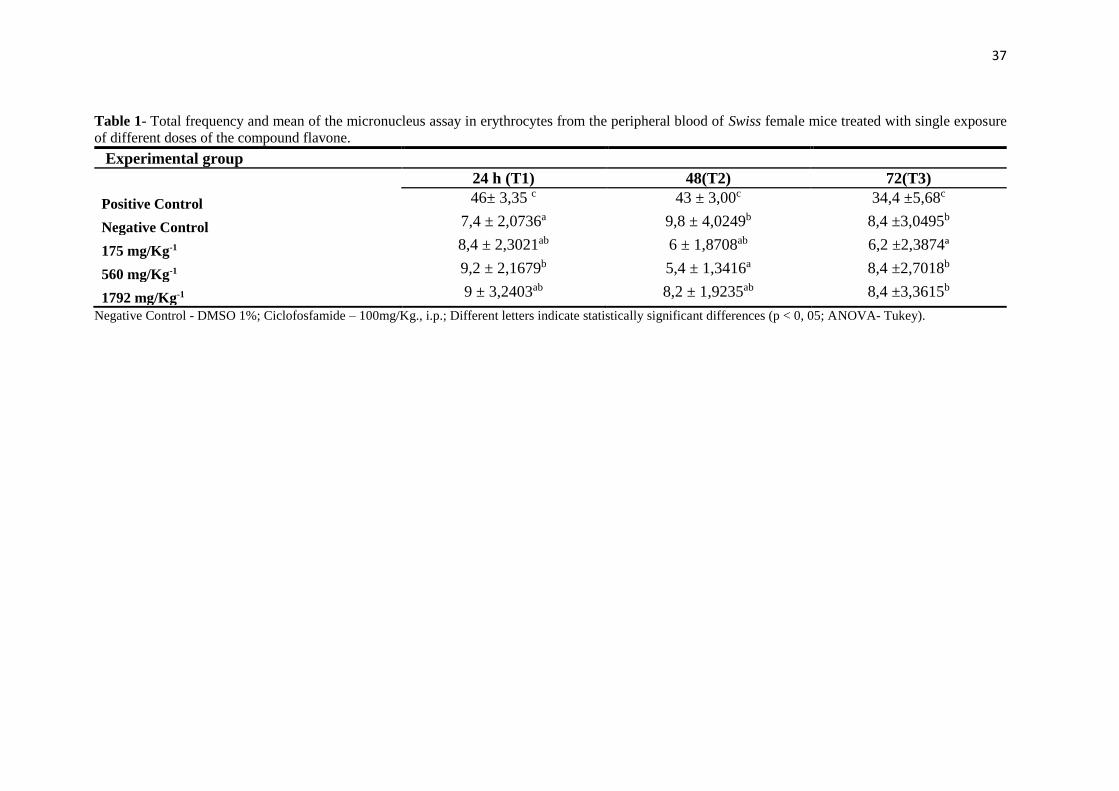
37
Table 1- Total frequency and mean of the micronucleus assay in erythrocytes from the peripheral blood of Swiss female mice treated with single exposure
of different doses of the compound flavone.
Experimental group
24 h (T1) 48(T2) 72(T3)
Positive Control 46± 3,35 c 43 ± 3,00c 34,4 ±5,68c
Negative Control 7,4 ± 2,0736a 9,8 ± 4,0249b 8,4 ±3,0495b
175 mg/Kg-1 8,4 ± 2,3021ab 6 ± 1,8708ab 6,2 ±2,3874ª
560 mg/Kg-1 9,2 ± 2,1679b 5,4 ± 1,3416a 8,4 ±2,7018b
1792 mg/Kg-1 9 ± 3,2403ab 8,2 ± 1,9235ab 8,4 ±3,3615b
Negative Control - DMSO 1%; Ciclofosfamide – 100mg/Kg., i.p.; Different letters indicate statistically significant differences (p < 0, 05; ANOVA- Tukey).

38
Table 2- Mean, ± S.E.M, total of injured cells and mean frequency between different classes for the anti-genotoxic test of flavones, comet assay in
Swiss female mice.
Groups Injured Cells Classes of damage Score
0 1 2 3
Negative Control 13,60±0,51a 86,40±0,51 13,00±0,45 0,60±0,25 0,00±0,00 14,20±0,66a
Positive Control 85,40 ±0,68c 14,60±0,68 68,20±0,58 13,80±0,66 3,40±0,40 106,00 ±1,64c
175 mg/Kg-1 17,20±0,66 ab 82,80±0,66 17,20±0,66 0,00±0,00 0,00±0,00 17,20±0,66 ab
560 mg/Kg-1 18,60±0,24b 81,40±0,24 17,60±0,40 1,00±0,31 0,00±0,00 19,60±0,40b
1792 mg/Kg-1 17,00±1,58ab 82,20±0,86 0,00±0,00 0,00±0,00 0,00±0,00 17,00±1,58ab
Negative Control - DMSO 1%; Ciclofosfamide – 100mg/Kg., i.p.; Different letters indicate statistically significant differences (p < 0, 05; ANOVA- Tukey).
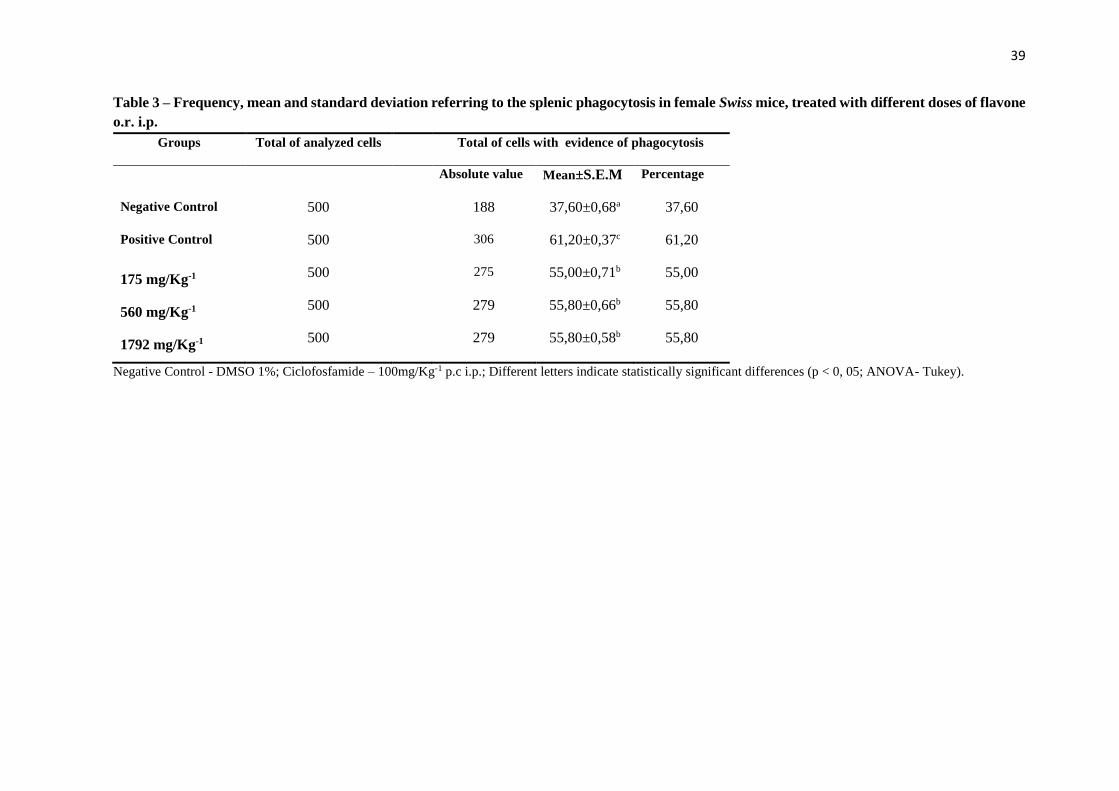
39
Table 3 – Frequency, mean and standard deviation referring to the splenic phagocytosis in female Swiss mice, treated with different doses of flavone
o.r. i.p.
Groups Total of analyzed cells Total of cells with evidence of phagocytosis
Absolute value Mean±S.E.M Percentage
Negative Control 500 188 37,60±0,68a 37,60
Positive Control 500 306 61,20±0,37c 61,20
175 mg/Kg-1 500 275 55,00±0,71b 55,00
560 mg/Kg-1 500 279 55,80±0,66b 55,80
1792 mg/Kg-1 500 279 55,80±0,58b 55,80
Negative Control - DMSO 1%; Ciclofosfamide – 100mg/Kg-1 p.c i.p.; Different letters indicate statistically significant differences (p < 0, 05; ANOVA- Tukey).

40
Table 4 - Differential count of groups treated with a single dose of flavone.
Experimental Group Red cells Hematocrit Hemoglobin Neutrophil Lymphocyte Monocyte Platelets
Positive Control 9,66±0,134a 46,6±1,341 a 15,4±0,547 a 53±6,89 a 45,6±7,02 a 1,40±0,54 a 348,8±71,394 b
Negative Control 9,24±0,391a 44,4±4,827 a 14,46±1,599 a 43,8±3,49 a 56,2±3,49 a 0,2±0,447 a 326±69,932 a
175 mg/Kg-1 9,28±0,192a 42,8±1,923 a 14,2±0,836 a 42±2,54 a 57,6±3,04 a 0,4±0,894 a 360,8±30,474 c
560 mg/Kg-1 9,56±0,357a 44,8±3,346 a 15±1,224 a 46±1,58 a 54±1,58 a 0,0±0,00 a 347±42,332 b
1792 mg/Kg-1 9,34±0,397a 43,4±3,974 a 14,6±1,140 a 49±2,0 a 49,8±2,94 a 1,2±1,30 a 392,2±44,650 c
Negative Control - DMSO 1%; Ciclofosfamide – 100mg/Kg-1 p.c i.p.; Different letters indicate statistically significant differences (p < 0, 05; ANOVA- Tukey).

41
Table 5- Biochemical parameters of groups treated with flavone
Experimental group Urea Creatinine Alkaline Phosphatase AST ALT
Positive Control 69±7,1763 a 0,5 ±0,070c 115,8 ±1,483b 75 ±3,033c 70,4 ±7,765c
Negative Control 47,8±4,061b 0,36±0,054b 117,41,673c 74,4±1,341b 51,6±3,781b
175 mg/Kg-1 44,4±3,872b 0,28±0,083b 115,8±3,114b 73,2±1,788b 48±2,549a
560 mg/Kg-1 44±2,543c 0,3±0,070b 114,6±3,781a 72,8±2,049b 48,21,7888b
1792 mg/Kg-1 44,6±271b 0,26±0,054a 117±2,915b 72,4±1,140a 49,8±2,489b
Negative Control - DMSO 1%; Ciclofosfamide – 100mg/Kg-1 p.c i.p.; Different letters indicate statistically significant differences (p < 0, 05; ANOVA- Tukey).

42
Table 6. Initial and final body weight and weight gain.
Experimental group Initial weight (g) Final weight (g) Weight gain (g)
Positive Control 28,742 ± 2,8688 29,616 ± 3,3262 0,87b Negative Control 25,588 ± 1,5529 27,088 ± 1,9822 1,50a
175 mg/Kg-1 27,658 ± 1,8828 28,248 ± 1,9313 0,59b
560 mg/Kg-1 25,660 ± 1,1828 27,010 ± 2,0212 1,35b
1792 mg/Kg-1 27,006 ± 1,8500 27,170 ± 2,0385 0,16c
Negative Control - DMSO 1%; Ciclofosfamide – 100mg/Kg-1 p.c i.p.; Different letters indicate statistically significant differences (p < 0, 05; ANOVA- Tukey).
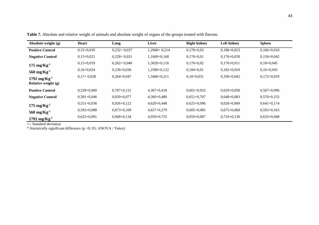
43
Table 7. Absolute and relative weight of animals and absolute weight of organs of the groups treated with flavone.
+/- Standard deviation
* Statistically significant difference (p <0, 05; ANOVA / Tukey)
Absolute weight (g) Heart Lung Liver Right kidney Left kidney Spleen
Positive Control 0,15+0,019 0,232+ 0,037 1,2960+ 0,214 0,178+0,03 0,186+0,023 0,166+0,018
Negative Control 0,15+0,021 0,228+ 0,031 1,1660+0,168 0,176+0,01 0,176+0,028 0,156+0,042
175 mg/Kg-1 0,15+0,019 0,262+ 0,040 1,3020+0,116 0,176+0,02 0,176+0,011 0,18+0,045
560 mg/Kg-1 0,16+0,024 0,236+0,036 1,2580+0,122 0,184+0,01 0,182+0,019 0,16+0,045
1792 mg/Kg-1 0,17+ 0,028 0,264+0,047 1,3460+0,211 0,18+0,031 0,196+0,042 0,172+0,010
Relative weight (g)
Positive Control 0,528+0,060 0,787+0,131 4,367+0,418 0,601+0,933 0,629+0,058 0,567+0,096
Negative Control 0,581+0,046 0,839+0,077 4,300+0,480 0,651+0,707 0,648+0,083 0,576+0,155
175 mg/Kg-1 0,551+0,036 0,926+0,122 4,620+0,448 0,625+0,096 0,626+0,069 0,641+0,174
560 mg/Kg-1 0,593+0,088 0,873+0,109 4,657+0,279 0,685+0,085 0,675+0,068 0,593+0,163
1792 mg/Kg-1 0,625+0,091 0,968+0,134 4,959+0,725 0,659+0,087 0,716+0,130 0,635+0,048

44
ANEXO II – NORMAS DA REVISTA

45
1.Format Requirements
PLOS ONE does not consider presubmission inquiries. All submissions should be prepared with
the following files:
Cover letter
Manuscript, including tables and figure legends
Figures (guidelines for preparing figures can be found at the Figure and Table Guidelines)
Prior to submission, authors who believe their manuscripts would benefit from professional editing
are encouraged to use language-editing and copyediting services. Obtaining this service is the
responsibility of the author, and should be done before initial submission. These services can be
found on the web using search terms like "scientific editing service" or "manuscript editing
service." Submissions are not copyedited before publication.
Submissions that do not meet the PLOS ONE Publication Criterion for language standards may
be rejected.
Cover Letter
You should supply an approximately one page cover letter that:
Concisely summarizes why your paper is a valuable addition to the scientific literature
Briefly relates your study to previously published work
Specifies the type of article you are submitting (for example, research article, systematic review,
meta-analysis, clinical trial)
Describes any prior interactions with PLOS regarding the submitted manuscript
Suggests appropriate PLOS ONE Academic Editors to handle your manuscript (view a complete
listing of our academic editors)
Lists any recommended or opposed reviewers
Your cover letter should not include requests to reduce or waive publication fees. Should your
manuscript be accepted, you will have the opportunity to include your requests at that time.
See PLOS ONE Editorial Policy for more information regarding publication fees.
Manuscript Organization
PLOS ONE considers manuscripts of any length. There are no explicit restrictions for the number
of words, figures, or the length of the supporting information, although we encourage a concise
and accessible writing style. We will not consider monographs.
All manuscripts should be double-spaced and include line numbers and page numbers.
Manuscripts should begin with the ordered sections:
Title
Authors
46
Affiliations
Abstract
Introductionand end with the sections of:
Acknowledgments
References
Figure Legends
Tables
Figures should not be included in the main manuscript file. Each figure must be prepared
and submitted as an individual file. Find more information about preparing figures here.
The title, authors, and affiliations should all be included on a title page as the first page of the
manuscript file.
There are no explicit requirements for section organization between these beginning and ending
sections. Articles may be organized in different ways and with different section titles, according
to the authors' preference. In most cases, internal sections include:
Materials and Methods
Results
Discussion
Conclusions (optional)
PLOS ONE has no specific requirements for the order of these sections, and in some cases it
may be appropriate to combine sections. Guidelines for individual sections can be found below.
Abbreviations should be kept to a minimum and defined upon first use in the text. Non-standard
abbreviations should not be used unless they appear at least three times in the text.
Standardized nomenclature should be used as appropriate, including appropriate usage of
species names and SI units.
PLOS articles do not support text footnotes. If your accepted submission contains footnotes, you
will be asked to move that material into either the main text or the reference list, depending on
the content.
Manuscript File Type Requirements
Authors may submit their manuscript files in Word (as .doc or .docx), LaTeX (as .pdf), or RTF
format. Only RTF and .doc files can be used during the production process. Word files must not
be protected.
LaTeX Submissions. If you would like to submit your manuscript using LaTeX, you must author
your article using the PLOS ONE LaTeX template and BibTeX style sheet. Articles prepared in
LaTeX may be submitted in PDF format for use during the review process. After acceptance,
however, .tex files and formatting information will be required as a zipped file. Please consult
our LaTeX guidelines for a list of what will be required.

47
Submissions with equations. If your manuscript is or will be in .docx format and contains
equations, you must follow the instructions below to make sure that your equations are editable
when the file enters production.
If you have not yet composed your article, you can ensure that the equations in your .docx file
remain editable in .doc by enabling "Compatibility Mode" before you begin. To do this, open a
new document and save as Word 97-2003 (*.doc). Several features of Word 2007/10 will now be
inactive, including the built-in equation editing tool. You can insert equations in one of the two
ways listed below.
If you have already composed your article as .docx and used its built-in equation editing tool, your
equations will become images when the file is saved down to .doc. To resolve this problem, re-
key your equations in one of the two following ways.
1. Use MathType to create the equation (recommended)
2. Go to Insert > Object > Microsoft Equation 3.0 and create the equation
If, when saving your final document, you see a message saying "Equations will be converted to
images," your equations are no longer editable and PLoS will not be able to accept your file.
Back to top
2. Guidelines for Standard Sections
Title
Manuscripts must be submitted with both a full title and a short title, which will appear at the top
of the PDF upon publication if accepted. Only the full title should be included in the manuscript
file; the short title will be entered during the online submission process.
The full title must be 250 characters or fewer. It should be specific, descriptive, concise, and
comprehensible to readers outside the subject field. Avoid abbreviations if possible. Where
appropriate, authors should include the species or model system used (for biological papers) or
type of study design (for clinical papers).
Examples:
Impact of Cigarette Smoke Exposure on Innate Immunity: A Caenorhabditis elegans Model
Solar Drinking Water Disinfection (SODIS) to Reduce Childhood Diarrhoea in Rural Bolivia: A
Cluster-Randomized, Controlled Trial
The short title must be 50 characters or fewer and should state the topic of the paper.
Back to top
Authors and Affiliations
All author names should be listed in the following order:
First names (or initials, if used),
Middle names (or initials, if used), and
48
Last names (surname, family name)
Each author should list an associated department, university, or organizational affiliation and its
location, including city, state/province (if applicable), and country. If the article has been submitted
on behalf of a consortium, all author names and affiliations should be listed at the end of the
article.
This information cannot be changed after initial submission, so please ensure that it is
correct.
To qualify for authorship, a researcher should contribute to all of the following:
1. Conception and design of the work, acquisition of data, or analysis and interpretation of data
2. Drafting the article or revising it critically for important intellectual content
3. Final approval of the version to be published
All persons designated as authors should qualify for authorship, and all those who qualify should
be listed. Each author must have participated sufficiently in the work to take public responsibility
for appropriate portions of the content. Those who contributed to the work but do not qualify for
authorship should be listed in the acknowledgments.
When a large group or center has conducted the work, the author list should include the
individuals whose contributions meet the criteria defined above, as well as the group name.
One author should be designated as the corresponding author, and his or her email address or
other contact information should be included on the manuscript cover page. This information will
be published with the article if accepted.
See the PLOS ONE Editorial Policy regarding authorship criteria for more information.
Back to top
Abstract
The abstract should:
Describe the main objective(s) of the study
Explain how the study was done, including any model organisms used, without methodological
detail
Summarize the most important results and their significance
Not exceed 300 words
Abstracts should not include:
Citations
Abbreviations, if possible Back to top
Introduction
The introduction should:

49
Provide background that puts the manuscript into context and allows readers outside the field to
understand the purpose and significance of the study
Define the problem addressed and why it is important
Include a brief review of the key literature
Note any relevant controversies or disagreements in the field
Conclude with a brief statement of the overall aim of the work and a comment about whether that
aim was achieved Back to top
Materials and Methods
This section should provide enough detail to allow suitably skilled investigators to fully replicate
your study. Specific information and/or protocols for new methods should be included in detail. If
materials, methods, and protocols are well established, authors may cite articles where those
protocols are described in detail, but the submission should include sufficient information to be
understood independent of these references.
We encourage authors to submit detailed protocols for newer or less well-established methods
as Supporting Information. Further information about formatting Supporting Information files, can
be found here.
Methods sections of papers on research using human or animal subjects and/or tissue or field
sampling must include required ethics statements. See theReporting Guidelines for human
research, clinical trials, animal research, and observational and field studies for more information.
Methods sections of papers with data that should be deposited in a publicly available
database should specify where the data have been deposited and provide the relevant accession
numbers and version numbers, if appropriate. Accession numbers should be provided in
parentheses after the entity on first use. If the accession numbers have not yet been obtained at
the time of submission, please state that they will be provided during review. They must be
provided prior to publication. A list of recommended repositories for different types of data can be
found here.
Methods sections of papers using cell lines must state the origin of the cell lines used. See
the Reporting Guidelines for cell line research for more information.
Methods sections of papers adding new taxon names to the literature must follow the Reporting
Guidelines below for a new zoological taxon, botanical taxon, or fungal taxon.
Back to top
Results, Discussion, and Conclusions
These sections may all be separate, or may be combined to create a mixed Results/Discussion
section (commonly labeled "Results and Discussion") or a mixed Discussion/Conclusions section
(commonly labeled "Discussion"). These sections may be further divided into subsections, each
with a concise subheading, as appropriate. These sections have no word limit, but the language
should be clear and concise.

50
Together, these sections should describe the results of the experiments, the interpretation of
these results, and the conclusions that can be drawn. Authors should explain how the results
relate to the hypothesis presented as the basis of the study and provide a succinct explanation of
the implications of the findings, particularly in relation to previous related studies and potential
future directions for research.
PLOS ONE editorial decisions do not rely on perceived significance or impact, so authors should
avoid overstating their conclusions. See the PLOS ONEPublication Criteria for more information.
Back to top
Acknowledgments
People who contributed to the work but do not fit the PLOS ONE authorship criteria should be
listed in the acknowledgments, along with their contributions. You must ensure that anyone
named in the acknowledgments agrees to being so named.
Funding sources should not be included in the acknowledgments, or anywhere in the manuscript
file. You will provide this information during the manuscript submission process.
Back to top
References
General guidelines
Authors may cite any and all available works in the reference list.
Authors may not cite unavailable and unpublished work, including manuscripts that have been
submitted but not yet accepted (e.g., “unpublished work,” “data not shown”).
If an article is submitted to a journal and also publicly available as a pre-print, the pre-print may
be cited.
If related work has been submitted to PLOS ONE or elsewhere, authors should include a copy
with the submitted article as confidential supplementary information, for review purposes only.
Authors should not state 'unpublished work' or 'data not shown,' but instead include those data
as supplementary material or deposit the data in a publicly available database.
Authors should not state 'unpublished work' or 'data not shown,' but instead include those data
as supplementary material or deposit the data in a publicly available database.
Reference formatting
References must be listed at the end of the manuscript and numbered in the order that they
appear in the text. In the text, citations should be indicated by the reference number in brackets.
Journal name abbreviations should be those found in the NCBI databases. A number of reference
software companies supply PLOS style files (e.g., Reference Manager, EndNote).
References should be formatted as follows:
Published papers. Hou WR, Hou YL, Wu GF, Song Y, Su XL, et al. (2011) cDNA, genomic
sequence cloning and overexpression of ribosomal protein gene L9 (rpL9) of the giant panda
51
(Ailuropoda melanoleuca). Genet Mol Res 10: 1576-1588.
Note: Use of a DOI number for the full-text article is acceptable as an alternative to or in addition
to traditional volume and page numbers.
Accepted, unpublished papers. Same as above, but “In press” appears instead of the page
numbers.
Electronic journal articles. Huynen MMTE, Martens P, Hilderlink HBM (2005) The health
impacts of globalisation: a conceptual framework. Global Health 1: 14. Available:
http://www.globalizationandhealth.com/content/1/1/14. Accessed 25 January 2012.
Books. Bates B (1992) Bargaining for life: A social history of tuberculosis. Philadelphia: University
of Pennsylvania Press. 435 p.
Book chapters Hansen B (1991) New York City epidemics and history for the public. In: Harden
VA, Risse GB, editors. AIDS and the historian. Bethesda: National Institutes of Health. pp. 21-28.
Published media, not peer-reviewed. Examples: print or online newspapers and magazine
articles. Fountain H (29 Jan 2014). For Already Vulnerable Penguins, Study Finds Climate
Change Is Another Danger. The New York Times.
Available:http://www.nytimes.com/2014/01/30/science/earth/climate-change-taking-toll-on-
penguins-study-finds.html. Accessed 17 March 2014.
New media, unregulated. Examples: blogs, websites, and other written works. Allen L (01
Sept 2010) Announcing PLOS Blogs. Available:http://blogs.plos.org/plos/2010/09/announcing-
plos-blogs/. Accessed 17 March 2014.
Master of Science and Doctor of Philosophy theses. Wells A (1999) Exploring the
development of the independent, electronic, scholarly journal. M.Sc. Thesis, The University of
Sheffield. Available: http://cumincad.scix.net/cgi-bin/works/Show?2e09. Accessed 17 March
2014.
Databases and repositories. Examples: figshare, archive.com. Roberts SB (2013) QPX
Genome Browser Feature Tracks. Database:
figshare.http://figshare.com/articles/QPX_Genome_Browser_Feature_Tracks/701214. Accessed
17 March 2014.

52
Multimedia. Examples: videos, movies, and TV shows. Hitchcock A, producer and director
(1954) Rear Window [Film]. Los Angeles: MGM.
Back to top
Tables
Tables should be included at the end of the manuscript. All tables should have a concise title.
Footnotes can be used to explain abbreviations. Citations should be indicated using the same
style as outlined above. Tables occupying more than one printed page should be avoided, if
possible. Larger tables can be published as Supporting Information. Please ensure that table
formatting conforms to our Guidelines for table preparation.
Back to top
Figure Legends
Figures should not be included in the manuscript file, but figure legends should be. Guidelines
for preparing figures can be found here.
Figure legends should describe the key messages of a figure. Legends should have a short title
of 15 words or less. The full legend should have a description of the figure and allow readers to
understand the figure without referring to the text. The legend itself should be succinct, avoid
lengthy descriptions of methods, and define all non-standard symbols and abbreviations.
Further information about figure legends can be found in the Figure Guidelines.
Back to top
Accession Numbers
All appropriate datasets, images, and information should be deposited in public resources. Please
provide the relevant accession numbers (and version numbers, if appropriate). Accession
numbers should be provided in parentheses after the entity on first use. Suggested databases
include, but are not limited to:
ArrayExpress
BioModels Database
Database of Interacting Proteins
DNA Data Bank of Japan [DDBJ]
DRYAD
EMBL Nucleotide Sequence Database
GenBank
Gene Expression Omnibus [GEO]
Protein Data Bank
UniProtKB/Swiss-Prot
53
ClinicalTrials.gov
In addition, as much as possible, please provide accession numbers or identifiers for all entities
such as genes, proteins, mutants, diseases, etc., for which there is an entry in a public database,
for example:
Ensembl
Entrez Gene
FlyBase
InterPro
Mouse Genome Database (MGD)
Online Mendelian Inheritance in Man (OMIM)
PubChem
Providing accession numbers allows linking to and from established databases and integrates
your article with a broader collection of scientific information.
Back to top
Striking Images
Authors are encouraged to upload a "striking image" that may be used to represent their paper
online in places like the journal homepage or in search results. The striking image must be derived
from a figure or supporting information file from the paper, ie. a cropped portion of an image or
the entire image. Striking images should ideally be high resolution, eye-catching, single panel
images, and should ideally avoid containing added details such as text, scale bars, and arrows.
If no striking image is uploaded, a figure from the paper will be designated as the striking image.
Please keep in mind that PLOS's Creative Commons Attribution License applies to striking
images. As such, do not submit any figures or photos that have been previously copyrighted
unless you have express written permission from the copyright holder to publish under the CCAL
license. Note that all published materials in PLOS ONE are freely available online, and any third
party is permitted to read, download, copy, distribute, and use these materials in any way, even
commercially, with proper attribution.
Care should be taken with the following image types in particular:
1. PLOS ONE is unable to publish any images generated by Google software (Google Maps, Street
View, and Earth)
2. Maps in general are usually copyrighted, especially satellite maps
3. Photographs
4. Commercial or government images, slogans, or logos
5. Images from Facebook or Twitter
Authors must also take special care when submitting manuscripts that contain potentially
identifying images of people. Identifying information should not be included in the manuscript
unless the information is crucial and the individual has provided written consent by completing
the Consent Form for Publication in a PLOS Journal (PDF).
For license inquiries, e-mail license [at] plos.org.
54
Back to top
3. Specific Reporting Guidelines
Human Subject Research
Methods sections of papers on research using human subject or samples must include ethics
statements that specify:
The name of the approving institutional review board or equivalent committee(s). If approval was
not obtained, the authors must provide a detailed statement explaining why it was not needed
Whether informed consent was written or oral. If informed consent was oral, it must be stated in
the manuscript:
o Why written consent could not be obtained
o That the Institutional Review Board (IRB) approved use of oral consent
o How oral consent was documented
For studies involving humans categorized by race/ethnicity, age, disease/disabilities, religion,
sex/gender, sexual orientation, or other socially constructed groupings, authors should:
Explicitly describe their methods of categorizing human populations
Define categories in as much detail as the study protocol allows
Justify their choices of definitions and categories, including for example whether any rules of
human categorization were required by their funding agency
Explain whether (and if so, how) they controlled for confounding variables such as socioeconomic
status, nutrition, environmental exposures, or similar factors in their analysis
In addition, outmoded terms and potentially stigmatizing labels should be changed to more
current, acceptable terminology. Examples: "Caucasian" should be changed to "white" or "of
[Western] European descent" (as appropriate); "cancer victims" should be changed to "patients
with cancer."
For papers that include identifying, or potentially identifying, information, authors must download
the Consent Form for Publication in a PLOS Journal (PDF), which the individual, parent, or
guardian must sign once they have read the paper and been informed about the terms of PLOS
open-access license. The signed consent form should not be submitted with the manuscript, but
authors should securely file it in the individual's case notes and the methods section of the
manuscript should explicitly state that consent authorization for publication is on file, using
wording like:
The individual in this manuscript has given written informed consent (as outlined in PLOS
consent form) to publish these case details.
For more information about PLOS ONE policies regarding human subject research, see
the Publication Criteria and Editorial Policies.
Back to top
55
Clinical Trials
Authors of manuscripts describing the results of clinical trials must adhere to
the CONSORT reporting guidelines appropriate to their trial design, available on the CONSORT
Statement website. Before the paper can enter peer review, authors must:
1. Provide the registry name and number in the methods section of the manuscript
2. Provide a copy of the trial protocol as approved by the ethics committee and a
completed CONSORT checklist as Supporting Information (which will be published alongside the
paper, if accepted)
3. Include the CONSORT flow diagram as the manuscript's "Figure 1"
Any deviation from the trial protocol must be explained in the paper. Authors must explicitly
discuss informed consent in their paper, and we reserve the right to ask for a copy of the patient
consent form.
The methods section must include the name of the registry, the registry number, and the URL of
your trial in the registry database for each location in which the trial is registered.
For more information about PLOS ONE policies regarding clinical trials, see the Editorial Policies.
Back to top
Animal Research
Methods sections of manuscripts reporting results of animal research must include required ethics
statements that specify:
The full name of the relevant ethics committee that approved the work, and the associated permit
number(s) (where ethical approval is not required, the manuscript should include a clear
statement of this and the reason why)
Relevant details for efforts taken to ameliorate animal suffering
For example:
This study was carried out in strict accordance with the recommendations in the Guide for
the Care and Use of Laboratory Animals of the National Institutes of Health. The protocol
was approved by the Committee on the Ethics of Animal Experiments of the University of
Minnesota (Permit Number: 27-2956). All surgery was performed under sodium
pentobarbital anesthesia, and all efforts were made to minimize suffering.
The organism(s) studied should always be stated in the abstract. Where research may be
confused as pertaining to clinical research, the animal model should also be stated in the title.
We ask authors to follow the ARRIVE (Animal Research: Reporting of In Vivo Experiments)
guidelines for all submissions describing laboratory-based animal research and to upload a
completed ARRIVE Guidelines Checklist to be published as supporting information. Please note
that inclusion of a completed ARRIVE Checklist will be a formal requirement for publication at a
later date.
56
For more information about PLOS ONE policies regarding animal research, see the Publication
Criteria and Editorial Policies.
Back to top
Observational and Field Studies
Methods sections for submissions reporting on any type of field study must include ethics
statements that specify:
Permits and approvals obtained for the work, including the full name of the authority that approved
the study; if none were required, authors should explain why
Whether the land accessed is privately owned or protected
Whether any protected species were sampled
Full details of animal husbandry, experimentation, and care/welfare, where relevant
For more information about PLOS ONE policies regarding observational and field studies, see
the Publication Criteria and Editorial Policies.
Back to top
Cell Line Research
Authors reporting research using cell lines should state when and where they obtained the cells,
giving the date and the name of the researcher, cell line repository, or commercial source
(company) who provided the cells, as appropriate. Authors must also include the following
information for each cell line:
For de novo (new) cell lines, including those given to the researchers a gift, authors must follow
our policies for human subject research or animal research, as appropriate. The ethics statement
must include:
Details of institutional review board or ethics committee approval; AND
For human cells, confirmation of written informed consent from the donor, guardian, or next of kin
For established cell lines, the Methods section should include:
A reference to the published article that first described the cell line; AND/OR
The cell line repository or company the cell line was obtained from, the catalogue number, and
whether the cell line was obtained directly from the repository/company or from another laboratory
Authors should check established cell lines using the ICLAC Database of Cross-contaminated or
Misidentified Cell Lines to confirm they are not misidentified or contaminated. Cell line
authentication is recommended - e.g. by karyotyping, isozyme analysis, or short tandem repeats
(STR) analysis - and may be required during peer review or after publication.
Back to top
57
Blots and Gels
Authors of manuscripts reporting results from blots (including Western blots) and electrophoretic
gels should follow these guidelines:
In accordance with PLOS ONE's policy on image manipulation, the image should not be adjusted
in any way that could affect the scientific information displayed, e.g. by modifying the background
or contrast
All blots and gels that support results reported in the manuscript should be provided
Original uncropped and unadjusted blots and gels, including molecular size markers, should be
provided in either the figures or the supplementary files
Lanes should not be overcropped around the bands; the image should show most or all of the
blot or gel. Any non-specific bands should be shown and an explanation of their nature should be
given
The image should include all relevant controls, and controls should be run on the same blot or
gel as the samples
A figure panel should not include composite images of bands originating from different blots or
gels. If the figure shows non-adjacent bands from the same blot or gel, this should be clearly
denoted by vertical black lines and the figure legend should provide details of how the figure was
made Back to top
Antibodies
Manuscripts reporting experiments using antibodies should include the following information:
The name of each antibody, a description of whether it is monoclonal or polyclonal, and the host
species
The commercial supplier or source laboratory
The catalogue or clone number and, if known, the batch number
The antigen(s) used to raise the antibody
For established antibodies, authors are encouraged to supply a stable public identifier from the
Antibody Registry (www.antibodyregistry.org).
Authors should also report the following experimental details:
The final antibody concentration or dilution
A reference to the validation study if the antibody was previously validated, and if not, details of
how the authors validated the antibody for the applications and species used. Authors should
consider adding information on new validations to a publicly available database such
as Antibodypediaor CiteAb. Back to top

58
Systematic Review/Meta-Analysis
A systematic review paper, as defined by The Cochrane Collaboration, is a review of a clearly
formulated question that uses explicit, systematic methods to identify, select, and critically
appraise relevant research, and to collect and analyze data from the studies that are included in
the review. These reviews differ substantially from narrative-based reviews or synthesis articles.
Statistical methods (meta-analysis) may or may not be used to analyze and summarize the results
of the included studies.
Reports of systematic reviews and meta-analyses must include a completed PRISMA (Preferred
Reporting Items for Systematic Reviews and Meta-Analyses) checklist and flow diagram to
accompany the main text. Blank templates are available here:
Checklist: PDF or Word document
Flow diagram: PDF or Word document
Authors must also state in their "Methods" section whether a protocol exists for their systematic
review, and if so, provide a copy of the protocol as Supporting Information and provide the registry
number in the abstract.
If your article is a Systematic Review or a Meta-Analysis you should:
State this in your cover letter
Select "Research Article" as your article type when submitting
Include the PRISMA flowchart as Figure 1 (required where applicable)
Include the PRISMA checklist as Supporting Information
Meta-Analysis of Genetic Association Studies
Manuscripts reporting a meta-analysis of genetic association studies must report results of value
to the field and should be reported according to the guidelines presented in “Systematic Reviews
of Genetic Association Studies” by Sagoo et al.
On submission, authors will be asked to justify the rationale for the meta-analysis and how it
contributes to the base of scientific knowledge in the light of previously published results. Authors
will also be asked to complete a checklist outlining information about the justification for the study
and the methodology employed. Meta-analyses that replicate published studies will be rejected if
the authors do not provide adequate justification.
Back to top
Paleontology and Archaeology Research
Manuscripts reporting paleontology and archaeology research must include descriptions of
methods and specimens in sufficient detail to allow the work to be reproduced. Data sets
supporting statistical and phylogenetic analyses should be provided, preferably in a format that
allows easy re-use.
Specimen numbers and complete repository information, including museum name and
geographic location, are required for publication. Locality information should be provided in the
manuscript as legally allowable, or a statement should be included giving details of the availability
of such information to qualified researchers.

59
If permits were required for any aspect of the work, details should be given of all permits that were
obtained, including the full name of the issuing authority. This should be accompanied by the
following statement:
All necessary permits were obtained for the described study, which complied with all
relevant regulations.
If no permits were required, please include the following statement:
No permits were required for the described study, which complied with all relevant
regulations.
See the PLOS ONE Editorial Policies for more information regarding manuscripts describing
paleontology and archaeology research.
Back to top
Software Papers
Manuscripts describing software should provide full details of the algorithms designed. Describe
any dependencies on commercial products or operating system. Include details of the supplied
test data and explain how to install and run the software. A brief description of enhancements
made in the major releases of the software may also be given. Authors should provide a direct
link to the deposited software from within the paper.
See the PLOS ONE Editorial Policies for more information about submitting manuscripts.
Back to top
Database Papers
For descriptions of databases, provide details about how the data were curated, as well as plans
for long-term database maintenance, growth, and stability. Authors should provide a direct link to
the database hosting site from within the paper.
See the PLOS ONE Editorial Policies for more information about submitting manuscripts
describing databases.
Back to top
New Zoological Taxon
For proper registration of a new zoological taxon, we require two specific statements to be
included in your manuscript.
In the Results section, the globally unique identifier (GUID), currently in the form of a Life Science
Identifier (LSID), should be listed under the new species name, for example:
Anochetus boltoni Fisher sp. nov. urn:lsid:zoobank.org:act:B6C072CF-1CA6-40C7-8396-
534E91EF7FBB
60
You will need to contact Zoobank to obtain a GUID (LSID). Please do this as early as possible to
avoid delay of publication upon acceptance of your manuscript. It is your responsibility to provide
us with this information so we can include it in the final published paper.
Please also insert the following text into the Methods section, in a sub-section to be called
"Nomenclatural Acts":
The electronic edition of this article conforms to the requirements of the amended
International Code of Zoological Nomenclature, and hence the new names contained
herein are available under that Code from the electronic edition of this article. This
published work and the nomenclatural acts it contains have been registered in ZooBank,
the online registration system for the ICZN. The ZooBank LSIDs (Life Science Identifiers)
can be resolved and the associated information viewed through any standard web browser
by appending the LSID to the prefix "http://zoobank.org/". The LSID for this publication is:
urn:lsid:zoobank.org:pub: XXXXXXX. The electronic edition of this work was published in
a journal with an ISSN, and has been archived and is available from the following digital
repositories: PubMed Central, LOCKSS [author to insert any additional repositories].
All PLOS ONE articles are deposited in PubMed Central and LOCKSS. If your institute, or those
of your co-authors, has its own repository, we recommend that you also deposit the published
online article there and include the name in your article.
Back to top
New Botanical Taxon
When publishing papers that describe a new botanical taxon, PLOS aims to comply with the
requirements of the International Code of Nomenclature for algae, fungi, and plants (ICN). In
association with the International Plant Names Index (IPNI), the following guidelines for
publication in an online-only journal have been agreed such that any scientific botanical name
published by us is considered effectively published under the rules of the Code. Please note that
these guidelines differ from those for zoological nomenclature, and apply only to seed plants,
ferns, and lycophytes.
Effective January 2012, "the description or diagnosis required for valid publication of the name of
a new taxon" can be in either Latin or English. This does not affect the requirements for scientific
names, which are still to be Latin.
Also effective January 2012, the electronic PDF represents a published work according to the
ICN for algae, fungi, and plants. Therefore the new names contained in the electronic publication
of a PLOS ONE article are effectively published under that Code from the electronic edition alone,
so there is no longer any need to provide printed copies.
Additional information describing recent changes to the Code can be found here.
For proper registration of the new taxon, we require two specific statements to be included in your
manuscript.
In the Results section, the globally unique identifier (GUID), currently in the form of a Life Science
Identifier (LSID), should be listed under the new species name, for example:
Solanum aspersum S.Knapp, sp. nov. [urn:lsid:ipni.org:names:77103633-1] Type:
Colombia. Putumayo: vertiente oriental de la Cordillera, entre Sachamates y San

61
Francisco de Sibundoy, 1600-1750 m, 30 Dec 1940, J. Cuatrecasas 11471 (holotype,
COL; isotypes, F [F-1335119], US [US-1799731]).
PLOS ONE staff will contact IPNI to obtain the GUID (LSID) after your manuscript is accepted for
publication, and this information will then be added to the manuscript during the production phase
In the Methods section, include a sub-section called "Nomenclature" using the following wording:
The electronic version of this article in Portable Document Format (PDF) in a work with an
ISSN or ISBN will represent a published work according to the International Code of
Nomenclature for algae, fungi, and plants, and hence the new names contained in the
electronic publication of a PLOS ONE article are effectively published under that Code
from the electronic edition alone, so there is no longer any need to provide printed copies.
In addition, new names contained in this work have been submitted to IPNI, from where
they will be made available to the Global Names Index. The IPNI LSIDs can be resolved and
the associated information viewed through any standard web browser by appending the
LSID contained in this publication to the prefix http://ipni.org/. The online version of this
work is archived and available from the following digital repositories: [INSERT NAMES OF
DIGITAL REPOSITORIES WHERE ACCEPTED MANUSCRIPT WILL BE SUBMITTED
(PubMed Central, LOCKSS etc)].
All PLOS ONE articles are deposited in PubMed Central and LOCKSS. If your institute, or those
of your co-authors, has its own repository, we recommend that you also deposit the published
online article there and include the name in your article.
Back to top
New Fungal Taxon
When publishing papers that describe a new fungal taxon name, PLOS aims to comply with the
requirements of the International Code of Nomenclature for algae, fungi, and plants (ICN). The
following guidelines for publication in an online-only journal have been agreed such that any
scientific fungal name published by us is considered effectively published under the rules of the
Code. Please note that these guidelines differ from those for zoological nomenclature.
Effective January 2012, "the description or diagnosis required for valid publication of the name of
a new taxon" can be in either Latin or English. This does not affect the requirements for scientific
names, which are still to be Latin.
Also effective January 2012, the electronic PDF represents a published work according to the
ICN for algae, fungi, and plants. Therefore the new names contained in the electronic publication
of a PLOS ONE article are effectively published under that Code from the electronic edition alone,
so there is no longer any need to provide printed copies.
Additional information describing recent changes to the Code can be found here.
For proper registration of the new taxon, we require two specific statements to be included in your
manuscript.
In the Results section, the globally unique identifier (GUID), currently in the form of a Life Science
Identifier (LSID), should be listed under the new species name, for example:
Hymenogaster huthii. Stielow et al. 2010, sp. nov. [urn:lsid:indexfungorum.org:names:518624]

62
You will need to contact either Mycobank or Index Fungorum to obtain the GUID (LSID). Please
do this as early as possible to avoid delay of publication upon acceptance of your manuscript. It
is your responsibility to provide us with this information so we can include it in the final published
paper. Effective January 2013, all papers describing new fungal species must reference the
identifier issued by a recognized repository in the protologue in order to be considered effectively
published.
In the Methods section, include a sub-section called "Nomenclature" using the following wording
(this example is for taxon names submitted to MycoBank; please substitute appropriately if you
have submitted to Index Fungorum):
The electronic version of this article in Portable Document Format (PDF) in a work with an
ISSN or ISBN will represent a published work according to the International Code of
Nomenclature for algae, fungi, and plants, and hence the new names contained in the
electronic publication of a PLOS ONE article are effectively published under that Code
from the electronic edition alone, so there is no longer any need to provide printed copies.
In addition, new names contained in this work have been submitted to MycoBank from
where they will be made available to the Global Names Index. The unique MycoBank
number can be resolved and the associated information viewed through any standard web
browser by appending the MycoBank number contained in this publication to the prefix
http://www.mycobank.org/MB/. The online version of this work is archived and available
from the following digital repositories: [INSERT NAMES OF DIGITAL REPOSITORIES
WHERE ACCEPTED MANUSCRIPT WILL BE SUBMITTED (PubMed Central, LOCKSS etc)].
All PLOS ONE articles are deposited in PubMed Central and LOCKSS. If your institute, or those
of your co-authors, has its own repository, we recommend that you also deposit the published
online article there and include the name in your article.
Back to top
Qualitative Research
Qualitative research studies use non-quantitative methods to address a defined research
question that may not be accessible by quantitative methods, such as people's interpretations,
experiences, and perspectives. The analysis methods are explicit, systematic, and reproducible,
but the results do not involve numerical values or use statistics. Examples of qualitative data
sources include, but are not limited to, interviews, text documents, audio/video recordings, and
free-form answers to questionnaires and surveys.
Qualitative research studies should be reported in accordance to the Consolidated criteria for
reporting qualitative research (COREQ) checklist. Further reporting guidelines can be found in
the Equator Network's Guidelines for reporting qualitative research.

63
ANEXO III

64
1 - Artigo publicado
65
2- Artigo publicado