análise comparativa da morfologia gonadal de seis espécies ... · 4. peixe de água doce -...
TRANSCRIPT

PONTIFÍCIA UNIVERSIDADE CATÓLICA DE MINAS GERAIS
INSTITUTO DE CIÊNCIAS BIOLÓGICAS E DA SAÚDE
PROGRAMA DE PÓS-GRADUAÇÃO EM ZOOLOGIA DOS VERTEBRADOS
Análise comparativa da morfologia gonadal de seis espécies de peixes
pertencentes aos gêneros Incertae Sedis em Characidae de ocorrência na bacia
do rio São Francisco, Brasil.
Yuri Simões Martins
Orientador: Nilo Bazzoli
Belo Horizonte, março de 2010.

Yuri Simões Martins
Análise comparativa da morfologia gonadal de seis espécies de peixes
pertencentes aos gêneros Incertae Sedis em Characidae de ocorrência na bacia
do rio São Francisco, Brasil.
Dissertação apresentada ao Programa de Pós-
Graduação em Zoologia de Vertebrados como
pré-requisito para obtenção do título de mestre
em Zoologia de Vertebrados.
Belo Horizonte, março de 2010.

FICHA CATALOGRÁFICA Elaborada pela Biblioteca da Pontifícia Universidade Católica de Minas Gerais
Martins, Yuri Simões M386a Análise comparativa da morfologia gonadal de seis espécies
de peixes pertencentes aos gêneros Incertae Sedis em Characidae
de ocorrência na bacia do rio São Francisco, Brasil / Yuri Simões Martins. Belo Horizonte, 2010.
45f. : il. Orientador: Nilo Bazzoli Dissertação (Mestrado) – Pontifícia Universidade Católica de
Minas Gerais. Programa de Pós-Graduação em Zoologia de Vertebrados.
1. Ovários. 2. Lambari (Peixe) - São Francisco, Rio. 3.
Testículos. 4. Peixe de água doce - Morfologia. I. Bazzoli, Nilo. II. Pontifícia Universidade Católica de Minas Gerais. Programa de Pós-Graduação em Zoologia de Vertebrados. III. Título.
CDU: 597.5


O presente estudo foi realizado durante o mestrado turma 2008/2010 no Programa de Pós-
Graduação em Zoologia de Vertebrados da Pontifícia Universidade Católica de Minas
Gerais, sob orientação do Professor Doutor Nilo Bazzoli, com apoio financeiro e logístico
das seguintes instituições:
Estação de Hidrobiologia e Piscicultura de Três Marias- CODEVASF.
Fundação de Amparo à Pesquisa de Minas Gerais (FAPEMIG).
Conselho Nacional de Desenvolvimento Científico e Tecnológico (CNPq).

Trabalho, loucura e amor: O sol é o limite

AGRADECIMENTOS
A minha mãe, pelo amor incondicional, pelo carinho e pela paciência.
Ao Nilo pela oportunidade, paciência e pelos anos de ensinamentos impagáveis e amizade.
Ao Rogério pela grande amizade, pelos ensinamentos e pela maravilhosa confecção do
material histológico.
A Cledma pela amizade e por sempre ajudar a resolver as burocracias da pesquisa e da
academia.
Ao Yoshimi e a todo pessoal da CODEVASF pelos momentos de descontração,
ensinamentos e apoio logístico durante as coletas.
Ao Fábio e a todas outras pessoas que ajudaram na coleta do material.
Aos Doutores Hélio Batista dos Santos e Laércio dos Anjos Benjamin.
Aos amigos de profissão que deram oportunidades de trabalho.
A EQUIPE pela amizade, ensinamentos, trabalho e união durante os melhores anos da
minha vida.
A todos os colegas, alunos, estagiários e professores do Mestrado em Zoologia que de
alguma forma me ensinaram grande parte do meu conhecimento acadêmico e profissional.
A Débora pela amizade, paciência e companheirismo.
A vida por tudo.

RESUMO
O presente estudo analisou comparativamente a morfologia gonadal de seis espécies
de peixes dos gêneros Incertae Sedis na famíla Characidae. Ovários em maturação de
Astyanax fasciatus apresentaram coloração acizentada e nas outras espécies coloração
amarelada. Os alvéolos corticais das espécies analisadas são pequenos (5.20± 1.44 -
8.28±2.02 µm) e formados por várias camadas de vesículas descontínuas exceto em
Bryconops affinis onde eles são grandes (12.87±2.41 µm) e formando várias camadas de
vesículas contínuas. A zona pelúcida é delgada variando de 2.44± 0.90 a 5.79±1.90 µm e
espessa em B. affinis (10.57± 2.08 µm). Ondulações na zona pelúcida foram observadas em
Astyanax bimaculatus, A. fasciatus, Hemmigramus marginatus e Moenkhausia costae. As
células foliculares de B. affinis são prismáticas (28.19± 13.71 µm) enquanto nas outras
espécies elas são pavimentosas (1.52± 0.64 - 2.12±0.78 µm). O presente estudo mostrou nos
ovócitos vitelogênicos de B. affinis características morfológicas estatisticamente diferentes
daquelas dos outros Incertae Sedis da família Characidae analisadas Nos testículos em
maturação avançada de A. bimaculatus e A. fasciatus os espermatozóides encontravam-se
embebidos em secreção acidófila que foi gradativamente reabsorvida no estádio
parcialmente espermiado.
Palavras-chave: ovários, testículos, secreção testicular, ovócito de peixe, morfologia
comparativa

ABSTRACT
This study presents a comparative analysis of gonad morphology for six fish species from
the genera Incertae Sedis in the Characidae family. Maturing ovaries of Astyanax fasciatus
presented grayish color whilst the other species’ were yellowish in color. The cortical
alveoli of the analyzed species are small (5.20±1.44 - 8.28±2.02 µm) and are formed by
various discontinuous layers of vesicles except in the Bryconops affinis, where they are large
(12.87±2.41 µm) and form various continuous layers of vesicles. The pellucid zone is thin
and varies from 2.44±0.90 to 5.79±1.90 µm and is thickest in B. affinis (10.57±2.08 µm).
Folds in the pellucid zone were observed in Astyanax bimaculatus, A. fasciatus,
Hemigrammus marginatus and Moenkhausia costae. The follicular cells of B. affinis are
columnar (28.19±13.71 µm) while they are squamous in other species (1.52±0.64 -
2.12±0.78 µm). This study showed that the morphologic characteristics of the B. affinis
vitellogenic oocytes are statistically different than the analyzed other ones Incertae Sedis of
the Characidae family. In advanced maturing testicles of A. bimaculatus and A. fasciatus, the
spermatozoa were embedded in acidophilic secretion which was gradually reabsorbed in the
partially spent males.
Keywords: ovaries, testes, testicular secretion, fish oocyte, comparative morphology

LISTA DE TABELAS
Tabela 1: Biometria de seis espécies dos gêneros Incertae Sedis em Characidae da bacia do
rio São Francisco, Brasil ........................................................................................................37
Tabela 2: Morfologia de ovócitos vitelogênicos de seis espécies dos gêneros Incertae Sedis
em Characidae da bacia do rio São Francisco , Brasil............................................................38
Tabela 3: Histometria de ovócitos vitelogênicos de seis espécies de peixes dos gêneros
Incertae Sedis na família Characidae da bacia do rio São Francisco, Brasil
................................................................................................................................................39
LISTA DE FIGURAS
Figura 1: Morfologia de ovários.............................................................................................40
Figura 2: Morfologia de testículos..........................................................................................41

SUMÁRIO
Página
1. INTRODUÇÃO GERAL................................................................................................ 12
1.1 Rio São Francisco......................................................................................................... 12
1.2 Morfologia gonadal...................................................................................................... 13
1.3 Gêneros Incertae Sedis na família Characidae............................................................. 13
1.4 Biologia das espécies.................................................................................................... 14
2. JUSTIFICATIVA........................................................................................................... 19
3. OBJETIVOS................................................................................................................... 19
3.1 Objetivo Geral.............................................................................................................. 19
3.2 Objetivos Específicos................................................................................................... 19
4. ARTIGO SUBMETIDO................................................................................................. 20
Abstract............................................................................................................................. .. 21
Introduction......................................................................................................................... 22
Material and Methods......................................................................................................... 24
Results................................................................................................................................. 26
Discussion................................................................................................................... ........ 27
Acknownledgements .......................................................................................................... 32
References................................................................................................................... ........ 32
5. CONCLUSÕES.............................................................................................................. 42
REFERÊNCIAS (Introdução Geral)................................................................................... 43

12
1. INTRODUÇÃO GERAL
1.1 Rio São Francisco
A bacia do rio São Francisco, conhecido como rio da integração nacional, drena as
áreas dos estados de Minas Gerais, Bahia, Pernambuco, Alagoas, Sergipe e o Distrito
Federal, além de abranger os biomas Caatinga, Cerrado e Mata Atlântica. Esta bacia é
tradicionalmente dividida em quatro segmentos: alto, médio, submédio e baixo (Godinho &
Godinho, 2003). O alto compreende da nascente até Pirapora, com uma extensão de 630
Km. O trecho alto da bacia é aquele que se encontra em estado mais avançado de
degradação devido ao grande desenvolvimento antrópico, sendo a poluição e assoreamento
os maiores causadores de impacto (Alves & Pompeu, 2001).
No alto São Francisco encontra-se o reservatório de Três Marias, formado em 1961,
sendo o mais antigo dos grandes reservatórios brasileiros. Quando na sua cota máxima
apresenta área de inundação de 1.050 km2 e volume da ordem de 21 bilhões de m
3. Foi
formado para atender os seguintes objetivos principais: regularização do rio São Francisco,
facilitar a navegação entre Pirapora e Juazeiro, controle das cheias, viabilizar a implantação
de projetos de irrigação, melhorar o funcionamento das usinas hidrelétricas no Sub-Médio
São Francisco e produção de energia (Britski et al., 1988).
Com o enchimento do reservatório houve a formação da cidade de Três Marias, que é
conhecida pela grande quantidade de pessoas que vivem da pesca e turismo de pesca. Em
1986 encontrava-se em atividade no reservatório de Três Marias um total de
158 pescadores efetivos que capturavam cerca de 400 toneladas de pescado por ano,
principalmente corvina, piau-branco, curimatã-pioa, curimatã-pacu, mandi-amarelo, traíra,
trairão e piranha (Sato & Osório, 1988). Atualmente a pesca no reservatório de
Três Marias é praticada por cerca de 300 pescadores artesanais, que capturam cerca de
500 toneladas de pescado por ano, sendo as espécies mais abundantes curimatã-pacu,
Curimatã-pioa, tucunaré, corvina, mandi amarelo, trairão e pirambeba (Sato & Sampaio,
2005). A pesca esportiva (amadora), que anteriormente era inexpressiva, teve um grande
incremento com o crescimento econômico da região e atualmente representa geração de
emprego e renda para moradores da região (FUNDEP, 2006).
São listadas, para a bacia do rio São Francisco, 244 espécies de peixes, das quais 214
são nativas (Barbosa & Soares, 2009). Esta ictiofauna vem sendo estudada desde as
primeiras expedições científicas no Brasil a exemplo de Lutken (1875). E embora haja um
longo período de estudos da ictiofauna do rio São Francisco, estudos básicos sobre a

13
biologia de peixes são ainda incipientes nesta bacia à velocidade que a bacia é severamente
impactada e as espécies são extintas.
1.2 Morfologia Gonadal
A morfologia gonadal tem sido amplamente estudada em nível anatômico e
microscópico para identificar ciclo reprodutivo, freqüência de desova, fecundidade e outros
parâmetros da biologia reprodutiva que podem ser utilizados como ferramentas nos estudos
sobre biologia de peixes (Parenti & Grier, 2004). Entretanto, estes parâmetros têm sido
analisados independentemente e geralmente para uma espécie, havendo ausência de estudos
holísticos, que envolvam várias espécies e com caráter comparativo (DeFalco & Capel,
2009). Variações na morfologia gonadal de peixes refletem importantes adaptações
ecológicas e comportamentais durante a reprodução (Fishelson & Gon, 2008). Além disso,
esta plasticidade fenotípica da morfologia gonadal demonstra a adaptabilidade às mudanças
ambientais (DeFalco & Capel, 2009).
Apesar da morfologia gonadal ter grande importância evolutiva e filogenética,
pesquisas comparativas dessa natureza são ainda pouco elucidadas entre os vertebrados
(Parenti & Grier, 2004; DeFalco & Capel, 2009). Alguns estudos têm analisado
comparativamente aspectos reprodutivos em um amplo espectro de espécies pertencentes a
vários grupos (Pudney, 1993; Bazzoli & Godinho, 1994; Bazzoli & Godinho, 1995; Rizzo et
al., 2002; Sato et al., 2003; Parenti & Grier, 2004); entretanto, poucos estudos comparam a
morfologia gonadal de espécies intimamente relacionadas (Fishelson & Gon, 2008; Belova,
2008). Além disso, estudos sobre a morfologia gonadal são realizados principalmente com
espécies de grande porte ou que tenham interesse na pesca comercial (Parenti & Grier,
2004).
Desta forma, a morfologia gonadal de grupos taxonômicos de espécies de pequeno
porte tem sido pouco estudada na região neotropical fazendo com que a biologia e a
sistemática destas espécies sejam pouco conhecidas.
1.3 Gêneros Incertae Sedis na família Characidae
A família Characidae possui mais de 1000 espécies descritas, distribuídas desde o
sudeste dos Estados Unidos até a região da Patagônia na Argentina (Mirande, 2009). Uma
série de controvérsias sobre a biologia e agrupamento de gêneros e subfamílias de
Characidae ocorre devido à ampla distribuição e ausência de estudos básicos e de espécies
em bacias ainda não amostradas na região neotropical (Géry, 1977; Weitzman & Malabarba,

14
1998; Lima et al., 2003; Mirande, 2009). A subfamília Tetragonopterinae e seus gêneros
aliados foram anteriormente agrupados em um grande grupo dentro da família Characidae
(Géry, 1977). Recentemente, foi visto que Tetragonopterinae e seus grupos aliados, além de
outros gêneros em Characidae, não formam grupos monofiléticos (Weitzman & Malabarba,
1998), e devido a este fato foram incluídos em gêneros Incertae Sedis em Characidae, cuja
biologia é pouco estudada (Lima et al., 2003). Desta forma, apenas o gênero
Tetragonopterus compõe a subfamília Tetragonopterinae atualmente (Lima et al., 2003; Reis
et al., 2003).
No presente estudo foram utilizadas seis espécies dentro do grupo Incertae Sedis em
Characidae encontradas no reservatório de Três Marias no trecho alto-médio do rio São
Francisco: Astyanax bimaculatus (Linnaeus, 1758), Astyanax fasciatus (Cuvier, 1819),
Bryconops affinis (Günther, 1864), Hemmigrammus marginatus Ellis, 1911, Moenkhausia
costae (Steindachner, 1907) e Triportheus guentheri (Garman, 1890); espécies sedentárias,
forrageiras e de pequeno porte que ocorrem na bacia do rio São Francisco.
1.4 Biologia das espécies
Astyanax bimaculatus (Linnaeus, 1758) lambari-do-rabo-amarelo
O lambari-do-rabo-amarelo, como é conhecido na região de Três Marias-MG
(Bristski et al. 1986), possui ampla ocorrência geográfica, distribuindo-se em bacias
hidrográficas localizadas do Panamá até a Patagônia (Lima et al., 2003). Em reservatórios,
A. bimaculatus habita a região litorânea, preferencialmente na presença de macrófitas
(Arcifa et al., 1991). Em lagoas marginais do rio São Francisco, a espécie foi considerada
herbívora, alimentando-se principalmente de macrófitas e, eventualmente, peixes, escamas,
algas filamentosas, sementes e insetos aquáticos (Pompeu & Godinho, 2003). Em zonas
riverinas, A. bimaculatus é espécie onívora apresentando oportunismo trófico sazonal, sendo
vegetais e insetos os principais componentes de sua dieta, variando a importância do item

15
alimentar conforme disponibilidade temporal (Villella et al., 2002). Esta espécie possui o
pico reprodutivo durante a época chuvosa e apresenta desova parcelada (Miranda, 1996).
Seus ovos são levemente adesivos, de coloração amarelada, e medindo cerca de 1 mm (Sato
et al., 2003). Além disso, a espécie apresenta fecundidade absoluta de aproximadamente
18200 ovócitos extruídos (Sato et al., 2003). Outros aspectos reprodutivos de A. bimaculatus
foram estudados na bacia do rio São Francisco, como: morfologia do núcleo vitelínico
(Cangussu-Mariani et al., 1990; Bazzoli & Godinho, 1995), morfologia e conteúdo
histoquímico dos alvéolos corticais (Bazzoli & Godinho, 1994), atresia folicular (Miranda et
al., 1999); superfície de ovos (Rizzo et al., 2002) e parâmetros reprodutivos em cativeiro
(Miranda, 1996; Sato et al., 2003).
Astyanax fasciatus (Cuvier, 1819) lambari-do-rabo-vermelho
Astyanax fasciatus possui ampla distribuição na região neotropical, ocorrendo do
México até a Argentina (Lima et al., 2003). Na região de Três Marias a espécie é conhecida
como lambari-do-rabo-vermelho (Britski et al., 1986). A. fasciatus habita a região litorânea
em rios e lagos em associação com macrófitas aquáticas (Vilella et al., 2002). Por outro
lado, quando a espécie ocorre em simpatria com A. bimaculatus, como no reservatório de
Três Marias, adultos de A. fasciatus habitam a região limnética a fim de evitar-se a
competição interespecífica com o congenérico (Arcifa et al., 1991). Em lagoas marginais do
rio São Francisco A. fasciatus foi considerada insetívora, mas alimentando-se também de
algas filamentosas, frutos e sementes (Pompeu & Godinho, 2003). Estudos da dieta desta
espécie em reservatórios e riachos demonstraram que ela é zooplanctófaga, alimentando-se
principalmente de microcrustáceos (Barbosa & Matsumura-Tundisi, 1984; Graciolli et al.,
2003). A. fasciatus possui desova parcelada com o pico reprodutivo no final da estação
chuvosa (Carvalho et al., 2009), podendo ser bioindicador de qualidade de habitat, pois os

16
parâmetros reprodutivos sofrem mudanças significativas conforme o grau de degradação do
ambiente (Schulz & Martins-Júnior, 2001). Além do ciclo reprodutivo, são conhecidos
outros parâmetros da reprodução da espécie, como morfologia do núcleo vitelínico (Bazzoli
& Godinho, 1995), morfologia e conteúdo histoquímico dos alvéolos corticais (Bazzoli &
Godinho, 1994) e dinâmica da ovogênese (Garcia et al., 2001).
Bryconops affinis (Günther, 1864) piaba-verde
Bryconops affinis é popularmente conhecida como piaba-verde na região de Três Marias
(Britski et al., 1986). A espécie distribui-se pelas bacias costais das Guianas e Suriname
(Lima et al., 2003) e também é encontrada na bacia do rio São Francisco (Britski et al.,
1986). A piaba-verdre habita todos os tipos de ambientes nas bacias em que se encontra, mas
é mais comum em riachos e em ambientes onde o nível da água está em constante flutuação,
tais como lagoas marginais e desembocadura de rios e de riachos (Planquette et al., 1996).
No reservatório de Três Marias a espécie foi considerada insetívora, alimentando-se
principalmente de insetos terrestres encontrados na superfície (Gomes & Verani, 2003). B.
affinis apresenta desova parcelada com o pico reprodutivo no início da estação chuvosa
(Nogueira at al., 1997). Além do ciclo reprodutivo, outros parâmetros da reprodução, como
morfologia do núcleo vitelínico (Bazzoli & Godinho, 1995), morfologia e conteúdo
histoquímico dos alvéolos corticais (Bazzoli & Godinho, 1994), dinâmica da gametogênese
(Andrade et al., 2001) e a superfície de ovos (Rizzo et al., 2002) já foram estudados.

17
Hemigrammus marginatus Ellis, 1911 piaba
A piaba como é conhecida na região de Três Marias (Britski et al., 1986) encontra-se
distribuída nas bacias dos rios São Francisco, Itapicuru, Paraná, Guaporé, Amazonas e
Orinoco (Lima et al., 2003). Estudos que analisaram sua alimentação em lagoas marginais
do rio São Francisco, bem como em outras bacias, constataram o hábito alimentar insetívoro,
mas alimentando-se também de algas filamentosas e zooplankton (Pompeu & Godinho,
2003; Crippa et al., 2009). H. marginatus habita ambientes de margem em associação a
macrófitas aquáticas (Crippa et al., 2009). Esta espécie possui desova parcelada com o pico
reprodutivo acontecendo juntamente com o pico de chuvas (Bazzoli et al., 1997).
Excetuando a dinâmica da ovogênese (Bazzoli et al., 1996), outros parâmetros da
reprodução de H. marginatus ainda são pouco estudados.
Moenkhausia costae (Steindachner, 1907) piaba
A piaba-mantega como é conhecida na região de Três Marias (Britski et al., 1986),
distribui-se nas bacias dos rios São Francisco e Itapicuru (Lima et al., 2003). A espécie é
pouco conhecida quanto a sua história de vida, havendo na literatura apenas relatos de sua
ocorrência em estudos de estrutura de comunidade (Silva et al., 2006) e ciclo reprodutivo
(Bazzoli et al., 1997), mostrando que a espécie apresenta desova parcelada com o pico
reprodutivo ocorrendo juntamente com o pico de chuvas (Bazzoli et al., 1997).

18
Triportheus guentheri (Garman, 1890) piaba-facão
A piaba-facão como é conhecida na região de Três Marias (Bristki et al., 1986), é
endêmica da bacia do rio São Francisco (Lima et al., 2003). Esta espécie habita rios e lagos
em porções onde ocorrem macrófitas, possui atividade de forrageio intensa na superfície,
alimentando-se principalmente de insetos. Eventualmente, outros itens compõem sua dieta,
como peixes e algas filamentosas (Gomes & Verani, 2003; Pompeu & Godinho, 2003). T.
guenteri possui desova parcelada com o pico reprodutivo ocorrendo entre os meses de
novembro e fevereiro (Godinho, 1994). Além do ciclo reprodutivo, outros parâmetros
ligados à reprodução já foram estudados, como: morfologia do núcleo vitelínico (Bazzoli &
Godinho, 1995), morfologia e conteúdo histoquímico dos alvéolos corticais (Bazzoli &
Godinho, 1994) e superfície de ovos (Rizzo et al., 2002).

19
2. JUSTIFICATIVA
Ao contrário dos Siluriformes, importante ordem de peixes sul-americanos, não existem
estudos comparativos sobre a morfologia gonadal dos Characiformes, apesar da importância
dessa ordem. As espécies dos gêneros Incertae Sedis em Characidae (Characiformes)
analisadas no presente trabalho carecem de informação sobre sua biologia básica. E desta
forma, estudos comparativos sobre morfologia gonadal são necessários para esclarecer e
levantar novas informações sobre a biologia reprodutiva de peixes ósseos (Parenti & Grier,
2004; Fishelson & Gon, 2008). Considerando que a origem e agrupamento dos gêneros
Incertae Sedis em Characidae são incertas, denota-se a necessidade de elaborar estudos que
forneçam características morfológicas que elucidem as diferenças entre os gêneros desta
família, uma vez que os parâmetros utilizados para separação de subfamílias deste grupo
ainda não formam agrupamentos claramente distintos (Mirande, 2009).
3. OBJETIVO
3.1 Objetivo geral
Analisar comparativamente as principais diferenças morfológicas de ovários e de testículos
em seis espécies de peixes dos gêneros Incertae Sedis em Characidae de ocorrência na
represa de Três Marias, rio São Francisco, Minas Gerais.
3.2 Objetivos específicos
Verificar diferenças quanto à organização anatômica, forma, volume, coloração e
estruturas acessórias de ovários e de testículos;
Descrever a morfologia e organização de alvéolos corticais, zona pelúcida e células
foliculares;
Determinar, em ovócitos vitelogênicos, diâmetro ovocitário, diâmetro dos alvéolos
corticais, espessura da zona pelúcida e altura das células foliculares;
Avaliar prováveis diferenças estatísticas entre diâmetro dos ovócitos vitelogênicos e
alvéolos corticais, espessura da zona pelúcida e altura das células foliculares;
Determinar o conteúdo histoquímico de alvéolos corticais, glóbulos de vitelo, zona
pelúcida, células foliculares e secreção testicular;
Descrever a morfologia dos testículos quanto à distribuição das espermatogônias,
organização dos túbulos seminíferos e presença de secreção testicular;
Comparar entre as espécies os parâmetros analisados.

20
4. ARTIGO SUBMETIDO

21
Comparative analysis of gonadal morphology in six fish species of the Incertae Sedis
genera in Characidae of occurrence in the São Francisco River Basin, Brazil.
Yuri Simões Martins¹, Fábio Pereira Arantes², Yoshimi Sato³, José Enemir dos Santos¹,
Elizete Rizzo² and Nilo Bazzoli¹.
¹Graduate Program on Zoology of Vertebrates, Pontifical Catholic University of Minas
Gerais, Belo Horizonte, Minas Gerais, Brazil. ²Institute of Biological Sciences, Morphology
Department, Federal University of Minas Gerais, Belo Horizonte, Minas Gerais, Brazil.
³Hydrobiology and Fish Hatchery Station of Três Marias, Três Marias, Minas Gerais, Brazil.
Email correspondence: [email protected]
Condensed title: Gonad morphology in Incertae Sedis Characidae
Abstract
Martins, Y. S. Arantes, F.P., Sato, Y., Santos, J. E., Rizzo, E. and Bazzoli, N. 2010.
Comparative analysis of gonadal morphology in six fish species of the Incertae Sedis genera
in Characidae of occurrence in the São Francisco River Basin, Brazil.– Acta Zoologica
(Stockholm) xx: 00-00.
This study presents a comparative analysis of gonad morphology for six fish species from
the genera Incertae Sedis in the Characidae family. Maturing ovaries of Astyanax fasciatus
presented grayish color whilst the other species’ were yellowish in color. The cortical
alveoli of the analyzed species are small (5.20±1.44 - 8.28±2.02 µm) and are formed by
various discontinuous layers of vesicles except in the Bryconops affinis, where they are large
(12.87±2.41 µm) and form various continuous layers of vesicles. The pellucid zone is thin

22
and varies from 2.44±0.90 to 5.79±1.90 µm and is thickest in B. affinis (10.57±2.08 µm).
Folds in the pellucid zone were observed in Astyanax bimaculatus, A. fasciatus,
Hemigrammus marginatus and Moenkhausia costae. The follicular cells of B. affinis are
columnar (28.19±13.71 µm) while they are squamous in other species (1.52±0.64 -
2.12±0.78 µm). This study showed that the morphologic characteristics of the B. affinis
vitellogenic oocytes are statistically different than the analyzed other ones Incertae Sedis of
the Characidae family. In advanced maturing testicles of A. bimaculatus and A. fasciatus, the
spermatozoa were embedded in acidophilic secretion which was gradually reabsorbed in the
partially spent males.
Keywords: ovaries, testes, testicular secretion, fish oocyte, comparative morphology
Introduction
The Characiformes order comprises many families and subfamilies, and the
Characidae family is the largest and most complex of this order with about 250 south-
American genera (Nelson, 2006). The old subfamily Tetragonopterinae, which is nowadays
included in the Incertae sedis genera of Characidae is, undoubtedly, the fish group with the
highest success rate in terms of colonization and diversification in neo-tropical waters,
having occupied practically every freshwater niches in the America (Géry, 1977; Lima et al.,
2003). Most species of the Incertae Sedis group are small in size, and is important in
aquariofily, as well as being important in the food chain as forragers fishes for various
ecosystems. Due to the lack of studies and to the complexity of the Characidae family,
clustering of these genera still presents taxonomic and phylogenetic issues (Lima et al.,
2003). Many small-sized species of this group used to be included in the Tetragonopterinae,
Triportheinae, Bryconinae and other alied groups (Géry, 1977) however today they are
included, until the last check-list of neotropical species, in the Incertae Sedis group of the
Characidae family (Reis et al., 2003). This is due to the lack of monophyly in amongst the

23
subfamilies, which were evidenced in phylogenetic studies, using osteological, biometric
and meristic parameters and others, showing that the origin, diversification, and clustering of
these taxa are still uncertain (Weitzman & Malabarba, 1998). The last check-list (Reis et al,
2003) listed 88 genera as being Incertae Sedis in the Characidae family, and their biology
and sistematics remain still largely unknown (Lima et al., 2003). On the other hand, a recent
paper on Characidae phylogeny pointed plus 4 new Incertae sedis genera within this family
(Mirande, 2009).
Analyses of gonad morphology are important for understanding the species biology
and have been extensively applied to the Teleostei. These analyses are useful tools,
encompassing various basic parameters and have been utilized in recent studies such as:
spermatogenesis (Nóbrega et al., 2008; Schulz et al., 2009), folliculogenesis (Lubzens et al.,
2010; Martins et al., 2010), reproductive cycle (Carvalho et al., 2009; Honorato-Sampaio et
al., 2009; Nuñes & Duponchelle, 2009; Vieira et al., 2009; Casali et al., 2010), fecundity
(Normando et al., 2009), reproductive strategies (Godinho et al., 2010), amongst others.
Despite the fact that gonad morphology is extensively used for the understanding of
reproduction and life story of the species, few studies compare reproductive parameters
between related species, since these characteristics may be phenotypically similar in some
groups (Godinho et al., 2010). Ultrastructure studies showed that the egg surface and
micropile patterns of Siluriformes and Characiformes follow a pattern in species of the same
family and subfamily (Rizzo et al., 2002). In a review, Parenti & Grier (2004) observed,
through the organization of semniferous lobules/tubules and the distribution of
spermatogonia throughout the germinal epithelium, an evolutionary net of the testis
morphology amongst groups of bonefish. Fishelson & Gon (2008) compare the ovarian
morphology related to reproductive strategies in Apogongidae species and observed that
such characteristics are common amongst closely related genera and species, contributing to

24
the understanding of the evolutionary reproductive history of this group. Through a
comparative analysis of the Myctophidae oogenesis, Belova (2008) observed similarities
during oocyte maturation which suggests that the Myctophidae should be divided in two
subfamilies.
Although the small-sized species of Characidae are the most successful and
diversified in the neotropical region (Géry, 1977), studies aimed at the understanding of this
group are still scarce, especially comparative ones. Furthermore, the classical parameters for
understanding one evolution of this group still do not form distinct clades that can clearly
separate the small-sized species of Characidae, and allocate them to genera Incertae Sedis in
this family (Weitzman & Malabarba, 1998; Lima et al., 2003; Mirande 2009).
Considering that the gonad morphology presents important data for reproductivce
biology, phylogeny and systematic studies, these kinds of analyses are important for
understanding the Characidae evolution. Therefore, this study comparatively assess the
gonad morphology of the species Astyanax bimaculatus (Linnaeus, 1758), Astyanax
fasciatus (Cuvier, 1819), Bryconops affinis (Günther, 1864), Hemigrammus marginatus
Ellis, 1911, Moenkhausia costae (Steindachner, 1907) and Triportheus guenteri (Garman,
1890), being sedentary species exhibiting fractioned spawning preferentially in lentic
environments (Godinho et al., 2010), included in Incertae Sedis genera of Characidae, and
found in the São Francisco River Basin, Brazil.
Material and Methods
A total of 362 specimens of forrager fish were captured in Três Marias reservoir in
the state of Minas Gerais (18-20º S, 44-46º W), in the southeastern Brazil, between March
2006 and February 2009. The fish were captured using gillnets and trawl. The captured
individuals belonged to six species and the sex, total length and body weight of each fish
were determined (Table 1). The captured specimens were taken to the CODEVASF/CEMIG

25
Hydrobiology and Fish Hatchery Station of Três Marias to be analyzed. The fish, which
were still alive in the nets, were handled in accordance with the Animal Experimentation
Guidelines established by the Brazilian College of Animal Experimentation (COBEA) and
were sacrificed with a cross section of the cervical medulla (Andersen et al., 2008).
After dissection, form and color of the testes and ovaries were analyzed and
photographies were taken. For microscopic analysis, gonad fragments were fixed in Bouin’s
liquid for 8-12 hours and were submitted to the routine histological techniques: embedded in
paraffin, sectioned with of 3-5 µm thickness and stained with hematoxylin-eosin. In
histological slides, the oocyte diameter, the cortical alveoli diameter, the pellucid zone
thickness and the follicular cell height were determined for 50 vitelogenic oocytes from each
species, with support of morphometric ocular coupled to a light microscope. In order to
compare the measurements obtained in the morphometric analysis, the Kruskal-Wallis test
was done, followed by Dunn test when required for the p≤ 0.05 significance.
In order to determine the content of the proteins and carbohydrates from testicular
secretion and the vitelogenic oocytes structures, the following histochemical techniques
were used (Pearse, 1985):
- Periodic acid Schiff (PAS) for neutral glycoproteins and sialomucins;
- Alcian Blue pH 2.5 for glyco-conjugated carboxylic and sulfated acids, including
sialomucins;
- Alcian Blue pH 0.5 for glyco-conjugated sulfated acids;
- Acidic hydrolysis with HCl 0.1 N (8 H at 60oC) to extract sialic acid, followed by PAS and
Alcian Blue pH 2.5.
26
Results
Ovaries
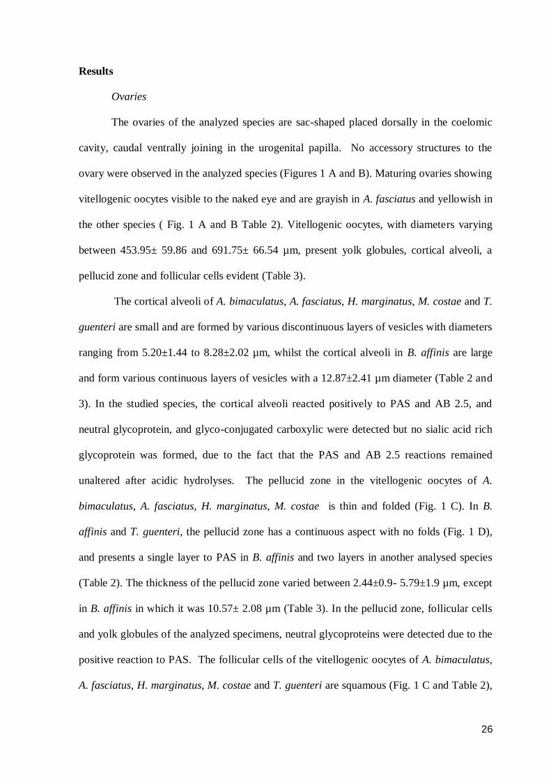
The ovaries of the analyzed species are sac-shaped placed dorsally in the coelomic
cavity, caudal ventrally joining in the urogenital papilla. No accessory structures to the
ovary were observed in the analyzed species (Figures 1 A and B). Maturing ovaries showing
vitellogenic oocytes visible to the naked eye and are grayish in A. fasciatus and yellowish in
the other species ( Fig. 1 A and B Table 2). Vitellogenic oocytes, with diameters varying
between 453.95± 59.86 and 691.75± 66.54 µm, present yolk globules, cortical alveoli, a
pellucid zone and follicular cells evident (Table 3).
The cortical alveoli of A. bimaculatus, A. fasciatus, H. marginatus, M. costae and T.
guenteri are small and are formed by various discontinuous layers of vesicles with diameters
ranging from 5.20±1.44 to 8.28±2.02 µm, whilst the cortical alveoli in B. affinis are large
and form various continuous layers of vesicles with a 12.87±2.41 µm diameter (Table 2 and
3). In the studied species, the cortical alveoli reacted positively to PAS and AB 2.5, and
neutral glycoprotein, and glyco-conjugated carboxylic were detected but no sialic acid rich
glycoprotein was formed, due to the fact that the PAS and AB 2.5 reactions remained
unaltered after acidic hydrolyses. The pellucid zone in the vitellogenic oocytes of A.
bimaculatus, A. fasciatus, H. marginatus, M. costae is thin and folded (Fig. 1 C). In B.
affinis and T. guenteri, the pellucid zone has a continuous aspect with no folds (Fig. 1 D),
and presents a single layer to PAS in B. affinis and two layers in another analysed species
(Table 2). The thickness of the pellucid zone varied between 2.44±0.9- 5.79±1.9 µm, except
in B. affinis in which it was 10.57± 2.08 µm (Table 3). In the pellucid zone, follicular cells
and yolk globules of the analyzed specimens, neutral glycoproteins were detected due to the
positive reaction to PAS. The follicular cells of the vitellogenic oocytes of A. bimaculatus,
A. fasciatus, H. marginatus, M. costae and T. guenteri are squamous (Fig. 1 C and Table 2),

27
whereas in B. affinis they are columnar (Fig. 1 D and Table 2). The follicular cells height in
B. affinis was 28.19±13.71 µm, and, in the other species it varied between 1.52±0.64-
2.12±0.78 µm (Table 3).
Testes
The testes of the analyzed specimens were placed dorsally in the coelomic cavity,
caudal ventrally joining in the urogenital papilla. There were no testicular accessory
structures in the analyzed species (Fig. 2A). Histologically, the testes are constituted by
seminiferous tubules, tubular compartment, and are organized in groups of germinative cells
surrounded by cytoplasmic extensions of the Sertoli cells forming cysts (Figures 2 B-F). In
the analyzed species, the seminiferous tubules branch out forming anastomosis (Fig. 2 B).
Between the seminiferous tubules a connective tissue matrix was observed, which contained
Leydig cells, the interstitial compartment. In each cyst, the germ cells were in the same
developmental stage, but during gonadal maturation the cysts were in different stages, and
presented spermatogonia throughout the testis. During the stage of advanced maturation, the
species A. bimaculatus and A. faciatus presented, in the lumen of the seminiferous tubules,
spermatozoa embedded in an acidophilic secretion (Figures 2 C and D) which was gradually
resorpted during the partially spent stage (Figs. 2 E and F). In this testicular secretion,
neutral glycoproteins were detected from the positive reaction to PAS.
Discussion
The ovarian color of the analyzed species varied between tones of gray in A.
fasciatus and tones of yellow in the other species. Pigments such as astaxanthin, lutein,
taraxanthin and other carotenoids on the yellow/orange yolk coloration have been largely
studied in species feed on Krill and temperate water copepods (Goodwin, 1986; Svensson et
al., 2010). On the other hand, there are no conclusive studies on neotropical fish species
about the origin, composition and function of the carotenoids on the yolk. The oocytes

28
coloration depends on diet and its important for offspring identification, selection of healthy
females and offspring selection for parental care (Blount & Houston, 2000). The carotenoid
derived coloration indicates health status, due to the fact that these substances are
antioxidant and immunostimulant (Olsen & Owens, 1998). Little is known about the
phylogenetic importance of the carotenoids, since it has been analyzed mainly between wide
vertebrate groups (Lubzens et al., 2003). Hence, the yolk color can has a role in sexual
selection, parental care and feeding (Blount & Houston, 2000). The diet and spatial
interaction analysis between A. bimaculatus and A. fasciatus in sympatry showed that the
species present habitat segregation and have feeding selectivity when adult (Arcifa et al.,
1991). Consequently, the color tone variation of the yolk observed in the present study may
be due to the feeding selectivity or to the differences in the carotenoids metabolic pathways.
In this study, larger diameters of cortical alveoli in vitellogenic oocytes were
observed in B. affinis when compared to the cortical alveoli of the other analyzed species.
Furthermore, no difference was found in the histochemical content of the cortical alveoli.
These resemblances in form and histochemical content of the cortical alveoli suggest that the
analyzed species may have a similar polyspermy block mechanism. This hypothesis is
reinforced by means of Bazzoli & Godinho (1994) who analyzed the morphology and
histochemical content of cortical alveoli in 102 species of neotropical fish and found that
species of the same family and subfamily have similar morphology and histochemical
content of cortical alveoli. The authors further suggest that these cortical alveoli
resemblances do not indicate phylogenetic relationships, although no similarity or
parsimony tests were done. Studies using phylogenetic analysis show that the cortical alveoli
morphology is an important parameter amongst the Myctophidae, Melamphaidae,
Bathylagidae and Platytroctidae families of Myctophiformes fish (Belova, 2008).

29
The pellucid zone of the analyzed species reacted positively to the PAS technique,
detecting neutral glycoprotein, a commonly content found in pellucid zone of the vertebrates
(Bazzoli & Rizzo, 1990; Lubzens et al., 2010). These glycoproteins are responsible for the
hardening of external layer of the pellucid zone after the release out of the egg on
environment and bactericidal properties (Hart, 1990; Lubzens et al., 2010). Histometric
analysis showed that the pellucid zone thickness of vitellogenic oocytes in B. affinis was
significantly larger than that of the other species. In teleostean fishes, the thickness of the
pellucid zone is directly related to the place of spawning (Riehl, 1996). Sedentary species
which cover eggs, as well as those which lay eggs in hostile habitats, have a thick pellucid
zone in order to better protect eggs against shocks and to endure the weight of materials
deposited over them (Groot & Alderdice, 1985; Riehl, 1996). On the other hand, sedentary
species which lay eggs in macrophytes, and close to the surface present a thinner pellucid
zone in order to facilitate gas exchange and excretion (Davenport, 1983; Riehl, 1996).
Furthermore, Rizzo et al. (2002) observed that sedentary species of Characidae which have
free or weakly adhesive eggs have a thin pellucid zone, with no specializations, having only
pore-canals on their surface, whilst sedentary species with strongly adhesive eggs possesses
specializations in the pellucid zone such as globular projections that contribute to attach
eggs to the substrate.
The follicular cells of the vitellogenic oocytes of B. affinis are columnar, and
statistically largest than the squamous follicular cells of the other species. The columnar
shape of the follicular cells in B. affinis may be related to egg adhesiveness when the this
cells produce mucosubstances and electron-dense globules which are transferred to the
pellucid zone to attach egg on substrate (Andrade et al., 2001; Rizzo et al¸ 2002), similar to
results observed in Silurus glanis Linnaeus 1758, where the mucosubstances are stored in
electron-dense vesicles, called mucosomes (Abraham et al, 1993). However vitellogenic

30
oocytes of A. bimaculatus have squamous follicular cells and their eggs are slightly adhesive
(Sato et al., 2003).
In this study, the morphologic characteristics of the testes showed a pattern similar to
that of the majority of Charciformes whose realize external fertilization, not possessing
accessory structures to their gonads ones (Isaac-Jr., 1999; Bazzoli, 2003; Gonçalves et al.,
2006). Histologically, spermatogonia were found throughout the testis length, which places
them in the unrestricted spermatogonial type (Grier, 1981). This testicular organization
allows for potential germinative cell production throughout the testis (Schulz & Miura,
2002). On analyzed species, seminiferous tubules forming anastomosis were observed,
which characterize the testes as being of the anastomosing tubular type, plesiomorphic
morphology amongst the bonefish (Parenti & Grier, 2004). This morphology has been
reported for other Characidae, (Grier et al., 1980; Martins et al., 2010), reinforcing the fact
that the testicular morphology is an important parameter in phylogenetic studies (Parenti &
Grier, 2004).
Despite the fact that the testicular morphology of the analyzed species does not
present remarkable differences, acidophilic secretion in the seminiferous tubules lumen of A.
bimaculatus and A. fasciatus was found, similar to others Astyanax spp (Carvalho et al.,
2009; Costa et al., 2009). The acidophilic secretion in the testes maturing stages of and its
resorption in the partially spent males of Astyanax spp may be done by the Sertoli cells,
since they present, besides phagocytic activity, a large quantity of synthesis organelles in the
cytoplasm (Pudney, 1993). Testicular secretion on fishes has been widely reported in
temperate water species (Lahnsteiner, 2003; Chowdhury & Joy, 2007). On the other hand,
in tropical fish, secretion in the seminiferous tubules is common in Siluriformes (Singh &
Joy, 1998; Santos et al., 2001; Guimarães-Cruz & Santos, 2004; Franceschini-Vicentini et
al., 2007) and rare in Characiformes (Isaac-Jr., 1999; Gonçalves et al., 2006). In temperate

31
water species, as well as Siluriformes, the testicular secretion is produced by the seminal
vesicles or tubules in the caudal region of the testes (Lahnsteiner et al., 1992; Lahnsteiner et
al., 1994; Sigh & Joy, 1998; Santos et al., 2001; Lahnsteiner, 2003; Guimarães-Cruz &
Santos, 2004; Franceschini-Vicentini et al., 2007). In A. bimaculatus and A. fasciatus,
however, the secretion observed in the seminiferous tubules seems to be secreted andr
esorpted throughout the testes, due to the fact that it is found in the seminiferous tubules of
the cranial, medium and caudal regions. In the testicular secretion of fishes, the presence of
monosaccharides, polysaccharides, mucopolysaccharides, enzymes, proteins, lipids and
hormones has been reported, doing the following functions across the storage and
spermatozoa maturation: maintaining pH of the seminiferous tubules, pheromonal function,
lytic activity and a probable role in the egg attach to the substrate (Lahnsteiner et al., 1992;
Lahnsteiner et al., 1994; Lahnsteiner, 2003; Chowdhury & Joy, 2007). The glycoproteic
acidophilic secretion observed in the seminiferous tubules of Astyanax spp may be related to
the energy supply to the spermatozoa, similar to Gonçalves et al. (2006) wich reported it on
the Characidae Brycon orthotaenia Günther, 1864.
The present comparative analysis shows phenotypic resemblances of the gonadal
morphology between the assessed species, which probably occurs by a phylogenetic
closeness. This study showed that the genera Astyanax, Hemigrammus, Moenkhausia and
Triporhteus have similar oocytes characteristics, but differ from Bryconops in relation to the
cortical alveoli organization and diameter, follicular cell height and pellucid zone thickness
and its number of layers. Rizzo et al. (2002) also observed differences in Bryconops eggs in
comparison to Asyanax and Triportheus through an ultrastructural analysis of the egg suface.
The phylogenetic analysis of Mirande (2009) also included Astyanax, Bryconops and
Triportheus in separated clades of Characidae family. The acidophilic secretion observed in
the seminiferous tubules of A. bimaculatus and A. fasciatus was also reported in other

32
Characidae genera, such as Salminus (Isaac-Jr, 1999) and Brycon (Gonçalves et al., 2006),
which presented different reproductive strategies those ones of Astyanax spp (Godinho et al.,
2010).
Acknowledgements
The authors thank the partnership of CEMIG-GT/CODEVASF and the Brazilian research
foundations CNPq, CAPES and FAPEMIG for financial support.
References
Abraham, M., Hilge, V., Riehl, R. and Iger, Y. 1993. Muco-follicle cells of the jelly coat in
the oocyte of the sheatfish (Silurus glanis L.).- Journal of Morphology 217 (1): 37-43.
Andersen, M. L., D’Almeida, V., Ko, G. M., Martins, P. J. F., Magalhães L. E. and Tufik, S.
2008. Princípios Éticos e Práticos do Uso de Animais de Experimentação, pp. 167, São
Paulo, Unifesp.
Andrade, R. F., Bazzoli, N., Rizzo, E., & Sato, Y. 2001. Continuous gametogenesis in the
neotropical freshwater teleost, Bryconops affinis (Pisces: Characidae).- Tissue and Cell 35
(5): 524-532.
Arcifa, M. S., Northcote, T. G. and Froelich, O. 1991. Interactive ecology of two cohabiting
characin fishes (Astyanax fasciatus and Astyanax bimaculatus) in eutrophicc brazilian
reservoir.- Journal of Tropical Ecology 7 (2): 257-268.
Bazzoli, N. & Rizzo, E. 1990. A comparative cytological and cytochemical study of the
oogenesis in ten Brazilian teleost fish species.- European Archives of Biology 101: 399-410.
Bazzoli, N. & Godinho, H. P. 1994. Cortical alveoli in oocytes of freshwater neotropical
teleost fish.- Bolletino di Zoology 61 (4): 301-308.
Bazzoli, N. 2003. Parâmetros reprodutivos de peixes de interesse comercial na região de
Pirapora In.: Godinho, H. P & Godinho, A. L. Águas, peixes e pescadores do São Francisco
das Minas Gerais. pp. 291-306, Belo Horizonte, Editora PUC Minas.
Belova, G. V. 2008. Oocyte morphology of several mesopelagic fishes in connection with
their taxonomic status and habitat conditions.- Russian Journal of Marine Biology 34 (2):
110-117.
Blount, J. D. & Houston, D. C. 2000. Why egg yolk is yellow.- Tree 15: 47-49.
Carvalho, P. A., Paschoalini, A. L., Santos, G. B., Rizzo, E. and Bazzoli, N. 2009.
Reproductive Biology of Astyanax fasciatus (Pisces: Characiformes) in a reservoir in
southeastern Brazil.- Journal of Applied Ichthyology 25: 306-313.
Casali, R. C. V., Vono, V., Godinho, H. P., Luz, R. K. and Bazzoli, N. 2010. Passage and
reproductive activity of fishes in the Igarapava fish ladder, Grande River, southeastern
Brazil.- River Research and Applications 26: 157-165.

33
Chowdhury, I. & Joy, K. P. 2007. Seminal vesicle and its role in the reproduction of
teleosts.- Fish Physiology and Biochemistry 33 (4): 383-398.
Costa, F. G., Adolfi, M. C., Andrade, C. G. T. J. and Borella, M. I. 2009. Changes of the
testis of Astyanax altiparanae during the reproductive cycle. A structural and ultrastructural
study.- Proceedings of XXII Congress of Brazilian Society of Microscopy and
Microanalysis: B02552.
Davenport, J. 1983. Oxygen and the developing eggs and larvae of the lumpfish,
Cyclopterus lumpus L. Journal of Marine Biology 63: 633-640.
Fishelson, L. & Gon, O. 2008. Comparative oogenesis in cardinal fishes (Apogonidae,
Perciformes), with special focus on the adaptative structures of the egg envelopes.-
Environmental Biology of Fishes 81 (4): 397-414.
Franceschini-Vicentini, I. B., Papa, L. P., Bombonato, M. T. S., Vicentini, C. A., Ribeiro, K.
and Orsi, A. M. 2007. A histological study of the seminal vesicle of the armoures catfish
Corydoras aeneus.- Anatomia, Histologia, Embryologia 36: 111-115.
Géry, J. 1977. Characoids of the world. pp. 672, Neptune City, T.F.H Publications Inc. Ltda.
Gonçalves, T. L., Bazzoli, N. and Brito, M. F. G. 2006. Gametogenesis and reproduction of
the matrinxã Brycon orthotaenia (Günther, 1864) (Pisces: Characidae) in the São Francisco
River, Minas Gerais, Brazil.- Brazilian Journal of Biology 66 (2A): 513-522.
Grier, H. J., Linton, J. R., Leatherland, J. F. & deVlaming, V. L. 1980. Structural evidence
for two difference testicular types in teleost fishes.- American Journal of Anatomy 159: 331–
345.
Grier, H. J. 1981. Cellular organization of the testis and spermatogenesis in fishes. -
American Zoologist 21 (2): 345-357.
Groot, E. P. & Alderice, D. F. 1985. Fine structure of the external egg membrane of five
species of pacific salmon and steelhead trout.- Canadian Journal of Zoology 63: 552-566.
Godinho, A. L., Lamas, I. R., and Godinho, H. P. 2010. Reproductive ecology of Brazilian
freshwater fishes.- Environmental Biology of Fishes 87: 143-162.
Goodwin, T. W. 1986. Metabolism, nutrition and function of carotenoids.- Annual Review of
Nutrition 6: 273-297.
Guimarães-Cruz, R. J. & Santos, J. E. 2004. Testicular structure of three species of
neotropical freshwater pimelodids (Pisces: Pimelodidae).- Revista Brasileira de Zoologia 21
(2): 267-271.
Hart, N. H. 1990. Fertilization in teleost fishes: mechanism of sperm – egg interactions. -
International Review of Cytology 121: 1-66.
Honorato-Sampaio, K., Santos, G. B., Bazzoli, N. and Rizzo, E. 2009. Observations on the
seasonal breeding biology and the fine structure of the egg surface in the white piranha

34
Serrasalmus Brandtii from the São Francisco River basin, Brazil.- Journal of Fish Biology
75: 1874-1882.
Isaac-Jr., J. B. 1999. Gametogênese e ciclo reprodutivo do dourado, Salminus brasiliensis
(Cuvier, 1817), do rio São Francisco, Minas Gerais. Msc., Dissertation pp. 89, Graduate
Program in Vertebrate Zoology from PUC Minas, Brazil.
Lahnsteiner, F., Seiwal, M., Patzner, R. A. and Ferrero, E. A. 1992. The seminal vesicles of
the male grass goby, Zosterisessor ophiocephalus (Teleostei, Gobidae).- Zoomorphology
111: 239-248.
Lahnsteiner, F., Patzner, R. A. and Weissman, T. 1994. The testicular main duct and
spermatic ducts in cyprinid fishes. I. Morphology, fine structure and histochemistry.-
Journal of Fish Biology 44: 937-951.
Lahnsteiner, F. 2003. Morphology, fine structure, biochemistry, and function of spermatic
ducts in marine fishes.- Tissue and Cell 35 (5): 363-373.
Lima, F. C. T., Malabarba, L. R., Buckup, P. A., Pezzi da Silva, J. F., Vari, R., Harold, P.
A., Benine, R., Oyakawa, O. T., Pavanelli, C. S., Menezes, N. A., Lucena, C. A. S.,
Malabarba, M. C. S. L., Lucena, Z. M. S., Reis, R. E., Langeani, F. L. Cassati, L., and
Bertaco, V. A. 2003. Genera Incertae Sedis in Characidae. In: R.E. Reis, S.O. Kullander
and C.J. Ferraris, Jr. (eds.). Checklist of the Freshwater Fishes of South and Central America
p. 106-168, Porto Alegre, EDIPUCRS.
Lubzens, E., Lissauer, L., Leavavi-Sivan, B., Avarre, J. C. and Sammar, M. 2003.
Carotenoid and retinoid transport to fish oocytes and eggs: What is the role of retinol
binding protein?- Molecular Aspects of Medicine 24 (6): 441-457.
Lubzens, E., Young, G., Bobe, J. & Cerdà, J. 2010. Oogenesis in teleosts: how fish eggs are
formed.- General Comparative Endocrinology 165: 367-389.
Martins, Y. S., Moura, D. F., Santos, G. B., Rizzo, E. & Bazzoli, N. 2010. Comparative
folliculogenesis and spermatogenesis of four teleost fish from a reservoir in south-eastern
Brazil.- Acta Zoologica in press.
Mirande, J. M. 2009. Weighted parsimony phylogeny of the family Characidae (Teleostei:
Characidormes).- Cladistics 25: 574-613.
Nelson, J. S. 2006. Fishes of the World. pp. 624, New York, John Wiley & Sons.
Nóbrega, R. H., Batlouni, S. R. and França, L. R. 2008. An overview of functional and
stereological evaluation of spermatogenesis and germ cell transplantation in fish. - Fish
Physiology and Biochemestry 35: 197-206.
Normando, F. T., Arantes, F. P., Luz, R. K., Thomé, R. G., Rizzo, E., Sato, Y and Bazzoli,
N. 2009. Reproduction and fecundity of tucunaré, Cichla kelberi (Perciformes: Cichlidae),
an exotic species in Três Marias reservoir, south eastern Brazil.- Journal of Applied
Ichthyology 25: 299-305.

35
Nuñez, K. & Duponchelle, F. 2009. Towards a universal scale to assess sexual maturation
and related life history traits in oviparous teleost fishes.- Fish Physiology and Biochemistry
35 (1): 167-180.
Olsen, V. A. & Owens, I. P. F. Costly sexual signals: are carotenoids, rare, risky or
required?- Trends in Ecology and Evolution 13 (2): 510-514.
Parenti, L. R. & Grier, H. J. 2004. Evolution and phylogeny of gonad morphology in bony
fishes. - Integrative and Comparative Biology 44 (5): 333-348.
Pearse, A. G. E. 1985. Histochemistry: Theoretical and Applied, pp.1055, London, Churchill
Livingstone.
Pudney, J. 1993. Comparative cytology of the non-mammalian vertebrate Sertoli cell. In.:
Russel, L. D. & M.D. Griswold (eds.) The Sertoli Cell, pp. 611-657, Clearwater, Cache
River Press.
Reis, R. E., Kullander, S. O. & Ferraris-Jr., C. J. 2003. Checklist of the Freshwater Fishes of
South and Central America, pp. 742, Porto Alegre, EDIPUCRS.
Riehl, R. 1996. The ecological significance of the egg envelope in teleosts with special
reference to limnic species.- Limnologica 26: 183-189.
Rizzo, E., Sato, Y., Barreto, B. P. and Godinho, H. P. 2002. Adhesiveness and surface
patterns of eggs in neotropical freshwater teleosts. - Journal of Fish Biology 61 (3): 615-
632.
Santos, J. E., Bazzoli, N., Rizzo, E. & Santos, G. B. 2001. Morphofunctional organization of
the male reproductive system of the catfish Iheringichthys labrosus (Lutken, 1874)
(Siluriformes: Pimelodidae).- Tissue and Cell 33 (5): 533-540.
Sato, Y., Fenerechi-Verani, N., Nuñer, A. P. O., Godinho, H. P. and Verani, J. R. 2003.
Padrões reprodutivos de peixes da bacia do São Francisco. In.: Godinho, H. P & A.L.
Godinho. Águas, peixes e pescadores do São Francisco das Minas Gerais. pp. 229-274, Belo
Horizonte, Editora PUC Minas.
Schulz, R. W. & Miura, T. 2002. Spermatogenesis and its endocrine regulation. - Fish
Physiology and Biochemestry 26 (1): 43-56.
Schulz, R. W., França, L. R., Lareyre, J. J., LeGac, F., Chiarini-Garcia, H., Nóbrega, R. H.
and Miura, T. 2009. Spermatogenesis in fish. - General and Conparative Endocrinology 165
(3): 390-411.
Singh, M. S. & Joy, K. P. 1998. A comparative study on histochemical distribution of some
enzymes related to steroid and glucuronide synthesis in seminal vesicle and testicular vesicle
and testis of catfish, Clarias batrachus.- Acta Biologica Hungarian 49: 143-154.
Svensson, P. A., Blount, J. D., Forsgren, E. and Tamudsen, T. 2010. Female ornamentation
and egg carotenoids of six sympatrics gobies.- Journal of Fish Biology 75 (10): 2777-2787.

36
Vieira, A. B. C., Salvador-Jr., L. F., Melo, R. M. C., Santos, G. B. and Bazzoli, N. 2009.
Reproductive Biology of the peacock bass Cichla piquiti (Perciformes: Cichlidae), an exotic
species in a Neotropical reservoir.- Neotropical Ichthyology 7 (4): 745-750.
Weitzman, S. H. & Malabarba, L. R. 1998. Perspectives about the phylogeny and
classification of the Characidae (Teleostei: Characiformes). In: Malabarba, L. R., R.E. Reis,
R.P. Vari, , Z.M.S. Lucena, C.A.S. Lucena (Eds.), Phylogeny and Classification of
Neotropical Fishes. pp., 161-170, Porto Alegre, EDIPUCRS.

37
Table 1: Biometrics of six species from Incertae Sedis genera in Characidae of the São
Francisco River Basin, Brazil.
Species n TL (cm) BW (g)
male female male female male female A. bimaculatus 22 41 7.4±2.6 11.3±1.2 7.5±4.1 11.3±5.6
A. fasciatus 36 30 8.3±2.1 9.2±1.2 7.2±3.6 10.5±6.3
B. affinis 32 45 10.2±1.4 11.4±2.4 9.2±3.4 12.6±4.1
H. marginatus 29 31 2.1±1.3 3.6±0.9 0.4±0.5 0.8±0.3
M. costae 18 24 3.2±0.8 4.1±2.1 1.2±0.3 1.9±0.6
T. guenteri 22 32 14.1±2.5 17.2±1.4 19.3±2.5 22.3±6.3
TL= total lenght; BW= body weight; n=number of collected individuals.

38
Table 2: Vitellogenic oocyte morphology of six species from Incertae Sedis genera in Characidae of
the São Francisco River, Brazil.
Species
Yolk
colour
Cortical alveloli Pellucid zone Follicular
cells height shape organization shape Number of layers
A. bimaculatus yellowish small discontinuous folded 2 squamous
A. fasciatus grayish small discontinuous folded 2 squamous
B. affinis yellowish large continuous continuous 1 columnar
H. marginatus yellowish small discontinuous folded 2 squamous
M. costae yellowish small discontinuous folded 2 squamous
T. guenteri yellowish small discontinuous continuous 2 squamous

39
Table 3: Vitellogenic oocyte histometry (µm) of six species from Incertae Sedis genera in
Characidae of the São Francisco River Basin, Brazil.
Structure
Species Diameter
Cortical alveoli
diameter
Zona pellucida
thickness
Follicular cells
height A. bimaculatus 563.90± 131.70ª 7.66±2.86
a, b 4.63± 1.06
a 1.95± 0.65
a
A. fasciatus 517.43± 43.51a, c
8.28±2.02b 5.79±1.90
a 2.12±0.78
a
B. affinis 691.75± 66.54b 12.87±2.41
c 10.57± 2.08
b 28.19± 13.71
b
H. marginatus 453.95± 59.86c 6.50± 1.98
a, d 2.44± 0.90
c 1.52± 0.64
a
M. costae 556.58± 64.40ª 6.57±2.08a, d
2.54± 0.75c 1.65± 0.70
a
T. guenteri 677.40± 91.29b 5.20± 1.44
d 5.43±0.94
a 1.57± 0.65
a
Different superscript letters indicate statistically significant differences (P < 0.05).

40
Figure 1: Ovaries of genera Incertae Sedis in Characidae: Mature ovaries of T. guenteri (A) and
A. fasciatus (B) with yellowish and grayish coloured vitellogenic oocytes. Detail of the vitellogenic oocytes of A. bimaculatus (C) and B. affinis (D). Hematoxylin-eosin. F= pellucid
zone folds; FC= follicular cells, YG= yolk globules, ZP= pellucid zone.

41
Figure 2: Testes of genera Incertae Sedis in Characidae: Mature testes of M. costae (A).
Anastomosing tubular testicle of H. marginatus (B). Acidophilic secretion in the testis of
A. bimaculatus (C and D). Resorption of acidophilic secretion in the testis of A. fasciatus
(E and F). *= anastomosis between seminiferous tubules, arrows = glycoproteic acidophilic secretion in the seminiferous tubules lumen, arrowheads= resorption vesicles,
S= Sertoli cell.
42
5. CONCLUSÕES
- As semelhanças fenotípicas observadas na morfologia gonadal das espécies analisadas,
provavelmente, deve-se ao fato das mesmas serem intimamente relacionadas.
- Macroscopicamente, não foram observadas diferenças marcantes na morfologia ovariana
macroscópica das espécies analisadas, exceto pela coloração acinzentada dos ovários de A.
fasciatus em relação à coloração amarelada das outras espécies.
- Histologicamente observaram-se diferenças morfológicas e estatísticas nos ovócitos
vitelogênicos de B. affinis em relação às outras espécies analisadas, apresentando maiores
valores de diâmetro do alvéolo cortical, espessura da zona pelúcida e altura das células
foliculares.
- Os testículos das espécies analisadas seguiram o padrão comum para Characiformes em
nível macroscópico e histológico, exceto pela presença de secreção acidófila glicoproteica
nos túbulos seminíferos de A. bimaculatus e A. fasciatus.

43
REFERÊNCIAS
Andrade, R. F., Bazzoli, N., Rizzo, E., & Sato, Y. 2001. Continuous gametogenesis in the
neotropical freshwater teleost, Bryconops affinis (Pisces: Characidae).- Tissue and Cell 35
(5): 524-532.
Arcifa, M. S., Northcote, T. G. and Froelich, O. 1991. Interactive ecology of two cohabiting
characin fishes (Astyanax fasciatus and Astyanax bimaculatus) in eutrophicc brazilian
reservoir.- Journal of Tropical Ecology 7 (2): 257-268.
Barbosa, P. M. M. & Matsumura-Tundisi, T. 1984. Consumption of zooplanktonic
organisms by Astyanax fasciatus Cuvier, 1818 (Osteichthyes, Characidae) in Lobo (Broa)
Reservoir, São Carlos, SP, Brazil.- Hydrobiologia 113 (1): 171-182.
Bazzoli, N. & Godinho, H. P. 1994. Cortical alveoli in oocytes of freshwater neotropical
teleost fish.- Bolletino di Zoology 61 (4): 301-308.
Bazzoli, N. & Godinho, H. P. 1995. Comparative morphology of yolk nucleus (Balbaniai
body) in freshwater neotropical teleost fish.- Revista Brasileira de Zoologia 55 (2): 207-214.
Bazzoli, N., Rizzo, E., Santos, J. E. and Sato, Y. 1996. Dinâmica da ovogênese de peixes
forrageiros da Represa de Três Marias, Minas Gerais: estudo histológico e histoquímico.-
Bios 4: 5-10.
Bazzoli, N., Rizzo, E. Santos, J. E. and Sato, Y. 1997. Biologia reprodutiva de quatro
espécies de peixes forrageiros da represa de Três Marias.- Bios 5 (5): 17-28.
Belova, G. V. 2008. Oocyte morphology of several mesopelagic fishes in connection with
their taxonomic status and habitat conditions.- Russian Journal of Marine Biology 34 (2):
110-117.
Britski, H. A., Sato, Y. and Rosa, A. B. S. 1986. Manual de identificação de peixes da
região de Três Marias: Com chaves de identificação para peixes da bacia do São
Francisco. Brasília, CODEVASF, 115p.
Carvalho, P. A., Paschoalini, A. L., Santos, G. B., Rizzo, E. and Bazzoli, N. 2009.
Reproductive Biology of Astyanax fasciatus (Pisces: Characiformes) in a reservoir in
southeastern Brazil.- Journal of Applied Ichthyology 25: 306-313.
Cangussu-Mariani, S. D., Rizzo, E. & Bazzoli, N. 1990. Morfologia e desenvolvimento do
núcleo vitelínico de lambari Astyanx bimaculatus (Linnaeus, 1758) (Osteichthyes,
Characidae).- Revista Brasileira de Zoologia 7: 207-213.
Crippa, V. E. L., Hahn, N. S. and Fugi, R. 2009. Food resource used by small-sized fish in
macrophyte patches in ponds of the upper Paraná River foodplain.- Acta Scientiarium:
Biological Sciences 32 (2): 119-125.
DeFalco, T. & Capel, B. 2009. Gonad morphogenesis in vertebrates: Divergent means to a
convergent end.- Annual review of Cell and Developmental biology 25: 457-482.

44
Fishelson, L. & Gon, O. 2008. Comparative oogenesis in cardinal fishes (Apogonidae,
Perciformes), with special focus on the adaptative structures of the egg envelopes.-
Environmental Biology of Fishes 81 (4): 397-414.
Garcia, J. A. D., Chini, H. A. S., Maistro, E. L. and Quagio-Grassioto, I. 2001. Dynamics
and cytochemistry of oogenesis in Astyanax fasciatus (Cuvier) (Teleostei, Characiformes,
Characidae) from Rio Sapucaí, Minas Gerais State, Brazil.- Revista Brasileira de Zoologia
18 (4): 1057-1064.
Géry, J. 1977. Characoids of the world. pp. 672, Neptune City, T.F.H Publications Inc. Ltda.
Godinho, A. L. 1994. Biologia reprodutiva da piaba-facão, Triportheus guenteri
(Characiformes, Characidae) e o manejo hidrológico da represa de Três Marias.- Revista
Brasileira de Biologia 54 (3): 515-524.
Gomes, J. H. C. & Verani, J. R. 2003. Alimentação de peixes do reservatório de Três
Marias. In.: Godinho, H. P & A.L. Godinho. Águas, peixes e pescadores do São Francisco
das Minas Gerais. pp.195-228, Belo Horizonte, Editora PUC Minas.
Graciolli, G., Azevedo, M. A., and Melo, F. A. G. 2003. Comparative studies of the diet of
Glandulocaudinae and Tetragonopterinae (Ostariophisi: Characidae) in a small stream in
Southern Brazil.- Studies on Neotropical Fauna Environment 38 (2): 95-103.
Lima, F. C. T., Malabarba, L. R., Buckup, P. A., Pezzi da Silva, J. F., Vari, R., Harold, P.
A., Benine, R., Oyakawa, O. T., Pavanelli, C. S., Menezes, N. A., Lucena, C. A. S.,
Malabarba, M. C. S. L., Lucena, Z. M. S., Reis, R. E., Langeani, F. L. Cassati, L., and
Bertaco, V. A. 2003. Genera Incertae Sedis in Characidae. In: R.E. Reis, S.O. Kullander
and C.J. Ferraris, Jr. (Eds.). Checklist of the Freshwater Fishes of South and Central
America p. 106-168, Porto Alegre, EDIPUCRS.
Miranda, A. C. L. 1996. Reprodução de Astyanax bimaculatus lacustris em viveiros e estudo
histológico e ultra-estrutural de atresia folicular de Astyanax bimaculatus lacustris e
Leporinus reinhardti em gaiolas de aqüicultura. UGMG, Belo Horizonte, (Dissertação
mestrado), pp. 138.
Miranda, A. C. L., Bazzoli, N., Rizzo, E. and Sato, Y. 1999. Ovarian follicular atresia in two
teleost species: a histological and ultrastructural study.- Tissue and Cell 31: 480-488.
Mirande, J. M. 2009. Weighted parsimony phylogeny of the family Characidae (Teleostei:
Characidormes).- Cladistics 25: 574-613.
Nelson, J. S. 2006. Fishes of the World. pp. 624, New York, John Wiley & Sons.
Nogueira, B. P., Bazzoli, N., Santos, J. E. and Barros, M. D. 1997. Biologia reprodutiva do
Bryconops cf affinis = Creatochanes affinis (Günther, 1864) (Teleostei: Characiformes) na
lagoa do Pantaninho, Lagoa da Prata, Minas Gerais.- Bios 5 (5): 43-51.
Parenti, L. R. & Grier, H. J. 2004. Evolution and phylogeny of gonad morphology in bony
fishes. - Integrative and Comparative Biology 44 (5): 333-348.
45
Planquette, P., Keith, P. and Le Bail, Y. 1996. Atlas of freshwater fishes of Guyana. 422p,
Paris, Natural Heritage Collection.
Pompeu, P. S. & Godinho, H. P. 2003. Dieta e estrutura trófica das comunidades de peixes
de três lagoas marginais do médio São Francisco. In.: Godinho, H. P & A.L. Godinho.
Águas, peixes e pescadores do São Francisco das Minas Gerais. pp.183-194, Belo
Horizonte, Editora PUC Minas.
Pudney, J. 1993. Comparative cytology of the non-mammalian vertebrate Sertoli cell. In.:
Russel, L. D. & M.D. Griswold (eds.) The Sertoli Cell, pp. 611-657, Clearwater, Cache
River Press.
Reis, R. E., Kullander, S. O. & Ferraris-Jr., C. J. 2003. Checklist of the Freshwater Fishes of
South and Central America, pp. 742, Porto Alegre, EDIPUCRS.
Rizzo, E., Sato, Y., Barreto, B. P. and Godinho, H. P. 2002. Adhesiveness and surface
patterns of eggs in neotropical freshwater teleosts. - Journal of Fish Biology 61 (3): 615-
632.
Sato, Y., Fenerechi-Verani, N, Nuñer, A. P. O., Godinho, H. P. and Verani, J. R. 2003.
Padrões reprodutivos de peixes da bacia do São Francisco. In.: Godinho, H. P & A.L.
Godinho. Águas, peixes e pescadores do São Francisco das Minas Gerais. pp. 229-274, Belo
Horizonte, Editora PUC Minas.
Schulz, U. H. and Martins-Júnior, H. 2001 Astyanax fasciatus as bioindicator of water
pollution of Rio dos Sinos, RS, Brasil. Brazilian Journal of Biology 61 (4): 615-622.
Silva, R. M., Bastos, G. B. and Ratton, T. 2006. Fish comunity structure of Juramento
reservoir, são francisco River basin, Minas Gerais, Brazil.- Revista Brasileira de Zoologia
23 (3): 832-840.
Vilella, F. S., Becker, F. G. and Hartz, S. M. 2002. Diet of Astyanax species (Teleostei,
Characidae) in an atlantic forest River in Southern Brazil.- Brazilian Archives in Biology
and Technology 45 (2): 223-232.
Weitzman, S. H. & Malabarba, L. R. 1998. Perspectives about the phylogeny and
classification of the Characidae (Teleostei: Characiformes). In: Malabarba, L. R., R.E. Reis,
R.P. Vari, , Z.M.S. Lucena, C.A.S. Lucena (Eds.), Phylogeny and Classification of
Neotropical Fishes. pp., 161-170, Porto Alegre, EDIPUCRS.