a case of incomplete atypical femoral fracture with ... · a case of incomplete atypical femoral...
TRANSCRIPT
A Case of Incomplete Atypical Femoral Fracture with Histomorphometrical Evidence of Osteomalacia
Hiroyuki Tsuchiea*, Naohisa Miyakoshib, Tomio Nishic, Hidekazu Abec, Toyohito Segawac, and Yoichi Shimadab
aDivision of Orthopedic Surgery, Nakadori General Hospital, Akita 010-8577, Japan, bDepartment of Orthopedic Surgery, Akita University Graduate School of Medicine, Akita 010-8543, Japan, and
cUgo Municipal Hospital, 44-5 Otomichi, Nishomonai, Ugo, Akita 012-1131, Japan
Roughly half of the femoral fracture patients diagnosed with AFF according to the criteria suggested by a task force of the American Society for Bone and Mineral Research (ASBMR) have not under-gone bisphosphonate (BP) therapy. One suspected cause of such fractures is severe bone loss due to osteomalacia, but the pathogenesis remains unknown. We report a case of an 84-year-old woman with AFF not treated by BP therapy, in whom underlying osteomalacia was histologically diagnosed. The involvement of femoral curvature and spino-pelvic malaligment in the fracture in the present case was considered.
Key words: osteomalacia, atypical fracture, femur, osteoporosis, kyphosis
lthough bisphosphonates (BPs) are the gold standard for osteoporosis pharmacotherapy,
several adverse effects related to their long-term use have recently been reported, such as osteonecrosis of the jaw (ONJ) and atypical low-energy subtrochanteric and diaphyseal femoral fractures due to severely sup-pressed bone turnover (SSBT) [1, 2]. These frac-tures are typically diagnosed as atypical femoral fracture (AFF). However, many patients have been reported [3] who fulfill the AFF criteria written by a task the American Society for Bone and Mineral Research (ASBMR) [4] but have not received BP treatment. One of the suspected causes of such frac-tures is severe bone loss due to osteomalacia, but the pathogenesis about the way that osteomalacia becomes AFF remains unknown.
We describe herein a unique case of AFF without BP therapy, in which underlying osteomalacia was histologically diagnosed. The involvement of femoral curvature and spino-pelvic malaligment in the fracture is also discussed. The patients and their families were informed that data from their case would be submitted for publication, and they provided their consent.
Case Report
An 84-year-old woman presented at our outpatient clinic who had been experiencing pain of the bilateral thighs for 2 months. There was no history of trauma. The pain was not debilitating at the onset but had gradually increased over the first month, such that she had not been able to walk independently without a walker thereafter. She had only been treated for
A
Acta Med. Okayama, 2015Vol. 69, No. 1, pp. 59ン63CopyrightⒸ 2015 by Okayama University Medical School.
Case Report http ://escholarship.lib.okayama-u.ac.jp/amo/
Received May 29, 2014 ; accepted September 3, 2014.*Corresponding author. Phone : +81ン18ン833ン1122; Fax : +81ン18ン831ン9418E-mail : [email protected] (H. Tsuchie)
Conflict of Interest Disclosures: No potential conflict of interest relevant to this article was reported.
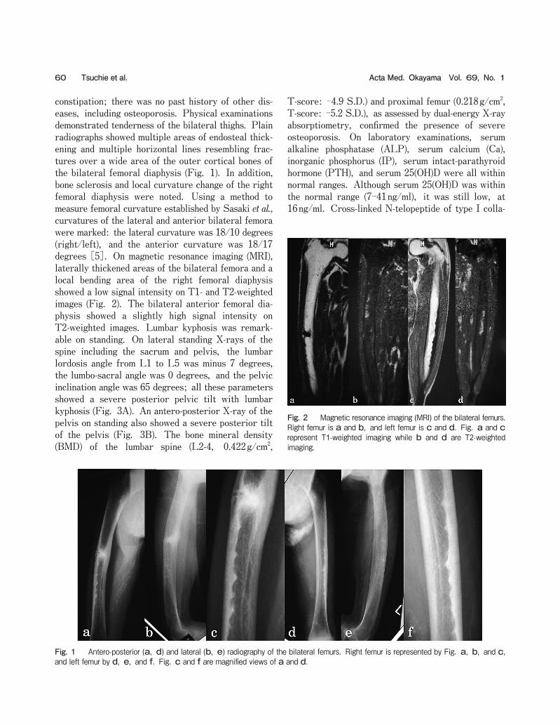
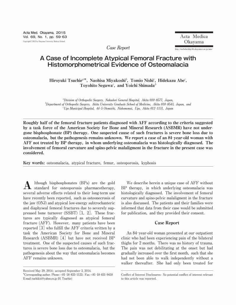
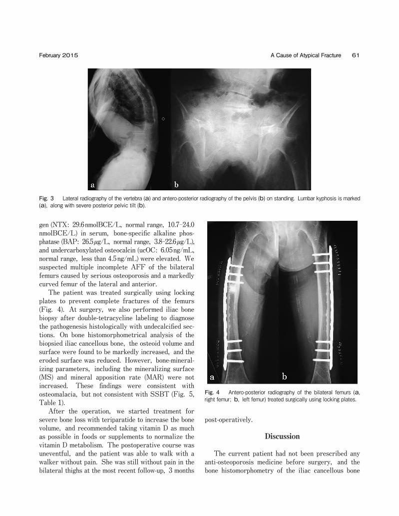
constipation; there was no past history of other dis-eases, including osteoporosis. Physical examinations demonstrated tenderness of the bilateral thighs. Plain radiographs showed multiple areas of endosteal thick-ening and multiple horizontal lines resembling frac-tures over a wide area of the outer cortical bones of the bilateral femoral diaphysis (Fig. 1). In addition, bone sclerosis and local curvature change of the right femoral diaphysis were noted. Using a method to measure femoral curvature established by Sasaki et al., curvatures of the lateral and anterior bilateral femora were marked: the lateral curvature was 18/10 degrees (right/left), and the anterior curvature was 18/17 degrees [5]. On magnetic resonance imaging (MRI), laterally thickened areas of the bilateral femora and a local bending area of the right femoral diaphysis showed a low signal intensity on T1- and T2-weighted images (Fig. 2). The bilateral anterior femoral dia-physis showed a slightly high signal intensity on T2-weighted images. Lumbar kyphosis was remark-able on standing. On lateral standing X-rays of the spine including the sacrum and pelvis, the lumbar lordosis angle from L1 to L5 was minus 7 degrees, the lumbo-sacral angle was 0 degrees, and the pelvic inclination angle was 65 degrees; all these parameters showed a severe posterior pelvic tilt with lumbar kyphosis (Fig. 3A). An antero-posterior X-ray of the pelvis on standing also showed a severe posterior tilt of the pelvis (Fig. 3B). The bone mineral density (BMD) of the lumbar spine (L2-4, 0.422g/cm2,
T-score: -4.9 S.D.) and proximal femur (0.218g/cm2, T-score: -5.2 S.D.), as assessed by dual-energy X-ray absorptiometry, confirmed the presence of severe osteoporosis. On laboratory examinations, serum alkaline phosphatase (ALP), serum calcium (Ca), inorganic phosphorus (IP), serum intact-parathyroid hormone (PTH), and serum 25(OH)D were all within normal ranges. Although serum 25(OH)D was within the normal range (7-41ng/ml), it was still low, at 16ng/ml. Cross-linked N-telopeptide of type I colla-
60 Acta Med. Okayama Vol. 69, No. 1Tsuchie et al.
Fig. 1 Antero-posterior (a, d) and lateral (b, e) radiography of the bilateral femurs. Right femur is represented by Fig. a, b, and c, and left femur by d, e, and f. Fig. c and f are magnified views of a and d.
Fig. 2 Magnetic resonance imaging (MRI) of the bilateral femurs. Right femur is a and b, and left femur is c and d. Fig. a and c represent T1-weighted imaging while b and d are T2-weighted imaging.
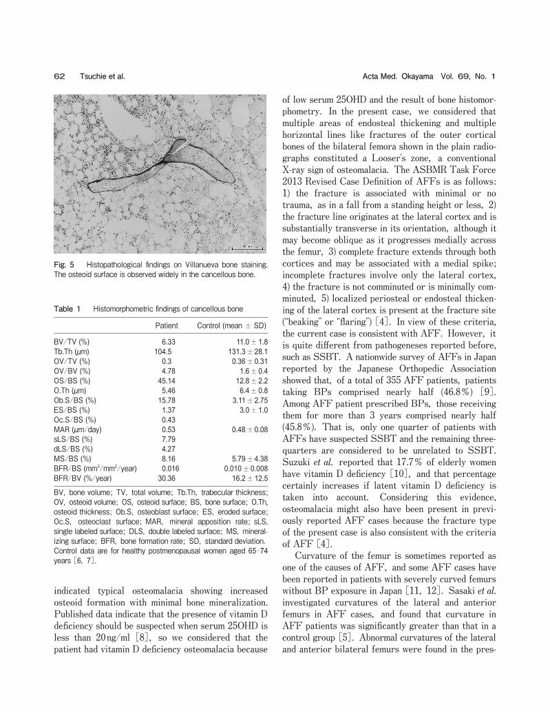
gen (NTX: 29.6nmolBCE/L, normal range, 10.7-24.0 nmolBCE/L) in serum, bone-specific alkaline phos-phatase (BAP: 26.5µg/L, normal range, 3.8-22.6µg/L), and undercarboxylated osteocalcin (ucOC: 6.05ng/mL, normal range, less than 4.5ng/mL) were elevated. We suspected multiple incomplete AFF of the bilateral femurs caused by serious osteoporosis and a markedly curved femur of the lateral and anterior. The patient was treated surgically using locking plates to prevent complete fractures of the femurs (Fig. 4). At surgery, we also performed iliac bone biopsy after double-tetracycline labeling to diagnose the pathogenesis histologically with undecalcified sec-tions. On bone histomorphometrical analysis of the biopsied iliac cancellous bone, the osteoid volume and surface were found to be markedly increased, and the eroded surface was reduced. However, bone-mineral-izing parameters, including the mineralizing surface (MS) and mineral apposition rate (MAR) were not increased. These findings were consistent with osteomalacia, but not consistent with SSBT (Fig. 5, Table 1). After the operation, we started treatment for severe bone loss with teriparatide to increase the bone volume, and recommended taking vitamin D as much as possible in foods or supplements to normalize the vitamin D metabolism. The postoperative course was uneventful, and the patient was able to walk with a walker without pain. She was still without pain in the bilateral thighs at the most recent follow-up, 3 months
post-operatively.
Discussion
The current patient had not been prescribed any anti-osteoporosis medicine before surgery, and the bone histomorphometry of the iliac cancellous bone
61A Cause of Atypical FractureFebruary 2015
Fig. 3 Lateral radiography of the vertebra (a) and antero-posterior radiography of the pelvis (b) on standing. Lumbar kyphosis is marked (a), along with severe posterior pelvic tilt (b).
Fig. 4 Antero-posterior radiography of the bilateral femurs (a, right femur; b, left femur) treated surgically using locking plates.
indicated typical osteomalacia showing increased osteoid formation with minimal bone mineralization. Published data indicate that the presence of vitamin D deficiency should be suspected when serum 25OHD is less than 20ng/ml [8], so we considered that the patient had vitamin D deficiency osteomalacia because
of low serum 25OHD and the result of bone histomor-phometry. In the present case, we considered that multiple areas of endosteal thickening and multiple horizontal lines like fractures of the outer cortical bones of the bilateral femora shown in the plain radio-graphs constituted a Looserʼs zone, a conventional X-ray sign of osteomalacia. The ASBMR Task Force 2013 Revised Case Definition of AFFs is as follows: 1) the fracture is associated with minimal or no trauma, as in a fall from a standing height or less, 2) the fracture line originates at the lateral cortex and is substantially transverse in its orientation, although it may become oblique as it progresses medially across the femur, 3) complete fracture extends through both cortices and may be associated with a medial spike; incomplete fractures involve only the lateral cortex, 4) the fracture is not comminuted or is minimally com-minuted, 5) localized periosteal or endosteal thicken-ing of the lateral cortex is present at the fracture site (“beaking” or “flaring”) [4]. In view of these criteria, the current case is consistent with AFF. However, it is quite different from pathogeneses reported before, such as SSBT. A nationwide survey of AFFs in Japan reported by the Japanese Orthopedic Association showed that, of a total of 355 AFF patients, patients taking BPs comprised nearly half (46.8オ) [9]. Among AFF patient prescribed BPs, those receiving them for more than 3 years comprised nearly half (45.8オ). That is, only one quarter of patients with AFFs have suspected SSBT and the remaining three-quarters are considered to be unrelated to SSBT. Suzuki et al. reported that 17.7オ of elderly women have vitamin D deficiency [10], and that percentage certainly increases if latent vitamin D deficiency is taken into account. Considering this evidence, osteomalacia might also have been present in previ-ously reported AFF cases because the fracture type of the present case is also consistent with the criteria of AFF [4]. Curvature of the femur is sometimes reported as one of the causes of AFF, and some AFF cases have been reported in patients with severely curved femurs without BP exposure in Japan [11, 12]. Sasaki et al. investigated curvatures of the lateral and anterior femurs in AFF cases, and found that curvature in AFF patients was significantly greater than that in a control group [5]. Abnormal curvatures of the lateral and anterior bilateral femurs were found in the pres-
62 Acta Med. Okayama Vol. 69, No. 1Tsuchie et al.
Fig. 5 Histopathological findings on Villanueva bone staining. The osteoid surface is observed widely in the cancellous bone.
Table 1 Histomorphometric findings of cancellous bone
Patient Control (mean ± SD)
BV/TV (%) 6.33 11.0±1.8Tb.Th (µm) 104.5 131.3±28.1OV/TV (%) 0.3 0.36±0.31OV/BV (%) 4.78 1.6±0.4OS/BS (%) 45.14 12.8±2.2O.Th (µm) 5.46 6.4±0.8Ob.S/BS (%) 15.78 3.11±2.75ES/BS (%) 1.37 3.0±1.0Oc.S/BS (%) 0.43MAR (µm/day) 0.53 0.48±0.08sLS/BS (%) 7.79dLS/BS (%) 4.27MS/BS (%) 8.16 5.79±4.38BFR/BS (mm3/mm2/year) 0.016 0.010±0.008BFR/BV (%/year) 30.36 16.2±12.5
BV, bone volume; TV, total volume; Tb.Th, trabecular thickness; OV, osteoid volume; OS, osteoid surface; BS, bone surface; O.Th, osteoid thickness; Ob.S, osteoblast surface; ES, eroded surface; Oc.S, osteoclast surface; MAR, mineral apposition rate; sLS, single labeled surface; DLS, double labeled surface; MS, mineral-izing surface; BFR, bone formation rate; SD, standard deviation.Control data are for healthy postmenopausal women aged 65-74 years [6, 7].
ent case. In addition, a local bending change of the right femoral diaphysis that increased the curvature was also found. If osteomalacia is a cause of femoral curvature, osteomalacia might also have been present in other previously reported AFF cases with marked curvature of the femur. In the present case, we also speculate that severe lumbar kyphosis with posterior pelvic tilt contributed to the increased femoral curvature. Spino-pelvic alignment on standing requires hip joint extension, knee joint flexion, and ankle joint dorsiflexion to maintain postural balance. Thus, the femur is posi-tioned obliquely to the ground, not vertically, and excessive muscular force on the thigh may be required. Goto conducted electromyography of muscles around the hip joint and femur in patients with lumbar kypho-sis in a standing position, and found that the load on the quadriceps muscles was elevated [13]. Increased load on the thigh may cause a curved femur. Hongo et al. reported that lumbar kyphosis in elderly women with osteoporosis was more common in Japanese than in Caucasians [14], such that a curved femur might be likely to occur in elderly Japanese women. Considering that vitamin D deficiency is often seen in elderly Japanese women [10], AFF-like fractures with a curved femur may be more common in Asians including Japanese than Caucasians. In conclusion, this was an interesting case of osteomalacia diagnosed by histological evidence, sus-pected as a cause of AFF. Osteomalacia may be present in AFF patients with marked curvature of the femur.
References
1. Bamias A, Terpos E and Dimopoulos MA: Avascular osteonecro-sis of the jaw as a side effect of bisphosphonate treatment. Onkologie (2010) 33: 288-289.
2. Odvina CV, Zerwekh JE, Rao DS, Maalouf N, Gottschalk FA and
Pak CY: Severely suppressed bone turnover: a potential compli-cation of alendronate therapy. J Clin Endocrinol Metab (2005) 90: 1294-1301.
3. Tan SC, Koh SBJ, Goh SK and Howe TS: Atypical femoral stress fractures in bisphosphonate-free patients. Osteoporos Int (2010) 22: 2211-2212.
4. Shane E, Burr D, Ebeling PR, Abrahamsen B, Adler RA, Cheung AM, Consman F, Curtis JR, Dell R, Dempster DW, Ebeling PR, Einhorn TA, Genant HK, Geusens P, Klaushofer K, Lane JM, McKiernan F, McKinney R, Ng A, Nieves J, OʼKeefe R, Papapoulos S, Howe TS, van der Meulen MC, Weinstein RS and Whyte MP: Atypical subtrochanteric and diaphyseal femoral fractures: Second report of a task force of the American Society for Bone and Mineral Research. J Bone Miner Res (2014) 1: 1-24.
5. Sasaki S, Miyakoshi N, Hongo M, Kasukawa Y and Shimada Y: Low-energy diaphyseal femoral fractures associated with bisphos-phonate use and severe curved femur: a case series. J Bone Miner Metab (2012) 30: 561-567.
6. Rehman MT, Hoyland JA, Denton J and Freemont AJ: Age related histomorphometric change in bone in normal British men and women. J Clin Pathol (1994) 47: 529-534.
7. Recker RR, Kimmel DB, Parfitt AM, Davies KM, Keshawarz N and Hinders S: Static and tetracycline-based bone histomorpho-metric date from 34 normal postmenopausal females. J Bone Miner Res (1988) 3: 133-144.
8. Ohata Y and Ozono K: Updates on rickets and osteomalacia: guidelines for diagnosis of rickets and osteomalacia. Clin Calcium (2013) 23: 1421-1428 (in Japanese).
9. Hagino H: Nationwide survey of the atypical femoral fractures in 2011. Nippon Seikeigeka Gakkai Zassi (J Jpn Orthop Assoc) (2013) 87: 733-738 (in Japanese).
10. Suzuki T, Kwon J, Kim H, Shimada H, Yoshida Y, Iwasa H and Yoshida H: Low serum 25-hydroxyvitamin D levels associated with falls among Japanese community-dwelling elderly. J Bone Miner Res (2008) 23: 1309-1317.
11. Ozawa H, Ishizuki M, Kitai A and Nagashima M: Fatigue fracture of the femoral shaft in elderly: a case report. Seikeigeka (2004) 55: 696-699 (in Japanese).
12. Hashimoto N, Naka K, Takino T and Muraoka H: Femoral shaft fracture with minor trauma: a report of 2 cases. Higashinihon-seisaikaisi (2000) 12: 362-365 (in Japanese).
13. Goto E: Biomechanical effect of lumber degenerative kyphosis on the hip joint: Study of electromyography of the muscles around hip joint in patients with lumber kyphosis. J Joint Surg (2004) 23: 504-509 (in Japanese).
14. Hongo M, Miyakoshi N, Shimada Y and Sinaki M: Association of spinal curve deformity and back extensor strength in elderly women with osteoporosis in Japan and the United States. Osteoporos Int (2012) 23: 1029-1034.
63A Cause of Atypical FractureFebruary 2015