a atuação da proteína quinase dependente de dsrna (pkr) no ... · inflamação e estresse de...
TRANSCRIPT
i
GUILHERME ZWEIG ROCHA
“A Atuação da Proteína Quinase Dependente de dsRNA (PKR) no Desenvolvimento de Tumor de
Cólon em Camundongos Obesos”
CAMPINAS
2014

ii

iii
UNIVERSIDADE ESTADUAL DE CAMPINAS
FACULDADE DE CIÊNCIAS MÉDICAS
GUILHERME ZWEIG ROCHA
“A Atuação da Proteína Quinase Dependente de dsRNA (PKR) no Desenvolvimento de Tumor de Cólon em
Camundongos Obesos”
Orientador: Prof. Dr. José Barreto Campello Carvalheira
Tese de Doutorado apresentada ao Programa de Pós-
Graduação em Fisiopatologia Médica da Faculdade de
Ciências Médicas da Universidade Estadual de Campinas,
para obtenção do título de Doutor em Ciências.
ESTE EXEMPLAR CORRESPONDE À VERSÃO FINAL DA TESE DEFENDIDA PELO ALUNO GUILHERME ZWEIG ROCHA E ORIENTADO PELO PROF.DR.JOSÉ BARRETO CAMPELLO CARVALHEIRA
Assinatura do Orientador
CAMPINAS
2014
iv
Ficha catalográfica Universidade Estadual de Campinas
Biblioteca da Faculdade de Ciências Médicas Maristella Soares dos Santos - CRB 8/8402
Informações para Biblioteca Digital
Título em outro idioma: The role of dsRNA dependent protein kinase (PKR) on colon
tumor development in obese mice
Palavras-chave em inglês: Neoplasms
Inflammation
Área de concentração: Fisiopatologia Médica
Titulação: Doutor em Ciências
Banca examinadora: Leonardo Oliveira Reis [Presidente]
Jose Andres Yunes
Carmen Verissima Ferreira Halder
Roger Chammas
Franco Battista Ennio Folli
Data de defesa: 09-05-2014
Programa de Pós-Graduação: Fisiopatologia Médica
Rocha, Guilherme Zweig, 1983- R582a A atuação da proteína quinase dependente de
dsRNA (PKR) no desenvolvimento de tumor de cólon em camundongos obesos / Guilherme Zweig Rocha. -- Campinas, SP : [s.n.], 2014.
Orientador : José Barreto Campello Carvalheira. Tese (Doutorado) - Universidade Estadual de
Campinas, Faculdade de Ciências Médicas. 1. Neoplasias. 2. Inflamação. I. Carvalheira, José
Barreto Campello,1971-. II. Universidade Estadual de Campinas. Faculdade de Ciências Médicas. III. Título.
v

vi
DEDICATÓRIA
Aos meus pais, Artur e Glacy, a toda a
minha Família e amigos por me darem
apoio para a realização deste trabalho.

vii
AGRADECIMENTOS
Agradeço inicialmente às primeiras pessoas que me apresentaram ao universo da pesquisa e foram
responsáveis pela minha formação.
Ao meu amor, Marília, sempre presente nos momentos bons e ruins, sempre melhorando a minha vida.
É a grande responsável por minha felicidade. Sem você, nada teria acontecido!
Ao professor e meu orientador José Barreto Campello Carvalheira, que me recebeu em seu laboratório,
me deu a oportunidade de realizar este trabalho, e, principalmente, por ter confiado em mim.
Aos meus amigos, de todos os lugares por onde passei. Rafael Camargo, Diogo Siqueira, vocês me
mostraram a real amizade. Aos amigos do IB, em quem sempre me espelhei para tentar ser melhor.
Vocês têm participação decisiva em todos os trabalhos por mim realizados.
Ao grupo de pesquisa, onde algumas pessoas realmente se destacaram por terem me ensinado algo
além do laboratório.
Ao Eduardo, que me ajudou muito, seja de maneira profissional ou então tornando a convivência mais
harmoniosa.
Ao Felipe, pela grande inteligência, amizade, humildade, paciência, senso de humor e por entender o
que é trabalhar em equipe.
Ao Gerson, que ao longo dos anos tornou-se um grande amigo, e por ser um profissional exemplar.
Ao Bruno e ao Tiago, por terem contribuído com enriquecimento cultural e por terem dado apoio.
viii
À Dioze e ao Alexandre, pelas dificuldades partilhadas e, acima de tudo, pela amizade que ficou.
Aos outros membros do laboratório, pela convivência diária.
Ao Prof. Mario Saad, que permitiu a utilização de seu laboratório e pelo conhecimento transmitido.
Aos meus pais, Artur e Glacy, que me deram suporte psicológico, pela dedicação, paciência e incentivo,
mostrando que tudo é possível e belo desde que você olhe pelo lado bom. Com eles aprendi, mais do
que tudo, a ter caráter. Vocês são meus grandes exemplos.
À FAPESP (Fundação de Amparo à Pesquisa do Estado de São Paulo), pela bolsa de estudo concedida.
ix
SUMÁRIO
PÁG.
LISTA DE FIGURAS ................................................................................................................ xi
RESUMO ................................................................................................................................... xii
ABSTRACT ............................................................................................................................... xiv
INTRODUÇÃO ......................................................................................................................... 16
Introdução ..................................................................................................................... 17
Obesidade e Câncer de Cólon ..................................................................................... 18
Interrelaçãoentre Inflamação e Câncer ..................................................................... 22
a)Aspectos Epidemiológicos .......................................................................... 22
b) Carcinogênese e Inflamação ..................................................................... 23
c) Câncer de Cólon associado à Inflamação ................................................ 25
Obesidade e Inflamação Subclínica e Resistência à Insulina ................................... 26
PKR: Características e Funções.................................................................................. 33
OBJETIVOS .............................................................................................................................. 38
Objetivo Geral .............................................................................................................. 39
Objetivos específicos .................................................................................................... 39
CAPÍTULO I ............................................................................................................................. 41
Obesity-Induced Increase in Tumor Necrosis Factor α Leads to Development of
Colon Cancer in Mice ..................................................................................................... 42
x
CAPÍTULO II ............................................................................................................................ 59
Absence of Double-Stranded RNA Dependent Protein Kinase Protects From
Development of Colitis-Associated Cancer .................................................................... 60
DISCUSSÃO .............................................................................................................................. 93
Discussão ....................................................................................................................... 94
CONCLUSÕES ......................................................................................................................... 98
Conclusão Geral ........................................................................................................... 99
REFERÊNCIAS BIBLIOGRÁFICAS .................................................................................. 100
Referências Bibliog’rficas .......................................................................................... 101
APÊNDICES ............................................................................................................................ 104
APÊNDICE I ................................................................................................................... 105
Insulin Receptor Substrate 1 Mediates Irinotecan-induced pPI3K/Akt/mTOR Pathway
Activation in Colon Cancer ....................................................................................................... 106
APÊNDICE II .................................................................................................................. 133
Lista de Publicações no período ............................................................................ 134
xi
LISTA DE FIGURAS
Figura 1. Fatores de Risco comuns para Resistência à Insulina e Câncer de Cólon
Figura 2. Modelo esquemático das vias de sinalização celular relacionadas com a inibição de síntese e degradação protéica e indução de apoptose dependentes da ativação da PKR.

xii
RESUMO
Embora a obesidade seja reconhecida como importante causa de diabetes e doença
cardiovascular, a associação entre obesidade e diferentes tipos de câncer tem recebido
muito menos atenção. A associação entre obesidade e o desenvolvimento de câncer de
cólon representa um dos principais avanços conceituais na patogênese do câncer de cólon
da última década. Recentemente a atuação da inflamação subclínica da obesidade na
carcinogênese ganhou destaque. Mecanisticamente acredita-se que a obesidade atue como
promotor tumoral, e seus efeitos pró-tumorigênicos dependam principalmente da resposta
inflamatória de baixo grau ocasionada pela obesidade que envolve a produção de citocinas
inflamatórias e pró-tumorigênicas (TNF e IL-6). Uma das principais características da
inflamação induzida por obesidade é a infiltração de macrófagos no tecido adiposo,
produzindo citocinas inflamatórias e outros mediadores que interferem na sinalização
insulínica. Inflamação e estresse de retículo que são conectadas em diversos níveis, são
sistemas adaptativos de curto período de expressão necessárias para a função e
sobrevivência do organismo, e ambas são prejudiciais quando ativadas cronicamente. Neste
sentido, a ativação da PKR durante a inflamação e posterior ativação de JNK pela PKR,
também interfere e prejudica a via de sinalização da insulina. A relação entre o câncer de
cólon e obesidade pode ser devido a ação, em nível molecular, da inflamação subclínica de
baixo grau e ao estresse celular causado por essa sinalização inflamatória. Sendo a PKR
responsiva à sinalização inflamatória e também à via insulínica em outros tecidos, e
relacionada à carcinogênese e à progressão em diversos tipos de câncer, a investigação de
sua participação é relevante a medida que propicia o entendimento da fisiopatologia
molecular de tumores de cólon. Assim, o objetivo principal do estudo foi avaliar o papel da

xiii
PKR no desenvolvimento de tumores de cólon em camundongos submetido a dieta
hiperlipídica. A ausência de PKR previne a formação de tumores. Além disso,
aparentemente a ausência de PKR em células mielóides também confere proteção contra a
resistência à insulina induzida por dieta hiperlipídica, reduzindo a inflamação induzida pela
obesidade. Essas observações demostram que a PKR pode ser um ponto principal durante a
carcinogênese associada à inflamação e pode representar um promissor alvo para a
intervenção terapêutica.

xiv
ABSTRACT
Although obesity is recognized as a major cause of diabetes and cardiovascular disease, the
association between obesity and different types of cancer has received much less attention.
The association between obesity and the development of colon cancer is one of the major
conceptual advances in the pathogenesis of colon cancer in the last decade. Recently the
role of subclinical inflammation in obesity and in carcinogenesis gained prominence.
Mechanistically it is believed that obesity acts as a tumor promoter, and their pro-
tumorigenic effects depend mainly on low-grade inflammatory response caused by obesity,
involving the production of inflammatory and pro-tumorigenic cytokines (TNF and IL-6).
A key feature of obesity-induced inflammation is the infiltration of macrophages in adipose
tissue, producing inflammatory cytokines and other mediators that interfere with insulin
signaling. Reticulum stress and inflammation are connected on many levels and work as
short period adaptive systems required for the function and survival of the organism, and
both are detrimental when chronically activated. In this regard, the activation of PKR
during inflammation and subsequent activation of JNK by PKR also interferes and impairs
insulin signaling pathway. Thus, PKR can form a metabolically active inflammatory
complex which then becomes part of the of insulin pathway and of the pathogens response
pathway and control of translation sensible to nutrients. The relationship between colon
cancer and obesity may be due to action at the molecular level, subclinical low-grade
inflammation and cellular stress caused by this inflammatory signaling. PKR is responsive
to inflammatory signaling and also to the insulin pathway in other tissues, and related to
carcinogenesis and progression in several types of cancer. Thus, investigation of it`s
participation is relevant as it provides the understanding of the molecular pathophysiology

xv
of colon tumors. Thus, the main objective of the study was to evaluate the role of PKR in
the development of colon tumors in mice subjected to a high-fat diet. The absence of PKR
prevents the formation of tumors. Moreover, apparently the absence of PKR in myeloid
cells also confers protection against resistance to insulin induced by a high-fat diet,
reducing inflammation induced by obesity. These observations demonstrate that PKR can
be a primary point during carcinogenesis associated with inflammation and may represent a
promising target for therapeutic intervention.

_ 16
Tese de Doutorado
INTRODUÇÃO
_ 17
Tese de Doutorado
INTRODUÇÃO
Estudos epidemiológicos da ultima década apontam a obesidade como importante condição
predisponente a maior morbidade e mortalidade. A prevalência da obesidade vem aumentando
em curtos períodos de tempo em todos os países desenvolvidos, com raras exceções, bem como
nos países em desenvolvimento 1. Atualmente, mais de 1 bilhão de adultos tem sobrepeso – e
pelo menos 300 milhões desses são clinicamente obesos. Os atuais níveis de obesidade variam de
menos de 5% na China, no Japão e em alguns países africanos, para mais de 75% na Samoa
urbana. Mas, mesmo em países com baixa prevalência como a China, as taxas são de quase 20%
em algumas cidades. Nos Estados Unidos cerca de 20% dos homens e 25% das mulheres são
obesas 2. No Brasil, em 2003 o excesso de peso afetava 41,1% dos homens e 40% das mulheres,
sendo que obesidade afetava 8,9% dos homens e 13,1% das mulheres adultas do país. Sendo
assim, os obesos representavam 20% do total de homens e um terço das mulheres com excesso
de peso.
Embora a obesidade seja reconhecida como importante causa de diabetes e doença
cardiovascular, a associação entre obesidade e diferentes tipos de câncer tem recebido muito
menos atenção. Em um grande estudo, envolvendo mais de 900.000 indivíduos acompanhados
prospectivamente por 16 anos, Calle et al. em 2003 3 observaram que a taxa de mortalidade
combinada por cânceres de diferentes localizações, em indivíduos com índice de massa corporal
(IMC) maior que 40, foi 52% (homens) e 62% (mulheres) superior a de pessoas com peso
normal. Tal tendência mostrou-se verdadeira também para tumores diretamente relacionados ao
tabagismo (pulmão, esôfago), mesmo quando comparada entre indivíduos não fumantes. Com
efeito, acredita-se que a obesidade atualmente seja a causa de 15-20% de todos os cânceres nos

_ 18
Tese de Doutorado
Estados Unidos, constituindo-se dessa maneira no principal fator de risco para o
desenvolvimento de câncer em indivíduos não fumantes.
OBESIDADE E CÂNCER DE CÓLON
A obesidade foi associada ao aumento no risco de câncer de cólon. No entanto, existem
diferenças importantes quanto à associação por gênero e sítio. A relação entre IMC e risco de
câncer de cólon foi positiva no sexo masculino, RR=1,24, embora as evidências no sexo
feminino sejam menos claras, RR=1,09 4.
Existem algumas hipóteses para explicar estas diferenças entre os gêneros. Uma delas,
reforçada por evidências epidemiológicas, é de que a adiposidade central, mais frequentemente
observada entre os homens, pode desempenhar um importante papel fisiopatológico, levando à
associação entre obesidade abdominal e aumento no risco de câncer de cólon. Esta associação
poderia ser explicada devido à maior implicação do aumento da circunferência abdominal com
alterações metabólicas quando comparado à obesidade gluteofemoral. Neste sentido, existem
consistentes resultados que associam resistência à insulina e hiperinsulinemia ao risco de câncer
de cólon.
A associação entre obesidade e o desenvolvimento de câncer de cólon representa um dos
principais avanços conceituais na patogênese do câncer de cólon da última década 5-6. A
incidência de sobrepeso e obesidade, condições freqüentemente associadas à diabetes melittus
tipo 2, é crescente e já alcançou níveis alarmantes no ocidente 7-9. Por outro lado, o câncer de
cólon é a segunda causa mais freqüente de câncer no mundo ocidental e sua incidência aumenta
marcadamente em indivíduos idosos. Portanto, a magnitude desse problema de saúde pública
_ 19
Tese de Doutorado
tende a crescer, particularmente porque pessoas idosas são mais resistentes à insulina e espera-se
que o número de pessoas idosas aumente significativamente nas próximas décadas.
A obesidade tem sido consistentemente associada ao aumento do risco de desenvolver
câncer de cólon em homens (RR aproximadamente 1,3) e em mulheres (RR aproximadamente
1,1) 10. Há vários anos sabe-se, através de estudos de carcinogênese em animais, que a restrição
alimentar e exercício físico inibem marcadamente o desenvolvimento de câncer de cólon 11-12, e
que dietas hipercalóricas e ricas em gordura promovem a carcinogênese colônica 13. Mc-Keown-
Eyessen 6 e Giovannucci 5 notaram que os fatores de riscos de câncer de cólon são muitos
semelhantes aos da resistência à insulina (Figura 1). Isso levou os autores a sugerirem que o
estilo de vida e os hábitos alimentares seriam causa da resistência à insulina e hiperinsulinemia, e
que esta agiria estimulando o crescimento das células do câncer de cólon. Atualmente muitas
evidências sustentam a associação entre câncer de cólon e resistência à insulina. Estas incluem:
(a) estudos de coorte de pacientes que têm evidência de resistência à insulina ou diabetes que
subseqüentemente reportaram maior incidência de câncer de cólon 14-17, (b) estudos de caso-
controle de pacientes com polipose colônica e câncer de cólon que mostram maior nível de
insulina, triglicérides, VLDL, maior obesidade abdominal ou tolerância anormal à glicose nos
pacientes afetados que no grupo controle 18-19 e (c) estudos prospectivos que mostram maior
mortalidade por câncer de cólon em indivíduos obesos 3.

_ 20
Tese de Doutorado
Figura 1: Fatores de Risco comuns para Resistência à Insulina e Câncer de Cólon
Atualmente, os mecanismos que conectam a obesidade com o risco aumentado de
desenvolver câncer de cólon envolvem os efeitos endócrinos e metabólicos da obesidade. Nesse
contexto, destacam-se a hiperinsulinemia crônica e a resistência à insulina 20.
A hiperinsulinemia crônica está associada com a patogênese do câncer de cólon 21. Esses
efeitos podem ser mediados diretamente pela presença de receptores de insulina nas células pré-
neoplásicas estimulando o crescimento 22, ou ter a sua gênese mediada por mecanismos comuns
que ocasionam a resistência à insulina como, por exemplo, a inflamação crônica subclínica com o
aumento do TNFa que agiria como agente promotor do crescimento tumoral 23.
A insulina é o principal hormônio na regulação da glicemia em mamíferos, que age
estimulando a captação de glicose em tecidos muscular e adiposo e inibindo a gliconeogênese no
tecido hepático, como também modificando a expressão ou atividades de uma série de enzimas e
sistemas de transporte 24. Desde a descoberta desse hormônio, muito tem sido feito no sentido de
Fatores de Risco Comuns para a Resistência à Insulina e Câncer de Cólon
Dieta
Peso Corporal
Gasto energético
Exercício
Inflamação
Risco aumentado Efeito Protetor
Ingestão de gordura
e carboidratos
Obesidade
Inatividade física
Ingestão de grãos
e vegetais
Peso normal
30 min/dia
_ 21
Tese de Doutorado
se entender os mecanismos moleculares de sua ação. Além disso, dados pré-clínicos e clínicos
implicam o eixo do fator análogo à insulina (IGF) à carcinogênese coloretal. A insulina exerce
diversas influências sobre este eixo, como aumento nos níveis de IGF-1, bem como redução da
síntese de IGFBP. Dessa forma, as implicações da hiperinsulinemia sobre o eixo do IGF podem
ser um dos mecanismos responsáveis pelo aumento no risco de câncer de cólon associado à
obesidade.
Em relação às citocinas e hormônios derivados do tecido adiposo, resultados recentes
associam estas adipocinas à carcinogênese do cólon. Quanto à leptina, cujos níveis circulantes
são intimamente relacionados ao volume de tecido adiposo e à resistência à insulina, dados pré-
clínicos a implicam na progressão desta neoplasia. É importante ressaltar que os níveis de leptina
encontram-se elevados em situações de alto consumo alimentar e estoque energético, por outro
lado quando há restrição calórica estes diminuem. Neste sentido, destacam-se consistentes dados
que associam restrição calórica à proteção ao câncer. Estas evidências sugerem que a leptina
possa influenciar a associação entre obesidade e câncer de cólon. Dois estudos de caso-controle
encontraram relação significativa para esta associação, reforçando a hipótese. Por outro lado, a
adiponectina é inversamente relacionada ao desenvolvimento de resistência à insulina, além de
apresentar propriedades antiinflamatórias. Seus níveis séricos costumam ser baixos em
indivíduos acima do peso. Até o momento, os dados que relacionam baixos níveis de
adiponectina ao aumento no risco de câncer coloretal são controversos e há necessidade de novos
estudos prospectivos para investigar esta hipótese.
Entretanto, recentemente a atuação da inflamação subclínica da obesidade na gênese do
câncer ganhou destaque. Em um estudo relacionando obesidade e hepatocarcinogênese, a

_ 22
Tese de Doutorado
obesidade atua como promotor tumoral, e seus efeitos pró-tumorigênicos dependem
principalmente da resposta inflamatória de baixo grau ocasionada pela obesidade que envolve a
produção de citocinas inflamatóriase pró-tumorigênicas TNF e IL-6 e ativação de STAT3 25.
INTER-RELAÇÃO ENTRE INFLAMAÇÃO E CÂNCER
a) Aspectos epidemiológicos
Estados de inflamação crônica provocados por vírus ou bactérias são responsáveis por
aproximadamente 1,2 milhões de casos novos de neoplasia ao ano 26-27. Diversos vírus e bactérias
como papilomavírus, vírus das hepatites B e C , Epstein-Barr vírus e Helicobacter pylori são
fatores de risco importantes para câncer cervical, hepatocarcinoma, doenças linfoproliferativas,
respectivamente. Mecanismos inflamatórios, bem como outros de inibição de proteínas de
supressão tumoral através dos quais infecções crônicas podem levar à promoção e progressão
tumoral são conhecidos28. Além disso, existem várias outras causas não-infecciosas de
inflamação crônica, como doença inflamatória intestinal e agentes externos (tabaco, asbestos,
sílica), que aumentam o risco de desenvolvimento de câncer. Os órgãos mais suscetíveis a
estados de inflamação crônica e conseqüente desenvolvimento neoplásico são: pulmões, bexiga,
e mais freqüentemente o trato gastrointestinal.
Além dos dados epidemiológicos, existem outras evidências que associam inflamação
crônica ao câncer. Polimorfismos no gene do fator de necrose tumoral (TNF) com promoção de
níveis aumentados de TNF estão associados com pior prognóstico em pacientes com Linfoma
não-Hodgkin 29. Ainda mais, o uso prolongado de anti-inflamatórios não-esteroidais, como a
aspirina, reduz a incidência de câncer de cólon, pulmão, estômago, esôfago, ovário, bem como
Linfoma de Hodgkin 30-33. Embora existam controvérsias quanto a esta relação devido ao relato

_ 23
Tese de Doutorado
do aumento do risco de desenvolvimento de câncer pancreático ou Linfoma não-Hodgkin,
através de mecanismos ainda não muito bem compreendidos.
b) Carcinogênese e inflamação
O processo de carcinogênese pode ser, convenientemente, dividido em três fases. Na
primeira delas, iniciação, o DNA da célula sofre mutações pela ação de carcinógenos físicos ou
químicos, promovendo ativação de oncogenes e/ou inativação de genes supressores tumorais. A
segunda fase da carcinogênese, promoção, é caracterizada por expansão clonal das células
geneticamente alteradas, através do aumento da proliferação celular e/ou redução de morte
celular. Finalmente, a terceira fase da carcinogênese (progresão) é caracterizada por invasão e
metástases, bem como crescimento no tamanho do tumor. Durante essa última fase, aquisições de
novas mutações que favoreçam um fenótipo maligno podem acontecer.
De forma geral, inflamação e imunidade podem afetar cada uma dessas fases de diferentes
maneiras. Comumente inflamação e imunidade inata costumam exercer efeitos pró-
tumorigênicos, enquanto que imunidade adaptativa apresenta ação potencialmente anti-
tumorigênica 34-36. Esses efeitos são mediados por diversos tipos de leucócitos, incluindo
macrófagos, macrófagos associados ao tumor, células dendríticas, neutrófilos, mastócitos e
células T, recrutados ao microambiente tumoral através de interações com células do estroma
local e células malignas. Esses leucócitos produzem citoquinas, fatores angiogênicos e de
crescimento, bem como metaloproteinases e seus inibidores, que possibilitam proliferação das
células malignas, invasão e disseminação à distância.
A expressão de diversas citoquinas pró-inflamatórias, como TNF, IL-1 e IL-8 é regulada
por genes alvo da via de ativação do NF-κB dependente do IKKβ. Ainda mais, muitos oncogenes
_ 24
Tese de Doutorado
e carcinógenos provocam ativação do NF-κB, enquanto que substâncias com conhecidas
propriedades quimiopreventivas podem interferir com sua ativação. Recentes estudos em modelo
animal trouxeram fortes e diretas evidências genéticas de que a via de ativação do NF-κB
dependente do IKKβ é um mediador crucial na promoção tumoral 37-38.
Existem duas vias de ativação do NF-κB 39. A via clássica é ativada por estímulos pró-
inflamatórios, dentre os quais: citoquinas (TNF, IL-1), proteínas da membrana celular bacteriana
(lipopolissacárides), vírus. Estas substâncias promovem fosforilação do IκB dependente do IKKβ
e do IKKg, resultando em ubiquitinação proteossomal e conseqüente liberação dos dímeros do
NF-κB (geralmente p50-REL-A) para migração nuclear e transcrição de genes alvo. A via
alternativa é ativada por membros da família do TNF e, de forma independente do IKKβ e IKKg,
promove fosforilação do p100 através do IKKα, resultando na translocação nuclear dos dímeros
p52-REL-B. A ativação da via clássica, que envolve os dímeros p50-REL-A, promove aumento
na transcrição de genes de três classes funcionais, todos eles contribuintes para promoção e
progressão tumoral. Os genes transcritos expressam proteínas envolvidas na regulação do ciclo
proliferação/morte celular como Bcl-XL (linfoma de célula B XL), cIAPs (inibidores celulares de
apoptose), GADD45β (bloqueador de proliferação e indutor de dano ao DNA 45β), BFL1
(proteína 2 relacionada ao linfoma de célula B), SOD2 (superoxido dismutase 2); proteínas do
sistema imune inato como citoquinas, proteases e moléculas de adesão, ou mediadores
inflamatórios como COX2, iNOS, TNF, IL-6.
A contribuição da via clássica de ativação do NF-κB na inflamação e proliferação celular é
bem aceita e a ativação sustentada do NF-κB foi descrita em diversas neoplasias 40. Devido à
variedade de genes alvo da via clássica, incluindo aqueles que expressam mediadores
inflamatórios, foi proposto que a via clássica de ativação do NF-κB seja determinante na
_ 25
Tese de Doutorado
associação entre inflamação e promoção e progressão tumoral 40. A utilização de camundongos
em estudos sobre câncer hepático e colônico associados à inflamação apóiam esta hipótese e
explicam como a inflamação impulsiona a promoção e progressão tumoral 37-38.
c) Câncer de cólon associado à inflamação
Em modelo animal de câncer de cólon associado à inflamação (CCAI), a deleção do IKKβ
(promovendo diminuição da atividade do NF-κB) nos enterócitos revelou que a atividade de
promoção tumoral do NF-κB resultou de sua propriedade anti-apoptótica sobre as células pré-
malignas. Neste modelo, camundongos eram expostos ao pró-carcinógeno azoximetano, que
sofre metabolização nos enterócitos, seguido por administração oral de dextran-sulfato de sódio,
indutor de colite crônica através da ruptura da barreira intestinal e exposição dos macrófagos da
lâmina própria às bactérias entéricas. A exposição dos macrófagos às bactérias resulta em
ativação do NF-κB nessas células através da sinalização de toll-like receptors, provocando,
assim, produção e secreção de citoquinas pró-inflamatórias que ativam NF-κB nas células
epiteliais intestinais. Ablação específica do IKKβ nos enterócitos mostrou diminuição na
incidência tumoral, embora não tenha afetado o tamanho e composição tumoral (progressão) ou
indução nas mutações em oncogenes (iniciação), o que indica que a ativação da via de ativação
do NF-κB dependente do IKKβ atua desde o início do processo de promoção tumoral 37.
Um segundo mecanismo pelo qual NF-κB pode afetar a promoção tumoral no CCAI resulta
de sua propriedade em induzir à produção de mediadores pró-inflamatórios pelas células
mielóides. Este mecanismo foi identificado através da ablação específica do IKKβ nas células
mielóides. Esta deleção diminuiu tanto a quantidade como o tamanho dos tumores, resultado não
observado quando foi realizada ablação enterócito específica 37. A diminuição no tamanho
tumoral teria acontecido como conseqüência da menor proliferação das células epiteliais
_ 26
Tese de Doutorado
malignas, uma vez que elas necessitam de fatores de crescimento produzidos pelas células
mielóides. Um desses fatores parece ser a IL-6, pois em estudo de CCAI em modelo animal a
administração de anticorpos específicos para o receptor da IL-6 também provocou diminuição na
quantidade e tamanho tumoral 41, resultado semelhante aquele da ablação mielóide específica do
IKKb. Além disso, em modelo de carcinoma hepático com camundongos knockout Mdr2 o
estroma tumoral também é importante fonte de citoquinas, neste caso o TNF, que favorecem a
proliferação das células malignas38. Dessa forma, em ambos os modelos animais, o NF-κB
promove proliferação de células tumorais através da oferta de fatores de crescimento pelo
estroma tumoral ou células inflamatórias associadas ao tumor.
OBESIDADE E INFLAMAÇÃO SUBCLÍNICA E RESISTÊNCIA À INSULINA
Acredita-se que a inflamação seja um mecanismo chave na patogenicidade de algumas
doenças como artrite reumatóide, doença de Crohn e aterosclerose, bem como câncer de fígado,
estômago e cólon 42. Hoje em dia já é bem estabelecido que obesidade é associada a um estado
de inflamação crônica subclínica (inflamação metabólica) e que possui um importante papel na
patogênese de diversas desordens metabólicas, incluindo diabetes tipo2 e a síndrome metabólica
43-44.As proteínas quinases ativadas por estresse, c-Jun N-terminal quinase 1 (JNK1) e quinase β
inibidora de IκB (IKKβ) são sinalizadoras centrais na imunidade inata e resposta a estresse que
controlam a expressão de diversos genes pró-inflamatórios. De maneira geral, JNK e o complexo
IKK estão situados em posições centrais de diferentes vias sinalizadoras envolvidas na
inflamação da imunidade inata e estresse, cuja principal função é a de ativar a defesa do
hospedeiro e manter a homeostase 45-47. Recentemente ficou evidente que a interferência na
atividade de JNK1 ou IKKβ melhora a sinalização de insulina em modelos murinos de
obesidade. Como quase todos os estressores metabólicos que causam resistência à insulina ou
_ 27
Tese de Doutorado
disfunção na função de ilhota ativam JNK1 e/ou IKKβ, estas proteínas quinases ocupam papéis
chave na relação entre obesidade e resistência à insulina, diabetes tipo 2 e síndrome metabólica.
A primeira evidência de uma função da inflamação em diabetes tipo 2 é datada de mais de
um século quando foi mostrado que a droga antiinflamatória salicilato causava efeitos benéficos
em pacientes diabéticos 48-49. A hipótese de inflamação metabólica foi revivida e ganhou nova
popularidade com a demonstração de que a interferência na sinalização normal de TNFα protegia
contra a resistência à insulina causada pela obesidade em modelos de roedores50-51. Além de
TNFα, outras citocinas pró-inflamatórias, como IL-6 e IL-1, são induzidas durante a obesidade e
foram mostradas ocasionando resistência à insulina em alguns modelos 52-53.
Uma das principais características da inflamação induzida por obesidade é a infiltração de
macrófagos no tecido adiposo 54-55. Na obesidade, os macrófagos do tecido adiposo (ATM) são
polarizados para a inflamação de fenótipo M1 e expressam o marcador CD11c, e a maioria deles
estão localizadas em torno de adipócitos necróticos ou próximos a gotículas de lipídio residuais
deixadas por adipócitos mortos e formam estruturas crown-like típicas 56-58. Acredita-se que os
macrófagos tenham uma importante função na resistência à insulina induzida por obesidade pelo
reconhecimento de acumulação de lipídios ou dano celular e produzindo citocinas e outros
mediadores que interferem na sinalização insulínica 43. A importância dos macrófagos e de
células hematopoiéticas em geral é apoiada por diversos estudos em camundongos portadores de
diferentes mutações genéticas em células hematopoiéticas (geradas pelo transplante de medula
óssea) ou em células mielóides (obtidas por combinação Cre-lox condicional). Estes estudos
mostraram que a interrupção do gene Ikkβ em células mielóides ou do gene Jnk1em células
hematopoiéticas reduz a inflamação metabólica e melhora a sensibilidade à insulina em modelos
_ 28
Tese de Doutorado
de obesidade induzida por dieta 59-61. Também foi mostrado que o recrutamento de macrófagos
para o tecido adiposo por superexpressão da quimiocina MCP-1 em tecido adiposo de
camundongos magros é suficiente para induzir resistência à insulina 62.
O mecanismo principal pelo qual os sinais inflamatórios interferem com a ação da insulina
envolvem modificações pós-traducionais das moléculas dos substratos do receptor de insulina,
principalmente através da fosforilação em serina. Essa modificação é essencialmente universal
para todas as formas de resistência à insulina sejam elas genética ou quimicamente induzidas nas
células, modelos animais ou doenças humanas42. Em esforços para identificar os mecanismos
que levam à resistência à insulina em geral e a fosforilaçãoem serina do substrato do receptor de
insulina, identificou-se JNK como um mediador central da resistência à insulina em cultura de
células e modelos de animais obesos 45. Esses estudos mostraram que a obesidade resulta em
ativação de JNK em tecidos sensíveis à insulina, como tecido adiposo e fígado. Além disso, o
bloqueio da atividade de JNK utilizando estratégias químicas, bioquímicas ou moleculares em
camundongos obesos resultou em aumento da sensibilidade à insulina e correção da
hiperglicemia, indicando um potencial para a utilização da inibição de JNK como estratégia
terapêutica contra diabetes tipo 2. Estudos recentes examinaram o papel da atividade de JNK em
células derivadas de medula óssea e como elas se relacionam com a inflamação do tecido
adiposo e hepático e a sensibilidade sistêmica à insulina 46, 63-64. Estes estudos mostraram que
enquanto a atividade de JNK das células derivadas da medula óssea contribui para a homeostase
metabólica, este efeito foi menor quando comparado com o de células do parênquima 46. Estes
resultados apóiam a hipótese de que a liberação de citocinas inflamatórias de células metabólicas
são reguladores dominantes das alterações inflamatórias, resistência à insulina e diabetes tipo 2
induzidas por dieta 46, 63. Como JNK representa um mecanismo central levando a doença
_ 29
Tese de Doutorado
metabólica, existe um aumento no interesse nas vias que levam a ativação de JNK na obesidade e
diabetes tipo 2, particularmente em tecido adiposo e hepático. Esses esforços levaram
recentemente à descoberta de disfunções de organelas, particularmente o estresse de retículo
endoplasmático como potencial mecanismo levando a ativação de JNK e resistência à insulina
em modelos de obesidade animal 65.
O retículo endoplasmático é uma rede membranosa responsável pelo tráfego de um grande
número de proteínas. O retículo endoplasmático é o principal sítio de síntese de proteínas,
maturação destas proteínas e, junto com o complexo de Golgi, o responsável pelo transporte e
liberação de proteínas corretamente montadas. Como o retículo endoplasmático possui um papel
central na integração de múltiplos sinais metabólicos críticos na homeostase celular, é de grande
importância para a célula a manutenção de um retículo endoplasmático com função normal 47.
Assim, em condições que desafiem o bom funcionamento do retículo endoplasmático,
principalmente sua capacidade de montagem de proteínas, a organela apresentou um sistema de
resposta adaptativa conhecida como resposta de proteínas mal-formadas (UPR). Condições que
podem ativar UPR incluem aumento da síntese de proteína, a presença de proteínas mutantes ou
mal-formadas, inibição da glicosilação de proteínas, desbalanceamento dos níveis de cálcio,
deprivação energética e de glicose, hipóxia, patógenos ou componentes ou toxinas associadas a
patógenos 47. Os três braços de resposta de UPR (PERK (PKR-like eukaryotic initiation factor 2a
kinase), IRE1 (inositol requiring enzyme 1), e ATF6 (activating transcription factor-6))
intersectam-se com uma variedade de sistemas de sinalização de estresse e de inflamação,
incluindo as vias NFκB/IKK e JNK-AP1, bem como vias ativadas por estresse oxidativo, que
podem influenciar o metabolismo. Inflamação e estresse de retículo que são conectadas em
diversos níveis, são sistemas adaptativos de curto período necessárias para a função e

_ 30
Tese de Doutorado
sobrevivência do organismo, e ambas são prejudiciais quando ativadas cronicamente.
Interessantemente, durante a UPR, também existe ativação de JNK. Dado que UPR é integrada
com sinalização de estresse, inflamação e ativação de JNK, bem como o fato de a obesidade
apresentar muitas condições que desafiem o bom funcionamento do retículo endoplasmático (de
aumento na demanda de síntese a disponibilidade energética e fluxos), hipotetiza-se que a
obesidade pode levar a uma condição de estresse de retículo em tecidos e órgãos
metabolicamente ativos. Em uma tentativa de se comprovar esta hipótese, mostrou-se que a
indução de estresse de retículo em células hepáticas utilizando tunicamicina ou tapsigargina,
agentes usados comumente para estimular o estresse de retículo, aumentou a fosforilação em
serina do substrato do receptor de insulina-1 de maneira dependente de IRE1α e de JNK e
bloqueou a ação da insulina 66. Em contraste, aumentando a capacidade de montagem de
proteínas pelo retículo endoplasmático por estratégias transgênicas protegem os camundongos
contra a resistência à insulina induzida por dieta.
Ativação de PERK resulta na fosforilação de eIF2α (eukaryotic translational initiation
factor 2a) em serina 51, que converte eIF2α em um competidor de eIF2B e reduz a taxa de
formação do complexo ternário, resultando em redução global da síntese protéica e subseqüente
redução no trabalho do retículo endoplasmático 67-68. PERK é uma das quatro proteínas que
podem mediar a fosforilação de eIF2α, as outras três quinases são PKR (doublestranded RNA-
activated protein kinase), GCN2 (general control non-derepressible kinase 2), e HRI (heme-
regulated inhibitor kinase). Apesar de estudos recentes mostrarem que PKR é ativada durante o
estresse de retículo e influenciar UPR e eventos sinalizatórios relacionados à inflamação 69. Além
da redução geral na síntese protéica este braço da UPR também está relacionado a regulação
ampla da transcrição através de mecanismos distintos, incluindo regulação transcricional de

_ 31
Tese de Doutorado
RNA ribossomal 70. Isto resulta em ativação de ATF4 (activating transcription factor-4), Nrf2
(nuclear erythroid 2 p45-related factor 2), e NF-κB (nuclear factor kappa b), um fator de
transcrição importante com inúmeras funções, incluindo regulação da resposta inflamatória..
Estudos recentes mostraram que NFκB pode ser ativado através desta via metabólica pela
supressão traducional de IκB, resultando na regulação de mediadores da inflamação como IL-6 e
TNFα. 71-72.
Se o retículo endoplamático funciona como uma organela sensora de nutrientes, outras vias
sensoras de nutrientes poderiam estar ligadas de maneira próxima a resposta gerada pelo retículo
endoplasmático. Ativação da via de sinalização da mTOR, que é uma via sensível a nutrientes,
estimula a síntese de proteínas e remodelamento no retículo endoplasmático 73. Assim, é possível
que na presença de excesso de nutrientes, esta via é estimulada adicionando à demanda
traducional e contribuindo para o estresse de retículo e disfunção metabólica. Esclerose tuberosa
é uma condição caracterizada por ativação constitutiva da via da mTOR devido a mutações nos
genes TSC-1 e TSC-2 74-76. Interessantemente, a esclerose tuberosa é associada com severa
resistência à insulina celular. Em estudos recentes, foi demonstrado que esta hiperatividade da
via da mTOR promove estresse de retículo e contribui para inibição da sinalização do receptor de
insulina, pelo menos em parte , através da fosforilação em serina do IRS-1 mediada por JNK 77.
Interessantemente, a atividade da mTOR é aumentada na obesidade 78, e este aumento na
sinalização pode ter papel na ação anormal da insulina associada com o estado de obesidade.
Previamente, a prórpia mTOR e a ativação de S6K1 dependente de mTOR, foram relacionadas
como potenciais mecanismos para a inibição da ação da insulina através da fosforilação direta de
IRS1 79-80. Trabalhos recentes também mostraram um papel para o estresse de retículo e a
ativação de JNK na regulação da ação da insulina, através da via da mTOR. É possível que o

_ 32
Tese de Doutorado
aumento da atividade da mTOR causado pela obesidade possa contribuir para a resposta do
estresse de retículo observada nesta condições, apesar de ser improvável que esta atividade por si
só seja um evento causal. Um estudo de angiogênese tumoral revelou que a proteína TSC-1 é um
alvo direto da quinase IKKβ, levando a sua degradação e conseqüente ativação da via da mTOR
81. Este achado levanta a possibilidade de que a ativação de quinases inflamatórias podem estar
relacionadas à via da mTOR e do estresse de retículo. De fato, inibição da via da mTOR em
vários tecidos resulta em um arranjo de anormalidades metabólicas, apesar de ainda ser
necessário mais estudos para estabelecer uma relação entre estas doenças e sua ligação com a
homeostase do retículo endoplasmático ou a alterações na resposta inflamatória 73. Assim, a falta
de nutrientes (e energia), bem como o excesso de nutrientes podem ser percebidos pelo retículo
endoplasmático de diferentes maneiras levando a montagem de diferentes respostas adaptativas.
Estas respostas adaptativas também estimulam diferentes sinais de estresse e vias inflamatórias.
Entretanto, a relação entre inflamação e estresse de retículo pode depender do tipo celular,
e muito trabalho ainda é necessário para o entendimento dos principais parâmetros que
determinam a susceptibilidade da célula a sinais inflamatórios e subseqüente efeitos na
homeostasia do retículo. Na verdade, muito do trabalho realizado até o presente momento é
baseado em experimentos in vitro. Entretanto, estudos recentes no cérebro proporcionam
evidências in vivo apoiando o modelo de que tanto estresse de retículo e inflamação são capazes
de ativar um ao outro e inibir o metabolismo normal da célula 82. Ativação de IKKβ pode levar a
estresse de retículo, e ativação de estresse de retículo pode estimular IKKβ. Dependendo do tipo
celular, estas respostas podem ser diferentes. No adipócito, por exemplo, sinais inflamatórios
podem não causar ativação de UPR (ou podem apenas ativá-la quando as células se encontram
em estresse metabólico), mas ativação de UPR pode permitir uma resposta inflamatória.

_ 33
Tese de Doutorado
Recentemente, descobriu-se uma função única para a PKR, uma quinase de eIF2α, na ligação de
nutrientes e estresse de retículo com inflamação e regulação metabólica 69. A PKR é ativada por
lipídeos e durante a obesidade tem um importante papel na ativação de JNK e na resposta
inflamatória. Interessantemente, PKR também interfere diretamente na ação da insulina pela
interação com IRS1. Assim, PKR pode formar um complexo infamatório metabolicamente ativo
– chamado inflamassomo ou metaflamassomo – que passa então a integrar a ação da insulina,
resposta a patógenos, controle da tradução com sensibilidade a nutrientes e estresse de retículo.
A formação de um metaflamossomo e a sua ativação por nutrientes e estresse de retículo pode
explicar a sobreposição funcional entre múltiplas vias de sinalização, como JNK, IKK e outras
na modulação do metabolismo.
PKR: CARACTERÍSTICAS E FUNÇÕES
A PKR é uma proteína formada por 551 aminoácidos (68 KDa) e possui dois domínios
funcionalmente distintos: domínio regulatório N-terminal para ligação de dsRNA e o domínio
catalítico C-terminal com atividade quinase. O domínio regulatório (dsRBD – ds RNA binding
domain) possui duas regiões para ligação de dsRNA com aproximadamente 70 resíduos de
aminoácidos cada. A homologia das seqüências de aminoácidos entre os dois dsRBDs da PKR é
de 49%, com 29% dos resíduos idênticos. O domínio quinase contém subdomínios conservados
de proteínas quinases e a homologia entre esses subdomínios identifica a PKR como uma
serina/treonina quinase
Esta proteína é constitutivamente expressa em baixos níveis na forma desfosforilada no
citoplasma celular, associada ao ribossomo. Uma pequena fração pode ser encontrada também no
núcleo. A PKR foi inicialmente descrita como um mediador da atividade antiviral e

_ 34
Tese de Doutorado
antiproliferativa de interferons 83. Sua ativação e/ou inibição podem ocorrer em resposta a uma
série de agentes que inclui dsRNA, compostos polianiônicos como a heparina e proteínas
celulares e virais 84. Assim, na presença destes compostos, a PKR sofre dimerização e
autofosforilação devido a um rearranjo estrutural do domínio catalítico C-terminal 85. Neste
domínio, foram identificados cinco possíveis sítios de fosforilação, sendo eles, Thr-451, Thr-446,
Thr-258, Thr-255 Ser-242. Uma vez ativada, esta enzima fosforila e ativa seu principal substrato
direto, o fator de inicialização da tradução eucariótica eIF2α, inibindo assim a atividade viral
(Figura 2).
O eIF2α é um fator essencial para a síntese protéica 86, formando um complexo terciário
com GTP e o iniciador Met-tRNA. Este complexo liga-se à subunidade ribossomal 40S que,
juntamente com o mRNA e fatores da mesma família como o eIF1, eIF1A, eIF3, eIF4 e eIF5,
formam um novo complexo denominado 48S. O início da tradução ocorre a partir da hidrólise do
GTP pelo fator eIF5 (GTPase) e liberação do eIF2-GDP do complexo 48S. Com isso ocorre o
recrutamento da subunidade ribossomal 60S e formação do complexo iniciador de tradução, 80S.
Finalmente, o GDP do eIF2α é reciclado a GTP com a ajuda do fator trocador de nucleotídeo de
guanina, eIF2B (GEF), com formação de um novo complexo terciário eIF2α-GTP-Met-tRNA,
necessário para uma nova etapa da tradução protéica. A subunidade α do eIF2 (eIF2α) contém
um sítio de fosforilação na Serina 51 e é considerada como subunidade regulatória desse fator.
Sua fosforilação aumenta a afinidade pelo GEF. No entanto, este último só é capaz de atuar como
trocador se o eIF2α estiver desfosforilado. Deste modo, o eIF2α fosforilado, apesar de sua alta
afinidade, atua como um inibidor do eIF2B. Com o trocador inibido, o eIF2α não retorna ao seu
estado ativo (ligado ao GTP) e, consequentemente, é bloqueado o processo de tradução protéica
em função da ausência de formação do complexo terciário.

_ 35
Tese de Doutorado
Sendo o eIF2α essencial para a inicialização da tradução protéica, sua alteração funcional
mais ampla torna a existência da vida inviável. Devido a isto é que sua estrutura permanece
conservada ao longo do processo evolucionário das espécies. É raro alguma doença estar
relacionada diretamente com mutações desta proteína, sendo mais comum uma disfunção de
proteínas quinases que regulam sua atividade, como, por exemplo, a PKR. Deste modo, a PKR se
torna um importante alvo de estudos para a descoberta de novos mecanismos de desenvolvimento
de várias doenças e potencial emprego terapêutico de drogas que possam modular sua atividade.
A PKR também é ativada por elementos diferentes de dsRNA, como fatores de
crescimento, citocinas, estímulos pró-inflamatórios e estresse oxidativo 87-88. Além disso, existe
ampla constatação experimental de que esta proteína integra e transmite sinais às vias que
envolvem uma série de fatores como STAT, IRF1, p53, JNK e p38 89-90. De maneira geral, a
ativação da PKR provoca inibição da síntese e aumento da degradação de proteínas. A inibição
da síntese protéica se dá em função de sua atividade modulatória sobre o eIF2α, descrita
anteriormente. O aumento da degradação protéica, por sua vez, é induzido pelo aumento da
expressão e atividade da via ubiquitina-proteassoma mediado pelo NF-κB 91-92. Através de
adaptadores, a PKR é capaz de ativar o complexo de quinases IKKα/IKKβ, que por sua vez,
fosforila o inibidor do NF-κB, IκB. Este, fosforilado, é degradado, liberando o fator NF-κB, o
qual, no núcleo, irá ativar vários genes relacionados com eventos da resposta inflamatória e
degradação protéica (Figura 2). Outro papel importante da PKR é o de indução de apoptose,
dependente ou não de infecção viral 93. A partir de sua ativação, a PKR atua sobre as vias de
caspases, com liberação de citocromo-c mitocondrial e consequente indução de apoptose celular
88.

_ 36
Tese de Doutorado
Figura 2. Modelo esquemático das vias de sinalização celular relacionadas com a inibição de
síntese e degradação protéica e indução de apoptose dependentes da ativação da PKR.
Os efeitos da PKR sobre a tradução e apoptose exercem grande impacto no crescimento
celular 94. Neste sentido, é sugerido que a PKR atue como um componente de supressão tumoral
através da ativação de múltiplos sinais que levam a inibição do crescimento celular e indução de
apoptose 95-97. No entanto, animais knockout para PKR não apresentam aumento espontâneo da
taxa de crescimento e desenvolvimento tumoral. Adicionalmente, muitos estudos demonstraram
que a expressão e atividade da PKR estão aumentadas em tumores humanos 98-100. Por exemplo,
experimentos conduzidos em câncer de colo de útero, mama, fígado e de cabeça e pescoço
mostraram uma correlação positiva entre aumento de expressão da PKR e aumento de células
tumorais diferenciadas 101-102. Paradoxalmente, tecidos normais tendem a ter baixos níveis desta
proteína 103. Essas observações sugerem que a proteína quinase dependente de ds-RNA não
possui papel de supressor tumoral e sim o de manter as propriedades tumorais de células 104.
_ 37
Tese de Doutorado
Outro estudo mostra, neste sentido, que o tratamento, in vitro, de células tumorais com o inibidor
específico da atividade da PKR (PKRi), {8-(imidazol-4-ilmetileno)-6H-azolidino[5,4-e]
benzotiazol-7-ona}, atenua a atrofia muscular e o crescimento tumoral em modelo de caquexia
em câncer 105. Até o presente momento, os dados acumulados a respeito do papel da PKR no
mecanismo de desenvolvimento e manutenção de células tumorais ainda são bastante
contraditórios e devem ser mais caracterizados.
Foi observado que a insulina e o IGF-I (Insulin-Like Growth Factor) atenuam a indução da
degradação protéica pelo PIF através da diminuição da atividade da PKR e que a utilização do
inibidor da fosfatase do eIF2α, salubrinal, reverte este efeito 105. Neste trabalho, sugere-se que
insulina e IGF-I exerçam papel modulatório sobre a atividade da proteína PKR, podendo, desta
forma, estar envolvida em doenças relacionadas com disfunções metabólicas. Portanto, as vias de
sinalização descritas atualmente como indutoras de estresse celular envolvendo, por exemplo, IL-
1, TNF, dsRNA, LPS (Lipopolissacárides) e IFNs são mediadas em grande parte pela PKR. Vias
da resposta inflamatória e apoptótica estão estreitamente conectadas e mudanças sutis em algum
fator determinante destas vias podem balançar o equilíbrio para um lado ou para outro.

_ 38
Tese de Doutorado
OBJETIVOS
_ 39
Tese de Doutorado
OBJETIVOS
Objetivo Geral
O objetivo principal do estudo será avaliar o papel da proteína quinase PKR no
desenvolvimento de tumores de cólon em camundongos submetido à dieta hiperlipídica.
Objetivos Específicos
· Caracterizar a importância da PKR no desenvolvimento de tumores cólon induzido por
azoximetano em camundongos C57 e knockout para PKR.
· Avaliar o efeito da inflamação induzida por dieta hiperlipídica na ativação da proteína
quinase PKR em cólon de camundongos C57 tratados ou não com azoximetano e dextran-
sulfato de sódio.
· Avaliar o efeito da inibição farmacológica da PKR no desenvolvimento de tumores de
cólon em camundongos C57 tratados com azoximentano e dextran-sulfato de sódio.
· Comparar o número de tumores, o índice de proliferação e de apoptose entre
camundongos C57 e knockout para PKR em dieta padrão após tratamento com azoximetano
e dextran-sulfato de sódio.
· Comparar o número de tumores, o índice de proliferação e de apoptose entre
camundongos C57 e knockout para PKR obesos induzidos por dieta hiperlipídica após
tratamento com azoximetano e dextran-sulfato de sódio.
· Avaliar a função da PKR de macrófagos no desenvolvimento de tumores de cólon em
animais knockout para PKR, através do transplante de medula óssea.
· Avaliar o desenvolvimento de tumores de cólon em animais C57 com macrófagos
knockout para PKR , através de transplante de medula óssea.

_ 40
Tese de Doutorado
· Caracterizar a resistência à insulina e avaliar a sinalização basal da via da insulina em
animais C57 com macrófagos knockout para PKR, através de transplante de medula óssea.
· Caracterizar a resistência à insulina e avaliar a sinalização basal da via da insulina em
animais knockout para PKR com macrófagos de C57, através de transplante de medula
óssea.
· Caracterizar a via de inflamação (JNK e IKKβ/IκBα/NFκB) e avaliar a sinalização basal
da via inflamatória em animais C57 e com macrófagos knockout para PKR, através de
transplante de medula óssea.
· Caracterizar a via de inflamação (JNK e IKKβ/IκBα/NFκB) e avaliar a sinalização basal
da via inflamatória em animais knockout para PKR e com macrófagos de C57, através de
transplante de medula óssea.

_ 41
Tese de Doutorado
CAPÍTULO I

Obesity-Induced Increase in Tumor Necrosis Factor-a Leads to
Development of Colon Cancer in Mice
MARCELO B. S. FLORES,* GUILHERME Z. ROCHA,* DANILO M. DAMAS–SOUZA,‡ FELIPE OSÓRIO–COSTA,*MARÍLIA M. DIAS,* EDUARDO R. ROPELLE,* JULIANA A. CAMARGO,* RITA B. DE CARVALHO,*HERNANDES F. CARVALHO,‡ MARIO J. A. SAAD,* and JOSÉ B. C. CARVALHEIRA*
*Department of Internal Medicine, Faculty of Medical Sciences, ‡Department of Anatomy, Cell Biology, Physiology and Biophysics, State University of Campinas,
Campinas, São Paulo, Brazil
BACKGROUND & AIMS: Epidemiology studies haveshown that obesity increases risk for colorectal cancer(CRC). We investigated the contribution of obesity-in-duced increases in levels of tumor necrosis factor (TNF)-aand hyperinsulinemia to the development of CRC in mice.METHODS: Lean and obese mice (C57BL6/J and ob/ob)were given a combination of azoxymethane and dextransulfate sodium, which led to the development of CRC;lean and obese severe combined immunodeficient micewere injected with HT-29 cells. We analyzed the roles ofTNF-a and insulin in the development of obesity-medi-ated CRC using immunoblot, immunohistochemical, andapoptosis assays. RESULTS: Genetic- and diet-inducedobesity increased the incidence and size of tumors thatdeveloped after administration of azoxymethane and dex-tran sulfate sodium, compared with lean mice. HT-29xenograft tumors grew more rapidly in obese than leanmice. Neutralization of TNF-a reduced activation of c-JunN-terminal kinase, IkB kinase, and the phosphatidylino-sitol 3-kinase–Akt–mammalian target of rapamycin sig-naling pathway; it also reduced the growth and develop-ment of tumors in obese mice. Reducing levels of insulinlevels had no effect on tumor growth in obese mice.CONCLUSIONS: TNF-a contributes to colon tumorgrowth in obese mice. Reagents that inhibit TNF-amight prevent the development or progression of CRCin obese individuals.
Keywords: JNK; Inflammation; Insulin Resistance; MouseModel.
Colorectal cancer (CRC) remains a major health bur-den with more than 1 million cases worldwide and a
disease-specific mortality of approximately 33% in thedeveloped world.1,2 The association between obesity andthe risk for CRC development is observed in both menand women (relative risk, 1.2–2.0).3,4 In addition to itsassociation with obesity, inflammation and hyperinsulin-emia also primarily may contribute to the risk for devel-opment of CRC.5–8
Among the major mediators of the inflammatory re-sponse is tumor necrosis factor (TNF)-a, whose overex-pression in adipose tissue is a common feature in humanand animal models of obesity.9,10 TNF-a contributes tothe deregulation of the insulin-signaling pathway, includ-
ing serine phosphorylation of insulin-receptor substrate(IRS) proteins by kinases such as c-jun N terminal kinase(JNK)11 and inhibitor of nuclear factor-kB kinase (IKK).12
JNK and IKK activation induce inhibitory serine 307(Ser307) phosphorylation of IRS-1,11,13,14 which decreasesinsulin-mediated phosphatidylinositol 3-kinase (PI3K)/Akt/mammalian target of rapamycin (mTOR) pathwayactivation.13
TNF-a, first identified as an antitumor agent, now alsois recognized as a tumor-promoting cytokine that linksinflammation and cancer.15 The importance of TNF-aand its intracellular mediators, such as JNK and IKK, ascolonic tumorigenic promoters is strengthened by knock-out and pharmacologic studies, using azoxymethane(AOM) or AOM combined with dextran sulfate sodium(DSS) as inducers of colorectal carcinogenesis.16–18 Fur-thermore, the increased TNF-a levels associated with obe-sity are a potent liver tumor promoter in mice.19
Although the evidence for the potential involvement ofinflammation and hyperinsulinemia in the developmentof obesity-mediated cancer is quite extensive, a systematicevaluation of the independent contribution of these fac-tors to CRC development is lacking. Here, we examinedwhether obesity modulates insulin signaling and inflam-mation in the colon and CRC. We show that the obesity-induced abnormal inflammatory response strongly pro-motes CRC.
Materials and Methods
Antibodies, Chemicals, and Buffers
All the reagents were from Sigma-Aldrich (St Louis,MO), unless otherwise specified. Octreotide was from NovartisPharmaStein AG (Stein, Switzerland), pioglitazone was fromTakeda Pharmaceutical (Osaka, Japan), infliximab was fromCentocor (Horsham, PA), and DSS was from MP Biochemicals(Solon, OH). Anti–phospho-mTOR, anti-mTOR, anti–phospho-
Abbreviations used in this paper: AOM, azoxymethane; CRC, colorec-
tal cancer; DSS, dextran sulfate sodium; HFD, high-fat diet; IKK, inhib-
itor of nuclear factor-kB kinase; IP, intraperitoneally; IRS, insulin recep-
tor substrate; ITT, insulin tolerance test; JNK, c-jun N terminal kinase;
mTOR, mammalian target of rapamycin; PBS, phosphate-buffered sa-
line; PI3K, phosphatidylinositol 3-kinase; SCID, severe combined im-
mune deficiency; TNF, tumor necrosis factor.
© 2012 by the AGA Institute
0016-5085/$36.00
http://dx.doi.org/10.1053/j.gastro.2012.05.045
BA
SIC
AN
D
TRA
NSLA
TIO
NA
LA
T
GASTROENTEROLOGY 2012;143:741–753
p70S6K, anti-p70S6K, anti–phospho-Akt, anti-Akt, and anti–b-tu-bulin antibodies were from Cell Signaling Technology (Beverly,MA). Anti–phospho-IR, anti-IR, anti–phospho-IRS-1 Tyr971, anti–phospho-IRS-1 Ser307, anti–IRS-1, anti–phospho-IKKb, anti-IKKb,anti-IkBa, anti–phospho-JNK, anti-JNK, anti–phospho-c-jun, anti–c-jun, anti–TNF-a, and anti-TNF receptor 1 antibodies were fromSanta Cruz Biotechnology (Santa Cruz, CA).
Animals
The Ethics Committee of the State University of Campi-nas approved all experiments. Mice were provided from theCentral Breeding Center of the State University of Campinas andwere divided randomly into 2 groups: control and high-fat diet(HFD). The diet composition is described in SupplementaryTable 1.
Insulin Tolerance Test, Serum Insulin, Leptin,and TNF-a Quantification
The mice were given an insulin tolerance test (ITT; 1.5IU insulin/kg body weight) as described previously.20 Plasma wasseparated by centrifugation (1100 3 g) for 15 minutes at 4°Cand stored at 280°C until the assay. Serum insulin and leptinwere measured by using a mouse enzyme-linked immunosorbentassay kit (Linco, St. Charles, MO). Serum TNF-a was measuredusing a mouse enzyme-linked immunosorbent assay kit(Thermo Scientific, Rockford, IL).
Tumor Induction and Analysis
Four-week-old male mice (C57BL6/J Unib and ob/ob)were placed on standard chow or on a high-fat diet for 1 weekand then injected intraperitoneally (IP) with 12.5 mg/kg AOM.After 5 days, 2.5% DSS (molecular weight, 36–50 kilodaltons)was given in the drinking water for 5 days, followed by 14 daysof regular water. This cycle was repeated twice and mice werekilled 10 days after the last cycle, at 16 weeks of age. Colons wereremoved, flushed with phosphate-buffered saline (PBS), fixed in4% paraformaldehyde, and paraffin-embedded. Sections (5 mm)were cut and stained with H&E. Tumor counts were performedin a blinded fashion. Tumor sizes were measured with calipers.Assessment of colitis disease scores was performed as previouslydescribed.21
Cell Culture
The human colon cancer cell line, HT-29, was purchasedfrom the Rio de Janeiro Cell Bank (Rio de Janeiro, Brazil), andcells were cultured in McCoy’s medium containing 10% fetalbovine serum with the addition of antibiotics and fungicides.Cells were maintained at 37°C in a humid atmosphere and 5%CO2.
Human Tumor Xenograft Models
Four-week-old male severe combined immune deficiency(SCID) mice (n 5 10 per group) were inoculated subcutaneouslyin the dorsal region with 1 3 106 HT-29 cells. Tumor volume (V)was calculated daily by measuring length (L) and width (W) ofthe tumor with calipers and using the following formula: V 5
{W 3 L 3 [(W 1 L)/2]} 3 0.52.Treatments with octreotide, pioglitazone, or infliximab began
with cell inoculation. Octreotide was given subcutaneously twicea day for a total dosage of 0.01 mg/animal/day. Pioglitazone wasgiven orally for a total dosage of 50 mg/kg/day. Infliximab wasgiven daily IP for a total dosage of 5 mg/kg body weight.
Tissue Extracts
Mice were anesthetized with sodium amobarbital (15mg/kg body weight, IP). Tumors were removed, minced coarsely,and homogenized in extraction buffer (1% Triton-X 100 [Sigma-Aldrich], 100 mmol/L Tris, pH 7.4, containing 100 mmol/Lsodium pyrophosphate, 100 mmol/L sodium fluoride, 10mmol/L EDTA, 10 mmol/L sodium vanadate, 2 mmol/L phe-nylmethylsulfonyl fluoride, and 0.1 mg of aprotinin/mL). Theextracts were centrifuged at 11,000 rpm and 4°C, and the su-pernatants of these tissues were used.
Isolation of the Stromal Vascular Fraction andAdipocyte of Adipose Tissue
Epididymal fat pads from mice were excised and mincedin PBS with calcium chloride and 0.5% bovine serum albumin.Collagenase type II was added at 1 mg/mL and incubated at37°C for 20 minutes with shaking. The cell suspension wasfiltered through a 100-mm filter and then centrifuged at 300 3
g for 5 minutes to separate floating adipocytes from the stromalvascular fraction pellet. Samples were digested until adipocytefractions were free of adherent cells by these 2 quality controlmethods to ensure recovery of the majority of the stromalvascular fraction population. Afterward, the 2 fractions wereused for protein analysis by immunoblotting.
Colonic Macrophage Isolation
The colons of mice were removed and washed in PBS.The intestines were opened longitudinally, washed in Hank’sbalanced salt solution, and cut into 0.5-cm sections. Tissue wasplaced in 10 mL fresh calcium/magnesium-free Hank’s balancedsalt solution containing 1 mmol/L EDTA and 1 mmol/L dithio-threitol, the tube was placed in a shaking water bath for 15minutes at 37°C and shaken vigorously, and the supernatantseparated. The remaining tissue was digested with 0.02% colla-genase V and 0.05% DNase (Roche Diagnostic, Indianapolis, IN)in complete RPMI 1640 for 30 minutes at 37°C in a shakingwater bath until complete digestion of the tissue. At the start ofand every 5–10 minutes during the incubation, the tube wasshaken vigorously, and the final supernatant was passedthrough a 100-mm filter. Afterward, the fraction was used forprotein analysis by immunoblotting.
Protein Analysis by Immunoblotting
Whole-tissue extracts were homogenized in extractionbuffer, treated with Laemmli sample buffer containing 100mmol/L dithiothreitol, and heated in a boiling water bath. Fortotal extracts, similar-sized aliquots (50 mg protein) were sub-jected to sodium dodecyl sulfate–polyacrylamide gel electropho-resis. Proteins were resolved in 8%–15% sodium dodecyl sulfategels and blotted onto nitrocellulose membranes (Bio-Rad, Her-cules, CA). Band intensities were quantified by optical densitom-etry of developed autoradiographs using Scion Image software(Scion Corporation, Frederick, MD).
Immunohistochemistry and TerminalDeoxynucleotidyl Transferase–MediatedDeoxyuridine Triphosphate Nick-End LabelingAssay
Ki67 staining was performed as described previously.22
Terminal deoxynucleotidyl transferase–mediated deoxyuridinetriphosphate nick-end labeling staining was performed using acommercial apoptosis detection kit (Roche Diagnostics GmbH,
BA
SIC
AN
D
TRA
NSLA
TIO
NA
LA
T
742 FLORES ET AL GASTROENTEROLOGY Vol. 143, No. 3
Mannheim, Germany), according to the manufacturer’s recom-
mendations. Analysis and documentation of results were per-
formed as described previously.22
Statistical Analysis
Data are presented as means 6 standard error of the
mean (SEM) of at least 3 independent experiments. Statistical
analysis was performed by using the analysis of variance test
with the Bonferroni post test. Significance was established at the
P , .05 level.
Results
Diet-Induced Obesity Engenders ColonicInflammation and Insulin SignalingImpairment
Four-week-old male mice received a single dose ofAOM and 3 cycles of DSS (AOM1DSS), as described inthe Materials and Methods section. Mice exposed to HFDpresented increased body weight, epididymal fat, seruminsulin, leptin, and TNF-a, compared with the controls(Supplementary Table 2). We observed decreased insulinsensitivity in the obese animals, as measured with the ITT(Supplementary Table 2).Colon sections from the obese mice showed higher
amounts of macrophages and leukocytes associated withepithelial cell damage and adenoma formation, leading toan inflammatory state in the colon (Figure 1A). To verifythe source of the increased TNF-a leading to the inflam-matory state observed in the colon, we evaluated theadipose tissue, with its two major components (the adi-pocytes and the stromal vascular fractions), or the colonictissue. As evidenced by the dosage of serum and tissueTNF-a, we observed that the majority of the TNF-a, isderived from the adipocytes and from the macrophagespresent in the adipose tissue (Figure 1B). The alterationsin colon tissue were accompanied by an increase in Ki67staining and diminished apoptosis (Figure 1C) when com-pared with control animals. In agreement with these re-sults, we observed an increase in the activation of proin-flammatory proteins in the obese mice. As shown inFigure 1D, HFD increases TNF-a expression, IKKb phos-phorylation, decreases IkBa expression, and also increasesJNK and c-jun phosphorylation in colonic tissues.We observed a slightly higher activation of insulin re-
ceptor b and IRS-1 with increased tyrosine phosphoryla-tion, accompanied by increased IRS-1 Ser307 phosphoryla-tion as well as higher levels of phosphorylated Akt,mTOR, and p70S6K in the colons of the obese mice, ascompared with control mice (Figure 1E). Next, we exam-ined the response of colonic tissue to insulin in controlmice. Insulin increased Akt phosphorylation in a time-dependent manner, with maximum phosphorylation at10 minutes (Supplementary Figure 1A). The HFD im-paired insulin-induced tyrosine phosphorylation of IRb
and IRS-1 (Figure 1F). As observed in Figure 1F, phos-phorylated Akt levels are similar in the insulin-stimulatedand nonstimulated colons of obese (HFD-fed) mice.
The Diet-Induced Obesity-MediatedInflammatory Microenvironment PromotesColon Cancer
Mice kept on an HFD showed increased tumorincidence and size compared with control mice (Figure 2Aand B). Similar to the colonic tissues, colon tumors showincreased TNF-a expression, IKKb phosphorylation, de-creased IkBa expression, and increased levels of phos-phorylated JNK and c-jun (Figure 2C).Next, we examined the effects of obesity on several
kinases involved in insulin signaling in carcinomas. Weobserved a slightly higher activation of IRb and IRS-1with increased tyrosine phosphorylation, accompanied byincreased IRS-1 Ser307 phosphorylation, as well as higherlevels of phosphorylated Akt, mTOR, and p70S6K on thetumors in the obese mice, when compared with the con-trol mice (Figure 2D). The HFD impaired insulin-inducedtyrosine phosphorylation of IRb and IRS-1 (Figure 2E). Asobserved in Figure 2E, phosphorylated Akt levels are sim-ilar in insulin-stimulated and nonstimulated colon tu-mors from HFD-fed obese mice.To rule out the effect of the inflammation caused by
DSS in the colon, we performed an experiment in whichmice were placed on an HFD or on a control diet, and asingle dose of AOM (12.5 mg/kg) was given IP. Twentyweeks after the AOM treatment, the mice were killed, andthe colon was examined for the presence of tumors. Asobserved previously, obese mice presented a higher in-flammatory state in the colon (Supplementary Figure 2A),owing to an obesity-related increase in circulating levels ofinflammatory cytokines, such as TNF-a, resulting in in-creased tumor incidence and size compared with controlmice (Supplementary Figure 2B).
Genetic Obesity Promotes ColonicInflammation and Insulin SignalingImpairment
We submitted 4-week-old male ob/ob and WT miceto the same AOM1DSS protocol. As expected, ob/ob micepresented increased body weight, epididymal fat, seruminsulin, and leptin when compared with controls (Supple-mentary Table 2). We observed decreased insulin sensitiv-ity in the obese animals, measured with the ITT (Supple-mentary Table 2).During the protocol, ob/ob mice showed significantly
more clinical signs of inflammatory response than con-trols, including rectal bleeding and prolapse (Figure 3A).Consistent with these signs, we observed a significantlyhigher level of histologic damage (Figure 3A). These alter-ations were accompanied by an increase in Ki67 stainingand diminished apoptosis (Figure 3B) when comparedwith lean animals.We next examined proinflammatory protein expression
and observed increased TNF-a expression, IKKb phos-phorylation, and decreased IkBa expression, associatedwith increased levels of phosphorylated JNK and c-jun inthe ob/ob colon, when compared with control mice (Figure3C). We examined the effects of obesity on several kinases
BA
SIC
AN
D
TRA
NSLA
TIO
NA
LA
T
September 2012 OBESITY–MEDIATED COLON CANCER 743

BA
SIC
AN
D
TRA
NSLA
TIO
NA
LA
T
744 FLORES ET AL GASTROENTEROLOGY Vol. 143, No. 3

4™™™™™™™™™™™™™™™™™™™™™™™™™™™™™™™™™™™™™™™™™™™™™™™™™™™™™™™™™™™™™™™™™™™™™™™™™™™
Figure 1. Diet-induced obesity promotes colonic inflammation and insulin signaling impairment. (A) Macroscopic changes in epithelial tissues, H&Estaining of histologic sections, and inflammatory index of colons from control (CTL) and HFD-fed mice. Results are means 6 SEM (n 5 8). (B) TNF-a dosageof serum, isolated adipocytes, adipose tissue stromal vascular fraction (SVF), enterocytes, and colonic SVF from CTL and HFD-fed mice. (C) Representativemicrophotographs and quantification of Ki67 and apoptotic (terminal deoxynucleotidyl transferase–mediated deoxyuridine triphosphate nick-end labeling[TUNEL]) staining on colonic tissue sections of CTL and HFD-fed mice, 4 fields per tumor section, mean 6 SEM. (D) Western blots showing colonic tumorlysates from C57BL6 mice. Colonic TNF-a, IKKb, JNK, and c-Jun expression and phosphorylation, and colonic expression of IkBa TNF receptor 1 (TNFR1)and b-tubulin. (E) Colonic IRb, IRS-1, IRS-1 Ser307, Akt, mTOR, and p70S6K expression and phosphorylation. (F) Insulin-induced IRb, IRS-1, and Aktphosphorylation in the colon. Data are the means 6 SEM. *P , .05 vs respective lean group. #P , .05 vs CTL2, ‡P , .05 vs HFD2, §vs CTL 1. CTL, white
bars; HFD, black bars.
Figure 2. Diet-induced obesitypromotes colon carcinogenesis.(A) Macroscopic changes in co-lonic tissues. Scale bars, 5 mm.(B) Tumor incidence and size ofcolons removed at the end ofprotocol from CTL and HFD-fedmice. Results are means 6 SEM(n 5 8). Western blots showingcolonic tumor lysates fromC57BL6/J control and HFD-fedmice. (C) Colonic tumor TNF-a,TNFR1, IKKb, IkBa, JNK, and c-Jun expression and phosphory-lation. (D) Colonic tumor IRb,IRS-1, IRS-1 Ser307, Akt, mTOR,and p70S6K expression andphosphorylation. (E) Insulin-in-duced IRb, IRS-1, and Akt phos-phorylation in colon tumors.Data are the means 6 SEM.*P , .05 vs respective controlgroup. #P , .05 vs CTL2. ‡P ,
.05 vs HFD2. §vs CTL1. CTL,white bars; HFD, black bars.
BA
SIC
AN
D
TRA
NSLA
TIO
NA
LA
T
September 2012 OBESITY–MEDIATED COLON CANCER 745

Figure 3. Genetic obesity promotes colonic inflammation and insulin signaling impairment. (A) Rectal prolapse presented in ob/ob mice duringprotocol treatment, H&E staining histologic sections, and inflammatory index of colons from CTL and ob/ob mice. Results are means 6 SEM (n 5
10). (B) Representative microphotographs and quantification of Ki67 and apoptotic (terminal deoxynucleotidyl transferase–mediated deoxyuridinetriphosphate nick-end labeling [TUNEL]) staining on colonic tissue sections of CTL and ob/ob mice, 4 fields per tumor section, mean 6 SEM. Westernblots of 6 independent experiments showing colonic lysates from control or ob/ob mice. (C) Colonic TNF-a, TNFR1, IKKb, IkBa, JNK, and c-Junexpression and phosphorylation. (D) Colonic IRb, IRS-1, IRS-1 Ser307, Akt, mTOR, and p70S6K expression and phosphorylation. (E) Insulin-inducedIRb, IRS-1, and Akt phosphorylation in the colon. Data are the means 6 SEM. *P , .05 vs respective control group. #P , .05 vs CTL2. ‡P , .05vs HFD2. §vs CTL1. CTL, white bars; ob/ob, black bars.
BA
SIC
AN
D
TRA
NSLA
TIO
NA
LA
T
746 FLORES ET AL GASTROENTEROLOGY Vol. 143, No. 3
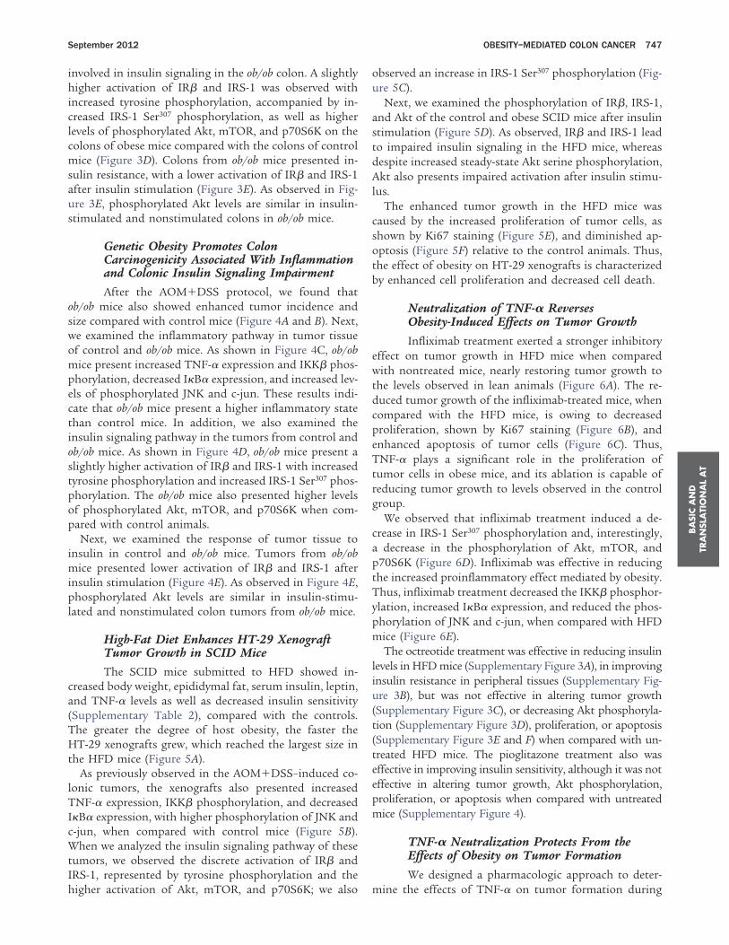
involved in insulin signaling in the ob/ob colon. A slightlyhigher activation of IRb and IRS-1 was observed withincreased tyrosine phosphorylation, accompanied by in-creased IRS-1 Ser307 phosphorylation, as well as higherlevels of phosphorylated Akt, mTOR, and p70S6K on thecolons of obese mice compared with the colons of controlmice (Figure 3D). Colons from ob/ob mice presented in-sulin resistance, with a lower activation of IRb and IRS-1after insulin stimulation (Figure 3E). As observed in Fig-ure 3E, phosphorylated Akt levels are similar in insulin-stimulated and nonstimulated colons in ob/ob mice.
Genetic Obesity Promotes ColonCarcinogenicity Associated With Inflammationand Colonic Insulin Signaling Impairment
After the AOM1DSS protocol, we found thatob/ob mice also showed enhanced tumor incidence andsize compared with control mice (Figure 4A and B). Next,we examined the inflammatory pathway in tumor tissueof control and ob/ob mice. As shown in Figure 4C, ob/obmice present increased TNF-a expression and IKKb phos-phorylation, decreased IkBa expression, and increased lev-els of phosphorylated JNK and c-jun. These results indi-cate that ob/ob mice present a higher inflammatory statethan control mice. In addition, we also examined theinsulin signaling pathway in the tumors from control andob/ob mice. As shown in Figure 4D, ob/ob mice present aslightly higher activation of IRb and IRS-1 with increasedtyrosine phosphorylation and increased IRS-1 Ser307 phos-phorylation. The ob/ob mice also presented higher levelsof phosphorylated Akt, mTOR, and p70S6K when com-pared with control animals.Next, we examined the response of tumor tissue to
insulin in control and ob/ob mice. Tumors from ob/obmice presented lower activation of IRb and IRS-1 afterinsulin stimulation (Figure 4E). As observed in Figure 4E,phosphorylated Akt levels are similar in insulin-stimu-lated and nonstimulated colon tumors from ob/ob mice.
High-Fat Diet Enhances HT-29 XenograftTumor Growth in SCID Mice
The SCID mice submitted to HFD showed in-creased body weight, epididymal fat, serum insulin, leptin,and TNF-a levels as well as decreased insulin sensitivity(Supplementary Table 2), compared with the controls.The greater the degree of host obesity, the faster theHT-29 xenografts grew, which reached the largest size inthe HFD mice (Figure 5A).As previously observed in the AOM1DSS–induced co-
lonic tumors, the xenografts also presented increasedTNF-a expression, IKKb phosphorylation, and decreasedIkBa expression, with higher phosphorylation of JNK andc-jun, when compared with control mice (Figure 5B).When we analyzed the insulin signaling pathway of thesetumors, we observed the discrete activation of IRb andIRS-1, represented by tyrosine phosphorylation and thehigher activation of Akt, mTOR, and p70S6K; we also
observed an increase in IRS-1 Ser307 phosphorylation (Fig-
ure 5C).
Next, we examined the phosphorylation of IRb, IRS-1,
and Akt of the control and obese SCID mice after insulin
stimulation (Figure 5D). As observed, IRb and IRS-1 lead
to impaired insulin signaling in the HFD mice, whereas
despite increased steady-state Akt serine phosphorylation,
Akt also presents impaired activation after insulin stimu-
lus.
The enhanced tumor growth in the HFD mice was
caused by the increased proliferation of tumor cells, as
shown by Ki67 staining (Figure 5E), and diminished ap-
optosis (Figure 5F) relative to the control animals. Thus,
the effect of obesity on HT-29 xenografts is characterized
by enhanced cell proliferation and decreased cell death.
Neutralization of TNF-a ReversesObesity-Induced Effects on Tumor Growth
Infliximab treatment exerted a stronger inhibitory
effect on tumor growth in HFD mice when compared
with nontreated mice, nearly restoring tumor growth to
the levels observed in lean animals (Figure 6A). The re-
duced tumor growth of the infliximab-treated mice, when
compared with the HFD mice, is owing to decreased
proliferation, shown by Ki67 staining (Figure 6B), and
enhanced apoptosis of tumor cells (Figure 6C). Thus,
TNF-a plays a significant role in the proliferation of
tumor cells in obese mice, and its ablation is capable of
reducing tumor growth to levels observed in the control
group.
We observed that infliximab treatment induced a de-
crease in IRS-1 Ser307 phosphorylation and, interestingly,
a decrease in the phosphorylation of Akt, mTOR, and
p70S6K (Figure 6D). Infliximab was effective in reducing
the increased proinflammatory effect mediated by obesity.
Thus, infliximab treatment decreased the IKKb phosphor-
ylation, increased IkBa expression, and reduced the phos-
phorylation of JNK and c-jun, when compared with HFD
mice (Figure 6E).
The octreotide treatment was effective in reducing insulin
levels in HFDmice (Supplementary Figure 3A), in improving
insulin resistance in peripheral tissues (Supplementary Fig-
ure 3B), but was not effective in altering tumor growth
(Supplementary Figure 3C), or decreasing Akt phosphoryla-
tion (Supplementary Figure 3D), proliferation, or apoptosis
(Supplementary Figure 3E and F) when compared with un-
treated HFD mice. The pioglitazone treatment also was
effective in improving insulin sensitivity, although it was not
effective in altering tumor growth, Akt phosphorylation,
proliferation, or apoptosis when compared with untreated
mice (Supplementary Figure 4).
TNF-a Neutralization Protects From theEffects of Obesity on Tumor Formation
We designed a pharmacologic approach to deter-
mine the effects of TNF-a on tumor formation during
BA
SIC
AN
D
TRA
NSLA
TIO
NA
LA
T
September 2012 OBESITY–MEDIATED COLON CANCER 747

obesity. Concomitantly with the AOM1DSS protocolwith the ob/ob mice, we started treatment with infliximaband observed that the infliximab treatment resulted indecreased levels of histologic damage in the colon sections(Figure 7A). Consistent with these signs, we observed thatinfliximab treatment decreased colon tumor incidenceand size in ob/ob mice (Figure 7B), an effect that was notobserved with pioglitazone (data not shown). In addition,
infliximab treatment exerted an inhibitory effect on tu-mor growth in the ob/ob mice, reducing proliferation,shown by Ki67 staining (Figure 7C), and enhancing apo-ptosis (Figure 7D). Furthermore, consistent with our pre-vious studies, infliximab treatment, besides neutralizingTNF-a, increased insulin sensitivity as measured by therate constant for glucose disappearance in the insulintolerance test9,23 (Supplementary Table 3).
Figure 4. Genetic obesity en-hances tumor formation. (A)Macroscopic changes in epithe-lial tissues. Scale bars, 5 mm. (B)Tumor incidence and size fromcolons removed at the end of theprotocol from CTL and ob/ob
mice. Results are means 6 SEM(n 5 8). Western blots of 5 inde-pendent experiments showingcolonic tumor lysates from con-trol and ob/ob mice. (C) Colonictumor TNF-a, TNFR1, IKKb,IkBa, JNK, and c-Jun expres-sion and phosphorylation. (D)Colonic tumor IRb, IRS-1, IRS-1Ser307, Akt, mTOR, and p70S6Kexpression and phosphoryla-tion. (E) Insulin-induced IRb,IRS-1, and Akt phosphorylationin the colon tumor. Data are themeans 6 SEM. *P , .05 vs therespective control group. #P ,
.05 vs CTL2. ‡P , .05 vs HFD2.§vs CTL1. CTL, white bars; ob/
ob, black bars.
BA
SIC
AN
D
TRA
NSLA
TIO
NA
LA
T
748 FLORES ET AL GASTROENTEROLOGY Vol. 143, No. 3
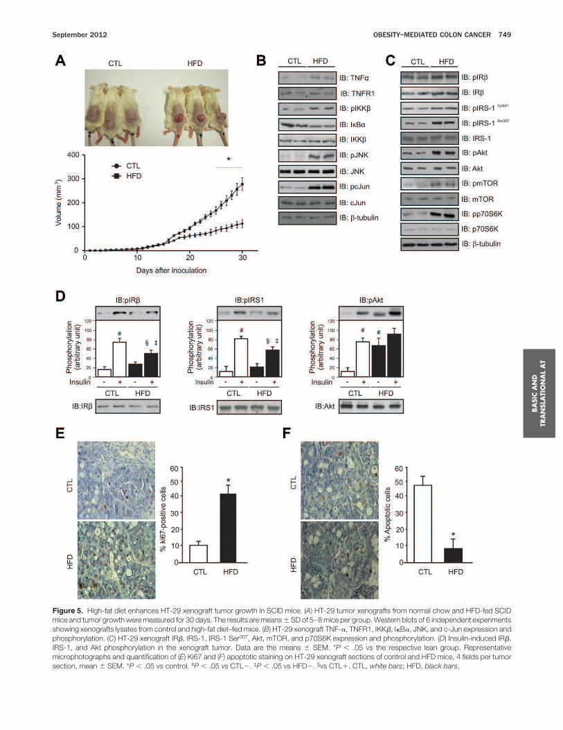
Figure 5. High-fat diet enhances HT-29 xenograft tumor growth in SCID mice. (A) HT-29 tumor xenografts from normal chow and HFD-fed SCIDmice and tumor growth were measured for 30 days. The results are means 6 SD of 5–8 mice per group. Western blots of 6 independent experimentsshowing xenografts lysates from control and high-fat diet–fed mice. (B) HT-29 xenograft TNF-a, TNFR1, IKKb, IkBa, JNK, and c-Jun expression andphosphorylation. (C) HT-29 xenograft IRb, IRS-1, IRS-1 Ser307, Akt, mTOR, and p70S6K expression and phosphorylation. (D) Insulin-induced IRb,IRS-1, and Akt phosphorylation in the xenograft tumor. Data are the means 6 SEM. *P , .05 vs the respective lean group. Representativemicrophotographs and quantification of (E) Ki67 and (F) apoptotic staining on HT-29 xenograft sections of control and HFD mice, 4 fields per tumorsection, mean 6 SEM. *P , .05 vs control. #P , .05 vs CTL2. ‡P , .05 vs HFD2. §vs CTL1. CTL, white bars; HFD, black bars.
BA
SIC
AN
D
TRA
NSLA
TIO
NA
LA
T
September 2012 OBESITY–MEDIATED COLON CANCER 749
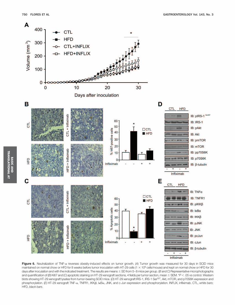
Figure 6. Neutralization of TNF-a reverses obesity-induced effects on tumor growth. (A) Tumor growth was measured for 30 days in SCID micemaintained on normal chow or HFD for 8 weeks before tumor inoculation with HT-29 cells (1 3 106 cells/mouse) and kept on normal chow or HFD for 30days after inoculation and with the indicated treatment. The results are means 6 SD from 5–8 mice per group. (B and C) Representative microphotographsand quantification of (B) Ki67 and (C) apoptotic staining on HT-29 xenograft sections, 4 fields per tumor section, mean 6 SEM. *P , .05 vs control. Westernblots showing HT-29 xenograft lysates from tumor-bearing SCID mice. (D) HT-29 xenograft IRS-1, IRS-1 Ser307, Akt, mTOR, and p70S6K expression andphosphorylation. (E) HT-29 xenograft TNF-a, TNFR1, IKKb, IkBa, JNK, and c-Jun expression and phosphorylation. INFLIX, infliximab. CTL, white bars;HFD, black bars.
BA
SIC
AN
D
TRA
NSLA
TIO
NA
LA
T
750 FLORES ET AL GASTROENTEROLOGY Vol. 143, No. 3
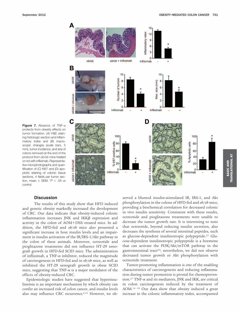
Discussion
The results of this study show that HFD inducedand genetic obesity markedly increased the developmentof CRC. Our data indicate that obesity-induced colonicinflammation increases JNK and IKKb expression andactivity in the colon of AOM1DSS–treated mice. In ad-dition, the HFD-fed and ob/ob mice also presented asignificant increase in host insulin levels and an impair-ment in insulin activation of the IR/IRS-1/Akt pathway inthe colon of these animals. Moreover, octreotide andpioglitazone treatments did not influence HT-29 xeno-graft growth in HFD-fed SCID mice. The administrationof infliximab, a TNF-a inhibitor, reduced the magnitudeof carcinogenesis in HFD-fed and in ob/ob mice, as well asinhibited the HT-29 xenograft growth in obese SCIDmice, suggesting that TNF-a is a major modulator of theeffects of obesity-induced CRC.Epidemiologic studies have suggested that hyperinsu-
linemia is an important mechanism by which obesity canconfer an increased risk of colon cancer, and insulin levelsalso may influence CRC recurrence.6,24 However, we ob-
served a blunted insulin-stimulated IR, IRS-1, and Aktphosphorylation in the colons of HFD-fed and ob/ob mice,providing a biochemical correlation for decreased colonicin vivo insulin sensitivity. Consistent with these results,octreotide and pioglitazone treatments were unable todecrease the tumor growth rate. It is interesting to notethat octreotide, beyond reducing insulin secretion, alsodecreases the synthesis of several intestinal peptides, suchas glucose-dependent insulinotropic polypeptide.25 Glu-cose-dependent insulinotropic polypeptide is a hormonethat can activate the PI3K/Akt/mTOR pathway in thegastrointestinal tract26; nevertheless, we did not observedecreased tumor growth or Akt phosphorylation withoctreotide treatment.Tumor-promoting inflammation is one of the enabling
characteristics of carcinogenesis and reducing inflamma-tion during tumor promotion is pivotal for chemopreven-tion.27 TNF-a and its mediators, JNK and IKK, are criticalin colon carcinogenesis induced by the treatment ofAOM.16–18 Our data show that obesity induced a greatincrease in the colonic inflammatory index, accompanied
Figure 7. Absence of TNF-aprotects from obesity effects ontumor formation. (A) H&E stain-ing histologic section and inflam-matory index and (B) macro-scopic changes (scale bars, 5mm), tumor incidence, and size ofcolons removed at the end of theprotocol from ob/ob mice treatedor not with infliximab. Representa-tive microphotographs and quan-tification of (C) Ki67 and (D) apo-ptotic staining of colonic tissuesections, 4 fields per tumor sec-tion, mean 6 SEM. *P , .05 vscontrol.
BA
SIC
AN
D
TRA
NSLA
TIO
NA
LA
T
September 2012 OBESITY–MEDIATED COLON CANCER 751

by an increase in IKK and JNK phosphorylation in diet-induced and genetic obesity, as well as the treatmentusing the TNF-a blocker, infliximab, showed a consistentdecrease in tumor growth in the HT-29 xenograft and incolitis-associated cancer development. Thus, the low-grade inflammatory state observed in obesity increasedplasma TNF-a levels and created fertile soil in the colonicepithelium that increased DSS and AOM action.TNF-a is an important component of the cancer net-
work of inflammatory mediators, such as cytokines andvascular endothelial growth factor.15 This network thenacts by modulating the cancer microenvironment andallowing cancer growth.28 It also has been shown that thePI3K/Akt pathway can be activated by TNF-a.29,30 More-over, the PI3K/Akt pathway is activated in patients withulcerative colitis31 and induces proinflammatory cytokineproduction.32 Our findings showing a reduction in theAkt/mTOR pathway, which is known to be an importantregulator of CRC growth,33,34 after infliximab treatmentof obese mice bearing colon cancer xenografts, suggests amajor role of this pathway in obesity-mediated coloncancer growth.Currently, infliximab is recommended for the treat-
ment of complicated Crohn’s disease and was approvedfor the treatment of ulcerative colitis that fails to respondto the standard therapies, such as corticosteroids andimmunosuppressors.35–37 In accordance with our study,neutralization of TNF-a is also an effective therapy invarious animal models of T-cell–mediated colitis.38,39
However, TNF-a neutralization in the DSS-induced coli-tis, an epithelial damage model, is still controversial.40–42
These different results may be related to the environmentthat the animals were bred in or to the differences in theinflammatory levels obtained with the different protocols.It is interesting to note that despite the contradictoryresults related to colitis, the effect of TNF-a neutraliza-tion or inhibition of IKKb was effective in reducing CRCin all studies.16,18,42 The results presented herein extendthese data, showing that infliximab also prevents obesity-mediated CRC. Altogether, these results suggest that theuse of anti–TNF-a therapy could decrease both insulinresistance and colon cancer risk; although the possiblecontribution of anti–TNF-a therapy to improve insulinsensitivity in human beings still is controversial and de-serves further investigation.35,43–45 Along this line, chemo-prevention of CRC in obese individuals with TNF-a neu-tralization should be evaluated.In summary, our findings establish a role for TNF-a in
obesity-mediated colon carcinogenesis and point toTNF-a blockers as attractive players in colon cancer che-moprevention for obese individuals.
Supplementary Material
Note: To access the supplementary materialaccompanying this article, visit the online version ofGastroenterology at www.gastrojournal.org, and at http://dx.doi.org/10.1053/j.gastro.2012.05.045.
References
1. Center MM, Jemal A, Smith RA, et al. Worldwide variations in
colorectal cancer. CA Cancer J Clin 2009;59:366–378.
2. Walther A, Johnstone E, Swanton C, et al. Genetic prognostic and
predictive markers in colorectal cancer. Nat Rev Cancer 2009;9:
489–499.
3. Calle EE, Rodriguez C, Walker-Thurmond K, et al. Overweight,
obesity, and mortality from cancer in a prospectively studied co-
hort of U.S. adults. N Engl J Med 2003;348:1625–1638.
4. Giovannucci E. Modifiable risk factors for colon cancer. Gastroen-
terol Clin North Am 2002;31:925–943.
5. Renehan AG, Zwahlen M, Minder C, et al. Insulin-like growth factor
(IGF)-I, IGF binding protein-3, and cancer risk: systematic review
and meta-regression analysis. Lancet 2004;363:1346–1353.
6. Sandhu MS, Dunger DB, Giovannucci EL. Insulin, insulin-like
growth factor-I (IGF-I), IGF binding proteins, their biologic interac-
tions, and colorectal cancer. J Natl Cancer Inst 2002;94:972–
980.
7. Sinicrope FA, Foster NR, Sargent DJ, et al. Obesity is an indepen-
dent prognostic variable in colon cancer survivors. Clin Cancer Res
2010;16:1884–1893.
8. Osorio-Costa F, Rocha GZ, Dias MM, et al. Epidemiological and
molecular mechanisms aspects linking obesity and cancer. Arq
Bras Endocrinol Metabol 2009;53:213–226.
9. Araujo EP, De Souza CT, Ueno M, et al. Infliximab restores glucose
homeostasis in an animal model of diet-induced obesity and dia-
betes. Endocrinology 2007;148:5991–5997.
10. Hotamisligil GS, Peraldi P, Budavari A, et al. IRS-1-mediated inhi-
bition of insulin receptor tyrosine kinase activity in TNF-alpha- and
obesity-induced insulin resistance. Science 1996;271:665–668.
11. Lee YH, Giraud J, Davis RJ, et al. c-Jun N-terminal kinase (JNK)
mediates feedback inhibition of the insulin signaling cascade.
J Biol Chem 2003;278:2896–2902.
12. Ghosh S, Karin M. Missing pieces in the NF-kappaB puzzle. Cell
2002;109(Suppl):S81–S96.
13. Aguirre V, Werner ED, Giraud J, et al. Phosphorylation of Ser307 in
insulin receptor substrate-1 blocks interactions with the insulin
receptor and inhibits insulin action. J Biol Chem 2002;277:1531–
1537.
14. Gao Z, Hwang D, Bataille F, et al. Serine phosphorylation of insulin
receptor substrate 1 by inhibitor kappa B kinase complex. J Biol
Chem 2002;277:48115–48121.
15. Balkwill F. Tumour necrosis factor and cancer. Nat Rev Cancer
2009;9:361–371.
16. Popivanova BK, Kitamura K, Wu Y, et al. Blocking TNF-alpha in
mice reduces colorectal carcinogenesis associated with chronic
colitis. J Clin Invest 2008;118:560–570.
17. Endo H, Hosono K, Fujisawa T, et al. Involvement of JNK pathway
in the promotion of the early stage of colorectal carcinogenesis
under high-fat dietary conditions. Gut 2009;58:1637–1643.
18. Greten FR, Eckmann L, Greten TF, et al. IKKbeta links inflamma-
tion and tumorigenesis in a mouse model of colitis-associated
cancer. Cell 2004;118:285–296.
19. Park EJ, Lee JH, Yu GY, et al. Dietary and genetic obesity promote
liver inflammation and tumorigenesis by enhancing IL-6 and TNF
expression. Cell 2010;140:197–208.
20. Ropelle ER, Pauli JR, Prada PO, et al. Reversal of diet-induced
insulin resistance with a single bout of exercise in the rat: the role
of PTP1B and IRS-1 serine phosphorylation. J Physiol 2006;577:
997–1007.
21. Kojouharoff G, Hans W, Obermeier F, et al. Neutralization of
tumour necrosis factor (TNF) but not of IL-1 reduces inflammation
in chronic dextran sulphate sodium-induced colitis in mice. Clin
Exp Immunol 1997;107:353–358.
22. Rocha GZ, Dias MM, Ropelle ER, et al. Metformin amplifies che-
motherapy-induced AMPK activation and antitumoral growth. Clin
Cancer Res 2011;17:3993–4005.
BA
SIC
AN
D
TRA
NSLA
TIO
NA
LA
T
752 FLORES ET AL GASTROENTEROLOGY Vol. 143, No. 3

23. Barbuio R, Milanski M, Bertolo MB, et al. Infliximab reverses
steatosis and improves insulin signal transduction in liver of rats
fed a high-fat diet. J Endocrinol 2007;194:539–550.
24. Ma J, Giovannucci E, Pollak M, et al. A prospective study of
plasma C-peptide and colorectal cancer risk in men. J Natl Cancer
Inst 2004;96:546–553.
25. Ranganath LR, Beety JM, Morgan LM. Inhibition of insulin, glu-
cose-dependent insulinotropic polypeptide (GIP) and glucagon-like
peptide-1 (GLP-1) secretion by octreotide has no effect on post-
heparin plasma lipoprotein lipase activity. Horm Metab Res 1999;
31:262–266.
26. Baggio LL, Drucker DJ. Biology of incretins: GLP-1 and GIP. Gas-
troenterology 2007;132:2131–2157.
27. Hanahan D, Weinberg RA. Hallmarks of cancer: the next genera-
tion. Cell 2011;144:646–674.
28. Li B, Vincent A, Cates J, et al. Low levels of tumor necrosis factor
alpha increase tumor growth by inducing an endothelial phenotype
of monocytes recruited to the tumor site. Cancer Res 2009;69:
338–348.
29. Chen Q, Casali B, Pattacini L, et al. Tumor necrosis factor-alpha
protects synovial cells from nitric oxide induced apoptosis through
phosphoinositide 3-kinase Akt signal transduction. J Rheumatol
2006;33:1061–1068.
30. Xu H, He Y, Yang X, et al. Anti-malarial agent artesunate inhibits
TNF-alpha-induced production of proinflammatory cytokines via
inhibition of NF-kappaB and PI3 kinase/Akt signal pathway in
human rheumatoid arthritis fibroblast-like synoviocytes. Rheuma-
tology (Oxford) 2007;46:920–926.
31. Huang XL, Xu J, Zhang XH, et al. PI3K/Akt signaling pathway is
involved in the pathogenesis of ulcerative colitis. Inflamm Res
2011;60:727–734.
32. Reddy SA, Huang JH, Liao WS. Phosphatidylinositol 3-kinase as a
mediator of TNF-induced NF-kappa B activation. J Immunol 2000;
164:1355–1363.
33. Stathopoulos GT, Kollintza A, Moschos C, et al. Tumor necrosis
factor-alpha promotes malignant pleural effusion. Cancer Res
2007;67:9825–9834.
34. Zoncu R, Efeyan A, Sabatini DM. mTOR: from growth signal inte-
gration to cancer, diabetes and ageing. Nat Rev Mol Cell Biol
2011;12:21–35.
35. Rutgeerts P, Sandborn WJ, Feagan BG, et al. Infliximab for induc-
tion and maintenance therapy for ulcerative colitis. N Engl J Med
2005;353:2462–2476.
36. Lugering A, Schmidt M, Lugering N, et al. Infliximab induces
apoptosis in monocytes from patients with chronic active Crohn’s
disease by using a caspase-dependent pathway. Gastroenterol-
ogy 2001;121:1145–1157.
37. Kim YJ, Hong KS, Chung JW, et al. Prevention of colitis-associated
carcinogenesis with infliximab. Cancer Prev Res (Phila) 2010;3:
1314–1333.
38. Neurath MF, Fuss I, Pasparakis M, et al. Predominant pathogenic
role of tumor necrosis factor in experimental colitis in mice. Eur
J Immunol 1997;27:1743–1750.
39. Powrie F, Leach MW, Mauze S, et al. Inhibition of Th1 responses
prevents inflammatory bowel disease in scid mice reconstituted
with CD45RBhi CD41 T cells. Immunity 1994;1:553–562.
40. Kojouharoff G, Hans W, Obermeier F, et al. Neutralization of
tumour necrosis factor (TNF) but not of IL-1 reduces inflammation
in chronic dextran sulphate sodium-induced colitis in mice. Clin
Exp Immunol 1997;107:353–358.
41. Naito Y, Takagi T, Handa O, et al. Enhanced intestinal inflamma-
tion induced by dextran sulfate sodium in tumor necrosis factor-
alpha deficient mice. J Gastroenterol Hepatol 2003;18:560–569.
42. Onizawa M, Nagaishi T, Kanai T, et al. Signaling pathway via
TNF-alpha/NF-kappaB in intestinal epithelial cells may be directly
involved in colitis-associated carcinogenesis. Am J Physiol Gas-
trointest Liver Physiol 2009;296:G850–G859.
43. Bernstein LE, Berry J, Kim S, et al. Effects of etanercept in
patients with the metabolic syndrome. Arch Intern Med 2006;
166:902–908.
44. Kiortsis DN, Mavridis AK, Vasakos S, et al. Effects of infliximab
treatment on insulin resistance in patients with rheumatoid arthri-
tis and ankylosing spondylitis. Ann Rheum Dis 2005;64:765–
766.
45. Paquot N, Castillo MJ, Lefebvre PJ, et al. No increased insulin
sensitivity after a single intravenous administration of a recombi-
nant human tumor necrosis factor receptor: Fc fusion protein in
obese insulin-resistant patients. J Clin Endocrinol Metab 2000;
85:1316–1319.
Received August 18, 2011. Accepted May 24, 2012.
Reprint requests
Address requests for reprints to: José B. C. Carvalheira, MD,
Department of Internal Medicine, Faculty of Medical Sciences-State
University of Campinas, 13083-887 Campinas, São Paulo, Brazil.
e-mail: [email protected]; fax: (55) 19-3521-8950.
Acknowledgments
The authors thank Dr Nicola Conran and Sherian Bowyer for
English language editing.
M.B.S.F. and G.Z.R. contributed equally to this work.
Conflicts of interest
The authors disclose no conflicts.
Funding
This study was supported by grants from the Fundação de Amparo
à Pesquisa do Estado de São Paulo and the Conselho Nacional de
Desenvolvimento Científico e Tecnológico.
BA
SIC
AN
D
TRA
NSLA
TIO
NA
LA
T
September 2012 OBESITY–MEDIATED COLON CANCER 753
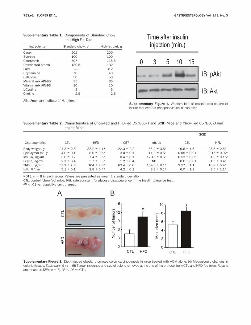
Supplementary Table 1. Components of Standard Chow
and High-Fat Diet
Ingredients Standard chow, g High-fat diet, g
Casein 202 200
Sucrose 100 100
Cornstarch 397 115.5
Dextrinated starch 130.5 132
Lard — 312
Soybean oil 70 40
Cellulose 50 50
Mineral mix AIN-93 35 35
Vitamin mix AIN-93 10 10
L-Cystine 3 3
Choline 2.5 2.5
AIN, American Institute of Nutrition.
Supplementary Table 2. Characteristics of Chow-Fed and HFD-Fed C57BL6/J and SCID Mice and Chow-Fed C57BL6/J and
ob/ob Mice
Characteristics CTL HFD C57 ob/ob
SCID
CTL HFD
Body weight, g 24.3 6 2.8 33.2 6 4.1a 22.3 6 2.2 55.2 6 3.5a 19.6 6 1.6 28.5 6 2.5a
Epididymal fat, g 4.0 6 0.1 8.0 6 0.5a 3.0 6 0.1 11.0 6 0.5a 0.05 6 0.01 0.15 6 0.02a
Insulin, ng/mL 3.8 6 0.2 7.4 6 0.5a 0.4 6 0.1 12.46 6 0.5a 0.53 6 0.05 1.0 6 0.15a
Leptin, ng/mL 2.1 6 0.4 3.7 6 0.5a 1.2 6 0.4 ND 0.6 6 0.01 1.5 6 0.4a
TNF-a, pg/mL 53.2 6 7.8 104 6 9.6a 63.4 6 0.6 169.6 6 9.1a 2.57 6 1.1 10.8 6 5.4a
Kitt, %/min 5.1 6 0.1 2.8 6 0.4a 4.2 6 0.1 3.3 6 0.1a 6.0 6 1.2 3.5 6 1.1a
NOTE. n 5 8 in each group. Values are presented as mean 6 standard deviation.
CTL, control (chow-fed) mice; Kitt, rate constant for glucose disappearance in the insulin tolerance test.aP , .01 vs respective control group.
Supplementary Figure 2. Diet-induced obesity promotes colon carcinogenesis in mice treated with AOM alone. (A) Macroscopic changes incolonic tissues. Scale bars, 5 mm. (B) Tumor incidence and size of colons removed at the end of the protocol from CTL and HFD-fed mice. Resultsare means 6 SEM (n 5 8). *P , .05 vs CTL.
Supplementary Figure 1. Western blot of colonic time-course ofinsulin-induced Akt phosphorylation in lean mice.
753.e1 FLORES ET AL GASTROENTEROLOGY Vol. 143, No. 3
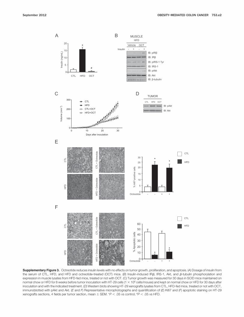
Supplementary Figure 3. Octreotide reduces insulin levels with no effects on tumor growth, proliferation, and apoptosis. (A) Dosage of insulin fromthe serum of CTL, HFD, and HFD and octreotide-treated (OCT) mice. (B) Insulin-induced IRb, IRS-1, Akt, and b-tubulin phosphorylation andexpression in muscle lysates from HFD-fed mice, treated or not with OCT. (C) Tumor growth was measured for 30 days in SCID mice maintained onnormal chow or HFD for 8 weeks before tumor inoculation with HT-29 cells (1 3 106 cells/mouse) and kept on normal chow or HFD for 30 days afterinoculation and with the indicated treatment. (D) Western blots showing HT-29 xenografts lysates from CTL, HFD-fed mice, treated or not with OCT,immunoblotted with pAkt and Akt. (E and F) Representative microphotographs and quantification of (E) Ki67 and (F) apoptotic staining on HT-29xenografts sections, 4 fields per tumor section, mean 6 SEM. *P , .05 vs control; #P , .05 vs HFD.
September 2012 OBESITY–MEDIATED COLON CANCER 753.e2
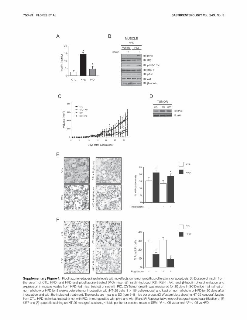
Supplementary Figure 4. Pioglitazone reduces insulin levels with no effects on tumor growth, proliferation, or apoptosis. (A) Dosage of insulin fromthe serum of CTL, HFD, and HFD and pioglitazone-treated (PIO) mice. (B) Insulin-induced IRb, IRS-1, Akt, and b-tubulin phosphorylation andexpression in muscle lysates from HFD-fed mice, treated or not with PIO. (C) Tumor growth was measured for 30 days in SCID mice maintained onnormal chow or HFD for 8 weeks before tumor inoculation with HT-29 cells (1 3 106 cells/mouse) and kept on normal chow or HFD for 30 days afterinoculation and with the indicated treatment. The results are means 6 SD from 5–8 mice per group. (D) Western blots showing HT-29 xenograft lysatesfrom CTL, HFD-fed mice, treated or not with PIO, immunoblotted with pAkt and Akt. (E and F) Representative microphotographs and quantification of (E)Ki67 and (F) apoptotic staining on HT-29 xenograft sections, 4 fields per tumor section, mean 6 SEM. *P , .05 vs control; #P , .05 vs HFD.
753.e3 FLORES ET AL GASTROENTEROLOGY Vol. 143, No. 3
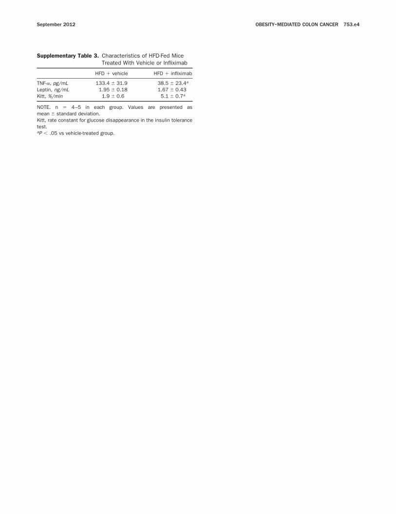
Supplementary Table 3. Characteristics of HFD-Fed Mice
Treated With Vehicle or Infliximab
HFD 1 vehicle HFD 1 infliximab
TNF-a, pg/mL 133.4 6 31.9 38.5 6 23.4a
Leptin, ng/mL 1.95 6 0.18 1.67 6 0.43
Kitt, %/min 1.9 6 0.6 5.1 6 0.7a
NOTE. n 5 4–5 in each group. Values are presented as
mean 6 standard deviation.
Kitt, rate constant for glucose disappearance in the insulin tolerance
test.aP , .05 vs vehicle-treated group.
September 2012 OBESITY–MEDIATED COLON CANCER 753.e4

_ 59
Tese de Doutorado
CAPÍTULO II

_ 60
Tese de Doutorado
ABSENCE OF DOUBLE –STRANDED RNA DEPENDENT PROTEIN KINASE
PROTECTS FROM DEVELOPMENT OF COLITIS-ASSOCIATED CANCER
Running title: PKR protects against CAC
Guilherme Z. Rocha, Marília M. Dias, Juliana A. Camargo, Andrey dos Santos, Mario J. A.
Saad, José B. C. Carvalheira.
Department of Internal Medicine, FCM, State University of Campinas (UNICAMP),
Campinas, SP, Brazil.
Financial support: Fundação de Amparo à pesquisa do Estado de São Paulo (FAPESP)
and Conselho Nacional de desenvolvimento científico e tecnológico (CNPq).
Please address correspondence to: José B. C. Carvalheira, M.D., Departamento de
Clínica Médica, FCM-UNICAMP, Cidade Universitária Zeferino Vaz, Campinas, SP,
Brazil, 13081-970, Fax: +55 19 5218950, e-mail: [email protected]
Conflict of interest: The authors declare no conflict of interests.
_ 61
Tese de Doutorado
ABSTRACT
The association between obesity and colon cancer development is a major
conceptual advance in the understanding of colon cancer pathogenesis in the last decade
and the role of obesity–related subclinical inflammation in carcinogenesis gained
significance. It is believed that obesity acts as a tumor promoter, and it`s pro-tumorigenic
effects depend mainly on low-grade inflammatory response caused by obesity-driven
production of inflammatory and pro-tumorigenic cytokines (TNF and IL -6). A key feature
of obesity-induced inflammation is the infiltration of macrophages in adipose tissue,
leading to production of inflammatory cytokines and other mediators that also interfere
with insulin signaling. Thus, the main objective of the study was to evaluate the role of
PKR in the development of colon tumors in wild type and PKR knockout mice subjected to
a high fat diet. In the present study, we manipulated the expression of PKR in myeloid cells
to better define the role of PKR during tumorigenesis induced by colitis associated cancer
and also their role in diet-induced inflammation and insulin resistance. We observe that
PKR absence prevents colon tumor formation. We define the function of PKR in myeloid
cells, where it mediates resistance to epithelial damage caused by DSS and prevents the
formation of tumors. Moreover, the absence of PKR in myeloid cells also confers
protection to insulin resistance induced by a high-fat diet, reducing the inflammation
induced by obesity. These observations demonstrate that PKR can be a primary point
during carcinogenesis associated with inflammation and may represent a promising target
for therapeutic intervention.

_ 62
Tese de Doutorado
INTRODUCTION
Colorectal cancer (CRC) remains a major health burden with more than one million
cases worldwide and a mortality of approximately 33% in the developed world (Center et
al., 2009; Walther et al., 2009). The association between obesity and the risk for CRC
development is observed in both men and women (relative risk: 1.2–2.0) (Calle et al., 2003;
Giovannucci, 2002). Among the major mediators of the inflammatory response is tumor
necrosis factor (TNF)-a, whose over expression in adipose tissue is a common feature in
human and animal models of obesity (Araújo et al., 2007; Osório-Costa et al., 2009).
TNF-a, first identified as an antitumor agent, is now also recognized as a tumor-promoting
cytokine that links inflammation and cancer (Balkwill, 2009). Furthermore, the increased
TNF-a levels associated with obesity are a potent liver tumor promoter in mice (Park et al.,
2010).
Chronic colon inflammation may release ROS and RNI (Hussain et al., 2003) that
cause DNA damage and affect cell proliferation or survival and this way promote
oncogenesis (Meira et al., 2008). Although, experimental evidences suggest that it acts
mainly as a tumor promoter instead as an initiator (Coussens and Werb, 2002; Karin and
Greten, 2005; Karin et al., 2006). Immune cells, frequently present in pre-neoplastic lesions
produce a variety of cytokines and chemokines that increase local inflammatory response
and increase pre-malignant cells survival and growth activating transcription factors such as
NF-kB (Lin and Karin, 2007; Pikarsky et al., 2004). NF-kB-stimulated cytokines
production by myeloid cells is essential for the colitis-associated cancer (CAC) growth,
while NF-kB activation in epithelial intestinal cells promotes survival of newly formed

_ 63
Tese de Doutorado
pre-malignant cells (Greten et al., 2004). These studies suggest that cytokines and growth
factors produced after NF-kB activation in intestinal myeloid cells stimulate proliferation
of pre-malignant intestinal epithelial cells in the first stages of CAC tumorigenesis. NF-kB
inactivation in myeloid cells through IKKb ablation inhibits production of inflammatory
mediators such as IL-6 and TNF-a and prevents intestinal epithelias cells proliferation
during CAC, decreasing the number of tumors formed and their size (Greten et al., 2004).
Metabolic diseases arise as groups including obesity, insulin resistance, type 2
diabetes, cardiovascular disease and cancer and are an important health problem
worldwide, with limited treatment options. In the last decade, we have observed that the
emergence of this group of diseases has strong inflammatory bases (Hotamisligil, 2006).
During the course of obesity, a wide range of stress and inflammatory responses are evoked
in metabolic tissues, leading to chronic local low grade inflammation which plays a central
role in the inhibition of insulin receptor signaling and disturbances of systemic metabolic
homeostasis. This atypical state involves immune and non-immune cells and pathways of
immune engagement with nutrients and metabolites answer.
PKR can regulate or act together with major inflammatory signaling pathways that
are involved in metabolic homeostasis, including the N-teminal kinase c-Jun (JNK) and
IkB kinase (IKK). Studies in PKR-/- animals have shown that these mice present a lean
phenotype and are protected against diet-induced obesity (Carvalho-Filho et al., 2012;
Nakamura et al., 2010). In addition to being more lean and responsive to insulin, high-fat
diet-induced PKR-/- mice exhibit reduced expression of proinflammatory cytokines such as
IL-6 and TNFα, compared to wild type animals (Carvalho-Filho et al., 2012; Nakamura et
_ 64
Tese de Doutorado
al., 2010). These data suggest that PKR may be involved in diet-induced inflammation. In
both mice and humans, there is a consistent and marked increase in the phosphorylation of
eIF2α and JNK activity in metabolic tissues of obese subjects (Carvalho-Filho et al., 2012;
Gregor et al., 2009; Nakamura et al., 2010). In diet-induced obese mice, the absence of
PKR protects against obesity and insulin resistance by preventing the activation of JNK and
IKKβ, indicating that PKR is an important modulator of the insulin signaling under normal
physiological conditions and in obesity (Carvalho-Filho et al., 2012; Nakamura et al.,
2010).
In the present study, we manipulated the expression of PKR in myeloid cells to
define the role of PKR during tumorigenesis induced by CAC and also their role in diet-
induced inflammation and insulin resistance. We define the function of PKR in myeloid
cells, where it mediates resistance to epithelial damage caused by DSS and prevents the
formation of tumors. Moreover, the absence of PKR in myeloid cells also confers
protection against insulin resistance induced by a high-fat diet, reducing the inflammation
induced by obesity. These observations demonstrate that PKR can be a primary point
during carcinogenesis associated with inflammation and may represent a promising target
for therapeutic intervention.
_ 65
Tese de Doutorado
METHODS
Antibodies, chemicals and buffers
All the reagents were from Sigma-Aldrich, unless otherwise specified. DSS was
from MP Biochemicals (Solon, OH, USA). Anti-phospho-PKR, anti-PKR, anti-phospho-
eIF2a, anti-eIF2a, anti-phospho-IRb, anti-phospho-IRS-1 Tyr941, anti-IkBa, anti-
phospho-JNK, anti-IL6 antibodies were from Santa Cruz Biotechnology (Santa Cruz, CA,
USA). Anti-phospho-Akt and anti-b-actin antibodies were from Cell Signaling
Biotechnology (Beverly, MA, USA).
Animals
The Ethics Committee of the State University of Campinas approved all
experiments. Mice were provided from Central Breeding Center of the State University of
Campinas and randomly divided into two groups, control and high-fat diet (HFD). The diet
composition is described in the Supplemental Table 1.
Insulin tolerance test, serum insulin, leptin, IL-6 and TNF-a quantification
The mice were given an insulin tolerance test (ITT; 1.5 IU insulin/kg body weight)
as described previously20. Plasma was separated by centrifugation (1,100xg) for 15 min at
4°C and stored at −80°C until the assay. Serum insulin and leptin were measured by using a
mouse ELISA kit (Linco, St. Charles, MO, USA). Serum IL-6 and TNF-a were measured
using a mouse ELISA kit (Thermo Scientific, Rockford, IL, USA).
_ 66
Tese de Doutorado
Tumor induction and analysis
Four-week-old male mice (C57BL6/J Unib (WT) and Pkr-/- (PKR-/-)) were placed
on standard chow or on a high-fat diet for one week and then injected intraperitoneally
(i.p.) with 12.5 mg/kg AOM. After five days, 2.5% DSS (MW 36–50 kDa) was given in the
drinking water for five days, followed by 14 days of regular water. This cycle was repeated
twice and mice were sacrificed 30 days after the last cycle, at 20 weeks of age. Colons were
removed, flushed with PBS, fixed in 4% paraformaldehyde and paraffin-embedded.
Sections (5 μm) were cut and stained with hematoxylin and eosin (H&E). Tumor counts
were performed in a blinded fashion. Tumor sizes were measured with calipers. Assessment
of colitis disease scores was performed as previously described (Kojouharoff et al., 1997).
Cell culture
The mouse melanoma cancer cell line, B16-F10, was purchased from the Rio de
Janeiro Cell Bank and cells were cultured in DMEM medium containing 10% fetal bovine
serum with the addition of antibiotics and fungicides. Cells were maintained at 37°C in a
humid atmosphere and 5% CO2.
Tumor graft models
Four-week-old male C57BL6/J Unib mice (n=10 per group) were inoculated
subcutaneously in the dorsal region with 1x10⁶ B16-F10 cells. Tumor volume (V) was
calculated daily by measuring length (L) and width (W) of the tumor with calipers and
using the formula V= {W x L x [(W+L)/2]} x 0.52.

_ 67
Tese de Doutorado
Treatment with PKR inhibitor (Merck) began 7 days after cells inoculation. PKR
inhibitor (20mg/kg) was given subcutaneously every two days.
Tissue extracts
Mice were anesthetized with sodium amobarbital (15 mg/kg body weight, i.p.).
Tumors and colons were removed, minced coarsely, and homogenized in extraction buffer
(1% Triton-X 100, 100 mM Tris, pH 7.4, containing 100 mM sodium pyrophosphate, 100
mM sodium fluoride, 10 mM EDTA, 10 mM sodium vanadate, 2 mM PMSF, and 0.1 mg of
aprotinin/ml). The extracts were centrifuged at 11,000 rpm and 4°C, and the supernatants of
these tissues used.
Bone marrow transplantation
5-week-old male mice were lethally irradiated (1050rad) and transplanted with 1 x
106 bone marrow cells from WT or PKR-/- mice.
Peritoneal macrophage isolation
Mice were killed by cervical dislocation, abdominal wall was exposed, and sterile
PBS injected into cavity. After a 10 seconds gentle shake, peritoneal lavage fluid
containing cells was withdrawn and plated on cell culture dishes in DMEM media,
supplemented with fetal bovine serum 10% at 37°C for one hour. Then, adherent
macrophages (>90% of the cells) were treated with 10ng/mL of LPS for three hours.
Afterward, cells were collected and mRNA was analyzed.
Protein analysis by immunobloting
_ 68
Tese de Doutorado
Whole tissue extracts were homogenized in extraction buffer, treated with Laemmli
sample buffer containing 100 mM DTT, and heated in a boiling water bath. For total
extracts, similar-sized aliquots (50 μg protein) were subjected to SDS-PAGE. Proteins were
resolved in 8-15% SDS gels and blotted onto nitrocellulose membranes (Bio-Rad). Band
intensities were quantified by optical densitometry of developed autoradiographs using UN-
SCAN-IT Gel (Silk Scientific, Inc. Orem, UT, USA).
Statistical analysis
Data are presented as means ± SEM of at least three independent experiments.
Statistical analysis was performed by employing the ANOVA test with the Bonferroni post
test. Significance was established at the p<0.05 level.
_ 69
Tese de Doutorado
RESULTS
Absence of PKR protects against tumor formation
It has been demonstrated that mice lacking PKR have lower levels of inflammation,
with reduced JNK activity and lower level of inflammatory cytokines such as TNF-a and
IL-6 under a high-fat diet (Nakamura et al., 2010). As TNF-a is involved in development
of colon cancer in mice (Flores et al., 2012), we sought to examine the effect of complete
absence of PKR on CAC development . We therefore injected wild-type (WT) and PKR-/-
mice with the procarcinogen azoxymethane (AOM) followed by three rounds of dextran
sodium sulfate (DSS) exposure to promote colitis. The PKR deficiency decreased tumor
frequency and numbers in mice (Figure 1A and B), although there was no statistically
difference in tumor size (Figure 1C).
Pharmacological inhibition of PKR protects against tumor formation
We also inhibited PKR activity with a pharmacological inhibitor and observed that
inhibition of PKR was able to reduce tumor growth of a subcutaneous tumor graft (Figure
2A, B and C). Furthermore, we submitted PKR inhibitor-treated WT mice to the AOM
+DSS tumor induction protocol and observed that when PKR is inhibited, tumor incidence

_ 70
Tese de Doutorado
and number are decreased (Figure 2 D and E), while there is no statistically difference in
tumor size (Figure 2 F). Taken together, these data indicate that PKR is important for tumor
development but not for tumor growth in CAC.
PKR activity in the colon of obese mice
Studies demonstrated that obesity activates the inflammatory pathway in the colon
(Flores et al., 2012) and that PKR is also active in liver and white adipose tissue of obese
mice (Carvalho-Filho et al., 2012; Nakamura et al., 2010). We next sought to evaluate PKR
activity in the colon of lean and obese WT and PKR-/- mice. As indicated in Figure 3A,
high-fat diet increased phosphorylation and activity of PKR in the colon. We also examined
the effect of AOM and DSS treatment on PKR activity in the colon of mice. DSS is known
to cause inflammation of the colon and we observe that in WT mice DSS treatment
increased PKR activity (Figure 3B).
PKR absence protects against obesity-induced colon tumor formation
We next examined the effect of obesity on WT and PKR-/- mice colon tumor
formation. Consistent with previous findings (Flores et al., 2012), WT obese mice exhibited
increased frequency and the number of colon tumors (Figure 4), although no difference was

_ 71
Tese de Doutorado
observed in tumor size. As expected, lack of PKR protected against tumor formation in lean
mice, as well as in high-fat diet treated mice (Figures 4A and B). As observed, high-fat diet
was not able to increase tumor formation in PKR-/- mice. We also analyzed circulating
cytokines levels of WT and PKR-/- mice in standard or high-fat diet. As observed in
Figures 4D and E, high-fat diet was only able to increase the level of TNF-a and IL-6 in
WT mice.
PKR is required for LPS-mediated induction of proinflammatory cytokines in macrophages
It was previously reported that PKR induces an inflammatory response in
macrophages (Carvalho-Filho et al., 2012). Therefore, we tested whether PKR directly
regulates expression of proinflammatory cytokines in macrophages exposed to LPS.
Incubation of WT peritoneal macrophages with LPS caused an increase in mRNA levels of
IL1b, IL-6 and TNF-a but not in iNOS (Figures 5A-D). Real-time PCR analysis showed
that LPS-induction of the inflammatory cytokines mRNA was attenuated in PKR-/-
macrophages (Figure 5A-D). Furthermore, anti-inflammatory IL-10 mRNA expression was
reduced in both WT and PKR-/- macrophages after LPS induction (Figure 5 E). Together,
these results support the hypothesis that PKR is an important component of the
inflammatory response triggered by exposure of macrophages to LPS, one of the molecules
associated with obesity.

_ 72
Tese de Doutorado
Deletion of PKR in myeloid cells decreases obesity-induced colon tumor formation
To address the role of PKR in myeloid cells, we injected PKR-/- bone marrow in
WT mice and WT bone marrow in PKR-/- mice. The effect of the deletion of PKR was
analyzed. We detected reduced expression of proinflammatory cytokines TNF-a and IL-6
on animals with PKR-/- bone marrow under a high-fat diet treatment than in WT bone
marrow mice with a high-fat diet (Figures 6 A-D).
Deletion of PKR in myeloid cells also reduced tumor incidence in the CAC model
(Figures 6E and F) of high-fat diet fed animals. Mice also showed a reduction in tumor
counts (Figures 6G and H) and even a protection in tumor size (Figures 6I and J). Mice
with PKR-/- bone marrow present similar number and size of tumors, independently of the
diet, while mice with WT bone marrow are much more susceptible to the inflammatory
effects of the high-fat diet in promoting tumor growth. These data support the notion that
PKR in myeloid cells can promote tumor growth and development through production of
tumor-promoting factors.
Resistance to diet-induced obesity is due to PKR deficiency in myeloid cells
Previous studies have shown that PKR-/- mice are resistant to diet-induced obesity
(Carvalho-Filho et al., 2012; Nakamura et al., 2010). To determine if myeloid PKR
_ 73
Tese de Doutorado
deficiency prevents obesity, WT and PKR-/- mice, as well as the bone marrow transplant
groups, were fed normal chow or high-fat diet for eight weeks starting three weeks
posttransplantation. As expected, no differences in body mass were observed in chow-fed
mice and PKR-/- mice on high-fat diet were protected from obesity (Figures 7A and B).
Importantly, mice reconstituted with PKR-/- bone marrow (PKR-/- à WT) also were
protected from high-fat diet weight gain.
PKR deficiency in myeloid cells improves insulin sensitivity
As expected, PKR-/- mice maintained in high-fat diet had improved glucose and
insulin tolerance compared to similarly fed WT mice. PKR-/- à WT on high-fat diet also
exhibited improved glucose and insulin tolerance compared to similarly fed WT à WT
mice (Figures 7C-F). To test whether PKR deficiency in the hematopoietic compartment
improves insulin signaling, we measured proteins from the insulin signaling pathway in
muscle of chow and high-fat diet fed mice with WT or PKR-/- bone marrow (Figures 7G
and H). Together, these data demonstrate that depletion of PKR in myeloid cells leads to
improved insulin sensitivity in both high-fat diet and chow fed mice.
We also assessed whether the inflammatory pathway also showed further
improvement in signaling. As shown in Figure 7I, high-fat diet in WT bone marrow
increased IL-6 and JNK phosphorylation and decrease IkBa expression.

_ 74
Tese de Doutorado
DISCUSSION
A connection between inflammation and cancer has been suspected for a long time.
Overweight and obesity greatly increases colon cancer risk, especially in men (Calle and
Kaaks, 2004; Calle et al., 2003). Despite the magnitude of the effect and the very large
number (900,000) of individuals on which the epidemiological study that provided this
insight was based, the mechanism by which obesity increases risk of death from colon
cancer remained unknown. Furthermore, the tumor-promoting effect of obesity in colon
cancer depends to a large extent on the low-grade inflammatory response it induces, which
involves elevated production of TNF-a and IL-6, both of which are tumor-promoting
cytokines (Lin and Karin, 2007). Notably, IL-6 signaling through ERK and STAT3 can
reduce epithelial cell apoptosis while stimulating cell proliferation, as recently found in
CAC (Grivennikov et al., 2009). Furthermore, expression of IL-6 is elevated in cirrhosis
and hepatocelularcarcinoma (HCC) (Tilg et al., 1992; Trikha et al., 2003). Elevated serum
IL-6 also correlates with a higher risk of progression from chronic viral hepatitis to HCC
(Nakagawa et al., 2009; Wong et al., 2009). Furthermore, IL-6 -/- male mice no more
responded to the tumor-promoting effect of obesity (Park et al., 2010). Although TNF-a
and IL-6 may not be the only mediators responsible for obesity-induced tumor promotion,
it is well-established that their production is elevated in obese mice due to the low-grade
inflammatory response caused by lipid accumulation (Arkan et al., 2005; Hotamisligil,
2006; Shoelson et al., 2007; Solinas et al., 2007). Regardless of its source, it is established
that IL-6 can propagate inflammation (Kamimura et al., 2003).
_ 75
Tese de Doutorado
Although cell-autonomous events such as proliferation and death evasion control
tumor development, the tumor microenvironment also makes a major contribution and
influences the physiology of malignant cells (Radisky and Bissell, 2004). Nearly all tumors
contain inflammatory and immune cells, such as dendritic cells, macrophages, and
lymphocytes, which produce cytokines and other factors that promote tumor growth and
survival (Balkwill et al., 2005; Coussens and Werb, 2002; Lin and Karin, 2007). The most
obvious tumor-promoting role of immune cells is manifested in inflammation-associated
cancers, where tumors arise and grow at sites of chronic inflammation. In previous work,
inhibition of NF-kB activation in myeloid cells, exerted through the cell-type-specific
ablation of IKKb, inhibits the proliferation of premalignant IECs in a mouse model of CAC
(Greten et al., 2004). Myeloid cells in the lamina propria of mice subjected to CAC
induction produce cytokines that stimulate the proliferation of adjacent premalignant IECs,
which harbor activating b-catenin mutations (Greten et al., 2004).
During early CAC induction, when it acts as a tumor promoter, IL-6 is produced
mainly by myeloid cells (Grivennikov et al., 2009). Correspondingly, mice lacking IL-6 in
bone marrow-derived cells exhibited fewer tumors, with a substantial reduction in overall
CAC tumor load (Grivennikov et al., 2009). Similarly, we also demonstrate that mice
lacking PKR in bone marrow derived cells also showed fewer and smaller tumors.
Besides its importance during early tumor promotion, IL-6 signaling also affects
tumor growth during late stages of CAC (Becker et al., 2004). Interestingly, IL-6 signaling
during that stage increases TNF-a production, and interference with TNF-a signaling
curtails tumor growth and reduces IL-6 production. Thus, the beneficial effect of TNF-a
_ 76
Tese de Doutorado
inhibition may be partially due to reduced IL-6 expression. While TNF-a can directly
induce IL-6 production by NF-kB-, NF-IL-6-, and AP-1-dependent mechanisms (Legrand-
Poels et al., 2000), it also can facilitate the recruitment and survival of proinflammatory
immune cells capable of IL-6 production (Kollias et al., 1999). Conversely, IL-6 can
directly induce TNF-a transcription and sustains chronic inflammation, in particular by
ensuring the continuous presence of TNF-a-producing cells (Atreya et al., 2000). Such
cross-regulation is not unique to IL-6 and TNF-a since, for example, IL-1 can also induce
IL-6 production (Legrand-Poels et al., 2000) and have an indirect impact on immune cell
recruitment (Dinarello, 1994). Indeed, IL-1a released by necrotic hepatocytes induces IL-6
synthesis (Sakurai et al., 2008), whereas IL-1b is involved in CAC development (Garlanda
et al., 2007; Xiao et al., 2007) and is also a well-established IL-6 inducer in immune cells
and even in IECs (Parikh et al., 1997). We previously established a role for TNF-a in
obesity-mediated colon carcinogenesis and that the use of TNF-a blockers should be
evaluated in colon cancer chemoprevention for obese individuals (Flores et al., 2012).
It has become apparent that the interaction between transformed cells and their
environment, including innate immune cells, fibroblasts and endothelial cells can be
instrumental in both tumor promotion and progression of many epithelial tumors, especially
in the context of chronic inflammation (Balkwill and Mantovani, 2001; Coussens and
Werb, 2002). The deletion of PKR in myeloid cells results in a marked decrease in
inflammation-induced proliferation and tumor size. Interestingly, the deletion of PKR in
myeloid cells had a more pronounced effect on tumor number than tumor size. This

_ 77
Tese de Doutorado
strongly suggests that PKR in myeloid cells is most likely involved in tumor development
than in tumor growth.
The role of macrophages in tumor development in contrast to tumor progression has
not been clearly established, although these cells are important components of the tumor
microenvironment (Balkwill and Mantovani, 2001; Coussens and Werb, 2002). Elimination
of macrophages using a CSF-1null-mutation illustrated that macrophage recruitment is
important for progression of mammary gland tumors, since invasive growth and metastasis
were significantly attenuated (Lin et al., 2001). In addition to identifying a key molecular
mechanism connecting inflammation and cancer, our results suggest that specific
pharmacological inhibition of PKR may be very effective in prevention of CAC.
Nevertheless, given the important role of PKR in promoting innate immune responses there
is a certain risk associated with long-term use of such drugs, a problem that needs to be
addressed before realizing the full benefit of PKR inhibitors in chemoprevention of CAC or
other cancers associated with chronic inflammation.

_ 78
Tese de Doutorado
REFERENCES
Araújo, E.P., De Souza, C.T., Ueno, M., Cintra, D.E., Bertolo, M.B., Carvalheira, J.B., Saad, M.J., and Velloso, L.A. (2007). Infliximab restores glucose homeostasis in an animal model of diet-induced obesity and diabetes. Endocrinology 148, 5991–5997.
Arkan, M.C., Hevener, A.L., Greten, F.R., Maeda, S., Li, Z.-W., Long, J.M., Wynshaw-Boris, A., Poli, G., Olefsky, J., and Karin, M. (2005). IKK-beta links inflammation to obesity-induced insulin resistance. Nat. Med. 11, 191–198.
Atreya, R., Mudter, J., Finotto, S., Müllberg, J., Jostock, T., Wirtz, S., Schütz, M., Bartsch, B., Holtmann, M., Becker, C., et al. (2000). Blockade of interleukin 6 trans signaling suppresses T-cell resistance against apoptosis in chronic intestinal inflammation: evidence in crohn disease and experimental colitis in vivo. Nat. Med. 6, 583–588.
Balkwill, F. (2009). Tumour necrosis factor and cancer. Nat. Rev. Cancer 9, 361–371.
Balkwill, F., and Mantovani, A. (2001). Inflammation and cancer: back to Virchow? Lancet 357, 539–545.
Balkwill, F., Charles, K.A., and Mantovani, A. (2005). Smoldering and polarized inflammation in the initiation and promotion of malignant disease. Cancer Cell 7, 211–217.
Becker, C., Fantini, M.C., Schramm, C., Lehr, H.A., Wirtz, S., Nikolaev, A., Burg, J., Strand, S., Kiesslich, R., Huber, S., et al. (2004). TGF-beta suppresses tumor progression in colon cancer by inhibition of IL-6 trans-signaling. Immunity 21, 491–501.
Calle, E.E., and Kaaks, R. (2004). Overweight, obesity and cancer: epidemiological evidence and proposed mechanisms. Nat. Rev. Cancer 4, 579–591.
Calle, E.E., Rodriguez, C., Walker-Thurmond, K., and Thun, M.J. (2003). Overweight, obesity, and mortality from cancer in a prospectively studied cohort of U.S. adults. N. Engl. J. Med. 348, 1625–1638.
Carvalho-Filho, M.A., Carvalho, B.M., Oliveira, A.G., Guadagnini, D., Ueno, M., Dias, M.M., Tsukumo, D.M., Hirabara, S.M., Reis, L.F., Curi, R., et al. (2012). Double-stranded RNA-activated protein kinase is a key modulator of insulin sensitivity in physiological conditions and in obesity in mice. Endocrinology 153, 5261–5274.
Center, M.M., Jemal, A., Smith, R.A., and Ward, E. (2009). Worldwide variations in colorectal cancer. CA. Cancer J. Clin. 59, 366–378.
Coussens, L.M., and Werb, Z. (2002). Inflammation and cancer. Nature 420, 860–867.

_ 79
Tese de Doutorado
Dinarello, C.A. (1994). The interleukin-1 family: 10 years of discovery. FASEB J. 8, 1314–
1325.
Flores, M.B.S., Rocha, G.Z., Damas-Souza, D.M., Osório-Costa, F., Dias, M.M., Ropelle, E.R., Camargo, J.A., de Carvalho, R.B., Carvalho, H.F., Saad, M.J.A., et al. (2012). Obesity-induced increase in tumor necrosis factor-α leads to development of colon cancer
in mice. Gastroenterology 143, 741–53.e1–4.
Garlanda, C., Riva, F., Veliz, T., Polentarutti, N., Pasqualini, F., Radaelli, E., Sironi, M., Nebuloni, M., Zorini, E.O., Scanziani, E., et al. (2007). Increased susceptibility to colitis-associated cancer of mice lacking TIR8, an inhibitory member of the interleukin-1 receptor family. Cancer Res. 67, 6017–6021.
Giovannucci, E. (2002). Modifiable risk factors for colon cancer. Gastroenterol. Clin. North Am. 31, 925–943.
Gregor, M.F., Yang, L., Fabbrini, E., Mohammed, B.S., Eagon, J.C., Hotamisligil, G.S., and Klein, S. (2009). Endoplasmic reticulum stress is reduced in tissues of obese subjects after weight loss. Diabetes 58, 693–700.
Greten, F.R., Eckmann, L., Greten, T.F., Park, J.M., Li, Z.-W., Egan, L.J., Kagnoff, M.F., and Karin, M. (2004). IKKβ Links Inflammation and Tumorigenesis in a Mouse Model of
Colitis-Associated Cancer. Cell 118, 285–296.
Grivennikov, S., Karin, E., Terzic, J., Mucida, D., Yu, G.-Y., Vallabhapurapu, S., Scheller, J., Rose-John, S., Cheroutre, H., Eckmann, L., et al. (2009). IL-6 and Stat3 are required for survival of intestinal epithelial cells and development of colitis-associated cancer. Cancer Cell 15, 103–113.
Hotamisligil, G.S. (2006). Inflammation and metabolic disorders. Nature 444, 860–867.
Hussain, S.P., Hofseth, L.J., and Harris, C.C. (2003). Radical causes of cancer. Nat. Rev. Cancer 3, 276–285.
Kamimura, D., Ishihara, K., and Hirano, T. (2003). IL-6 signal transduction and its physiological roles: the signal orchestration model. Rev. Physiol. Biochem. Pharmacol. 149, 1–38.
Karin, M., and Greten, F.R. (2005). NF-kappaB: linking inflammation and immunity to cancer development and progression. Nat. Rev. Immunol. 5, 749–759.
Karin, M., Lawrence, T., and Nizet, V. (2006). Innate immunity gone awry: linking microbial infections to chronic inflammation and cancer. Cell 124, 823–835.
Kojouharoff, G., Hans, W., Obermeier, F., Männel, D.N., Andus, T., Schölmerich, J., Gross, V., and Falk, W. (1997). Neutralization of tumour necrosis factor (TNF) but not of
_ 80
Tese de Doutorado
IL-1 reduces inflammation in chronic dextran sulphate sodium-induced colitis in mice. Clin. Exp. Immunol. 107, 353–358.
Kollias, G., Douni, E., Kassiotis, G., and Kontoyiannis, D. (1999). On the role of tumor necrosis factor and receptors in models of multiorgan failure, rheumatoid arthritis, multiple sclerosis and inflammatory bowel disease. Immunol. Rev. 169, 175–194.
Legrand-Poels, S., Schoonbroodt, S., and Piette, J. (2000). Regulation of interleukin-6 gene expression by pro-inflammatory cytokines in a colon cancer cell line. Biochem. J. 349 Pt 3, 765–773.
Lin, W.-W., and Karin, M. (2007). A cytokine-mediated link between innate immunity, inflammation, and cancer. J. Clin. Invest. 117, 1175–1183.
Lin, E.Y., Nguyen, A. V, Russell, R.G., and Pollard, J.W. (2001). Colony-stimulating factor 1 promotes progression of mammary tumors to malignancy. J. Exp. Med. 193, 727–
740.
Meira, L.B., Bugni, J.M., Green, S.L., Lee, C.-W., Pang, B., Borenshtein, D., Rickman, B.H., Rogers, A.B., Moroski-Erkul, C.A., McFaline, J.L., et al. (2008). DNA damage induced by chronic inflammation contributes to colon carcinogenesis in mice. J. Clin. Invest. 118, 2516–2525.
Nakagawa, H., Maeda, S., Yoshida, H., Tateishi, R., Masuzaki, R., Ohki, T., Hayakawa, Y., Kinoshita, H., Yamakado, M., Kato, N., et al. (2009). Serum IL-6 levels and the risk for hepatocarcinogenesis in chronic hepatitis C patients: an analysis based on gender differences. Int. J. Cancer 125, 2264–2269.
Nakamura, T., Furuhashi, M., Li, P., Cao, H., Tuncman, G., Sonenberg, N., Gorgun, C.Z., and Hotamisligil, G.S. (2010). Double-stranded RNA-dependent protein kinase links pathogen sensing with stress and metabolic homeostasis. Cell 140, 338–348.
Osório-Costa, F., Rocha, G.Z., Dias, M.M., and Carvalheira, J.B.C. (2009). Epidemiological and molecular mechanisms aspects linking obesity and cancer. Arq. Bras. Endocrinol. Metabol. 53, 213–226.
Parikh, A.A., Salzman, A.L., Kane, C.D., Fischer, J.E., and Hasselgren, P.O. (1997). IL-6 production in human intestinal epithelial cells following stimulation with IL-1 beta is associated with activation of the transcription factor NF-kappa B. J. Surg. Res. 69, 139–
144.
Park, E.J., Lee, J.H., Yu, G.-Y., He, G., Ali, S.R., Holzer, R.G., Osterreicher, C.H., Takahashi, H., and Karin, M. (2010). Dietary and genetic obesity promote liver inflammation and tumorigenesis by enhancing IL-6 and TNF expression. Cell 140, 197–
208.

_ 81
Tese de Doutorado
Pikarsky, E., Porat, R.M., Stein, I., Abramovitch, R., Amit, S., Kasem, S., Gutkovich-Pyest, E., Urieli-Shoval, S., Galun, E., and Ben-Neriah, Y. (2004). NF-kappaB functions as a tumour promoter in inflammation-associated cancer. Nature 431, 461–466.
Radisky, D.C., and Bissell, M.J. (2004). Cancer. Respect thy neighbor! Science 303, 775–
777.
Sakurai, T., He, G., Matsuzawa, A., Yu, G.-Y., Maeda, S., Hardiman, G., and Karin, M. (2008). Hepatocyte necrosis induced by oxidative stress and IL-1 alpha release mediate carcinogen-induced compensatory proliferation and liver tumorigenesis. Cancer Cell 14, 156–165.
Shoelson, S.E., Herrero, L., and Naaz, A. (2007). Obesity, inflammation, and insulin resistance. Gastroenterology 132, 2169–2180.
Solinas, G., Vilcu, C., Neels, J.G., Bandyopadhyay, G.K., Luo, J.-L., Naugler, W., Grivennikov, S., Wynshaw-Boris, A., Scadeng, M., Olefsky, J.M., et al. (2007). JNK1 in hematopoietically derived cells contributes to diet-induced inflammation and insulin resistance without affecting obesity. Cell Metab. 6, 386–397.
Tilg, H., Wilmer, A., Vogel, W., Herold, M., Nölchen, B., Judmaier, G., and Huber, C. (1992). Serum levels of cytokines in chronic liver diseases. Gastroenterology 103, 264–
274.
Trikha, M., Corringham, R., Klein, B., and Rossi, J.-F. (2003). Targeted anti-interleukin-6 monoclonal antibody therapy for cancer: a review of the rationale and clinical evidence. Clin. Cancer Res. 9, 4653–4665.
Walther, A., Johnstone, E., Swanton, C., Midgley, R., Tomlinson, I., and Kerr, D. (2009). Genetic prognostic and predictive markers in colorectal cancer. Nat. Rev. Cancer 9, 489–
499.
Wong, V.W.-S., Yu, J., Cheng, A.S.-L., Wong, G.L.-H., Chan, H.-Y., Chu, E.S.-H., Ng, E.K.-O., Chan, F.K.-L., Sung, J.J.-Y., and Chan, H.L.-Y. (2009). High serum interleukin-6 level predicts future hepatocellular carcinoma development in patients with chronic hepatitis B. Int. J. Cancer 124, 2766–2770.
Xiao, H., Gulen, M.F., Qin, J., Yao, J., Bulek, K., Kish, D., Altuntas, C.Z., Wald, D., Ma, C., Zhou, H., et al. (2007). The Toll-interleukin-1 receptor member SIGIRR regulates colonic epithelial homeostasis, inflammation, and tumorigenesis. Immunity 26, 461–475.
_ 82
Tese de Doutorado
FIGURE LEGENDS
Figure 1: Analysis of tumor formation in WT and PKR-/- mice after AOM and 3 doses of
DSS: (A) tumor incidence, (B) number of tumors and (C) tumor size in WT mice and
PKR-/-. Data shown are mean ± SEM. * p <0.05 vs. WT (n = 5 experiments).
Figure 2: Effect of pharmacological inhibition of PKR. (A) Growth curve of LLC tumors
in WT mice treated with pharmacological inhibitor of PKR (PKRi) or vehicle. (B)
Expression and phosphorylation of PKR and (C) expression and phosphorylation of eIF2a
protein in LLC tumor tissue of WT mice treated with pharmacological inhibitor of PKR
(PKRi) or vehicle. (D) tumor incidence, (E) number of tumors and (F) tumor size of AOM
and 3 doses of DSSin WT mice treated with pharmacological inhibitor of PKR (PKRi) or
vehicle. Data shown are mean ± SEM. * p <0.05 vs. vehicle (n = 8-10 animals per group).
Figure 3: Expression and phosphorylation of PKR and eIF2a proteins in colonic mucosa of
WT and PKR-/- mice. (A) Phosphorylation and expression of PKR and eIF2a proteins in
the colon of WT and PKR-/- mice subjected to the control diet (CTL) or high-fat diet
(HFD) for 8 weeks without treatment with AOM and DSS. (B) expression and
phosphorylation of PKR and eIF2a proteins in the colon of WT and PKR-/- mice subjected
to the control diet (CTL) or high-fat diet (HFD) for 8 weeks and treated with AOM and
DSS.
_ 83
Tese de Doutorado
Figure 4: Analysis of tumor formation in WT and PKR-/- mice after treatment with AOM
and DSS and control (CTL) or high-fat diet (HFD): (A) Incidence of tumors, (B) number of
tumors and (C) size of tumors in WT and PKR-/- mice treated with AOM and DSS in
animals subjected to control diet (CTL) or high-fat diet (HFD) for 8 weeks. Data shown are
mean ± SEM. (n = 4-7 experiments).
Figure 5: Analysis of mRNA expression in peritoneal macrophages of WT and PKR-/-
mice stimulated with LPS (A) IL-1b, (B) IL-6, (C) iNOS, (D) TNF-a and (E) IL-10. Data
shown are mean ± SEM. * p <0.05 vs WT without LPS and # p<0.05 vc PKR-/- without
LPS
Figure 6: PKR-/- myeloid cells protect against obesity induced colon tumor formation. (A-
D) serum dosage of TNF-a and IL-6, (E and F) tumor incidence, (G and H) tumor number
and (I and J) of the indicated groups. WT with WT bone marrow, WT → WT, PKR-/- with
PKR-/- bone marrow PKR-/- → PKR-/-, WT with PKR-/- bone marrow, PKR-/- → WT
and PKR-/- with WT bone marrow, WT → PKR-/-) after treatment with AOM and DSS
and control diet (CTL) or high-fat diet (HFD): Data shown are mean ± SEM. * p <0.05 vs
WTàWT CTL, # p<0.05 vs. PKR-/-àWT CTL and $ p<0.05 vs. WTàWT HFD.
Figure 7: Characterization of the insulin sensitivity of the bone marrow transplantation
animals (A) Weight of transplanted animals underwent control diet (CTL) or high fat diet

_ 84
Tese de Doutorado
(HFD) for 8 weeks. (B) Glucose Tolerance Test (GTT) of transplanted animals underwent
control diet (CTL) or high-fat diet (HFD) for 8 weeks. (C) insulin tolerance test (Kitt) of
transplanted animals in the control diet (CTL) or high-fat diet (HFD) for 8 weeks. (D)
protein expression and phosphorylation of the insulin signaling pathway in the colon of
transplanted animals subjected to control diet (CTL) or high-fat diet (HFD) for 8 weeks. (E)
Protein expression and phosphorylation of the insulin signaling pathway in the colon of
transplanted animals subjected to control diet (CTL) or high-fat diet (HFD) for 8 weeks and
stimulated with insulin at the time of extraction. (F) Phosphorylation and protein expression
of inflammatory pathway in colon transplanted animals underwent control diet (CTL) or
high-fat diet (HFD) for 8 weeks. WT with WT bone marrow, WT → WT, PKR-/- with
PKR-/- bone marrow PKR-/- → PKR-/-, WT with PKR-/- bone marrow, PKR-/- → WT
and PKR-/- with WT bone marrow, WT → PKR-/-) after treatment with AOM and DSS.
Data shown are mean ± SEM. * p <0.05 vs. WT → WT CTL # p <0.05 vs PKR -/- WT →
CTL $ p <0.05 vs. WT → WT HFD , † p <0.05 vs. WT → PKR -/- CTL ‡ p <0.05 vs PKR
-/- → PKR -/- CTL .

_ 85
Tese de Doutorado
SUPLEMENTAL MATERIAL
Supplemental Table1: Components of Standard Chow and High-fat Diet
Ingredients Standard Chow
(g)
High Fat Diet
(g)
Casein 202 200
Sucrose 100 100
Cornstarch 397 115.5
Dextrinated starch 130.5 132
Lard - 312
Soybean oil 70 40
Cellulose 50 50
Mineral mix American Institute of Nutrition
(AIN)-93 35 35
Vitamin mix AIN-93 10 10
L-Cystine 3 3
Choline 2.5 2.5

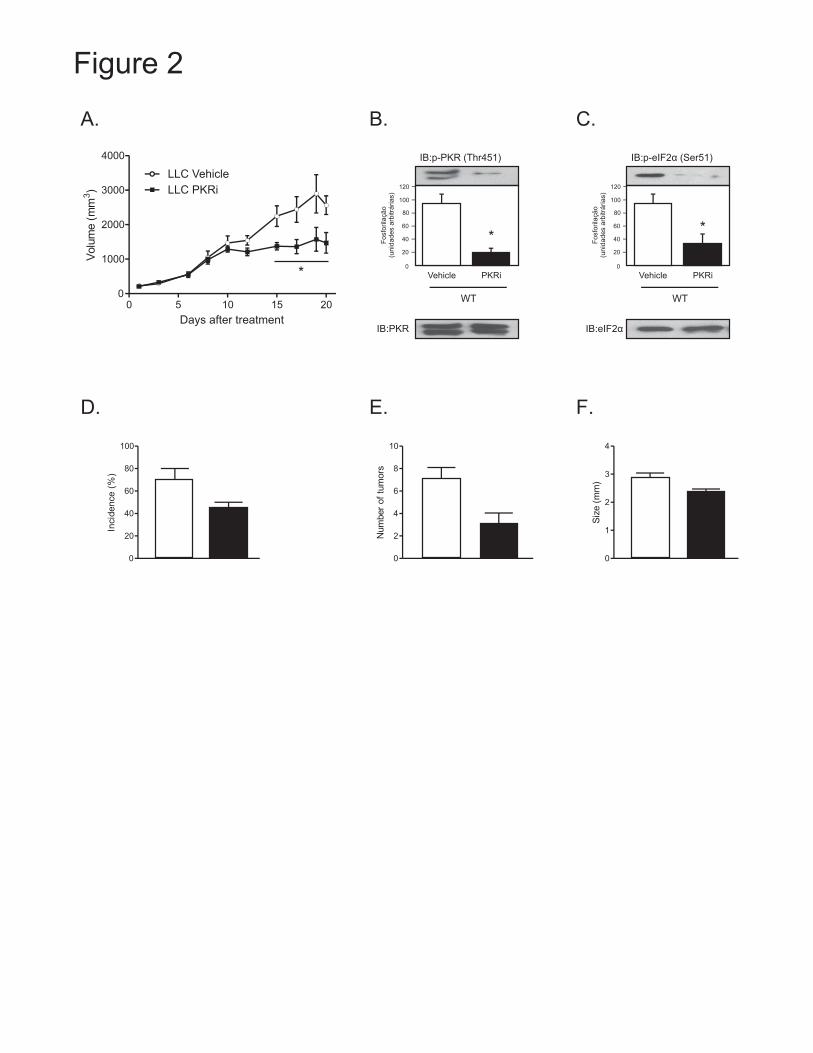

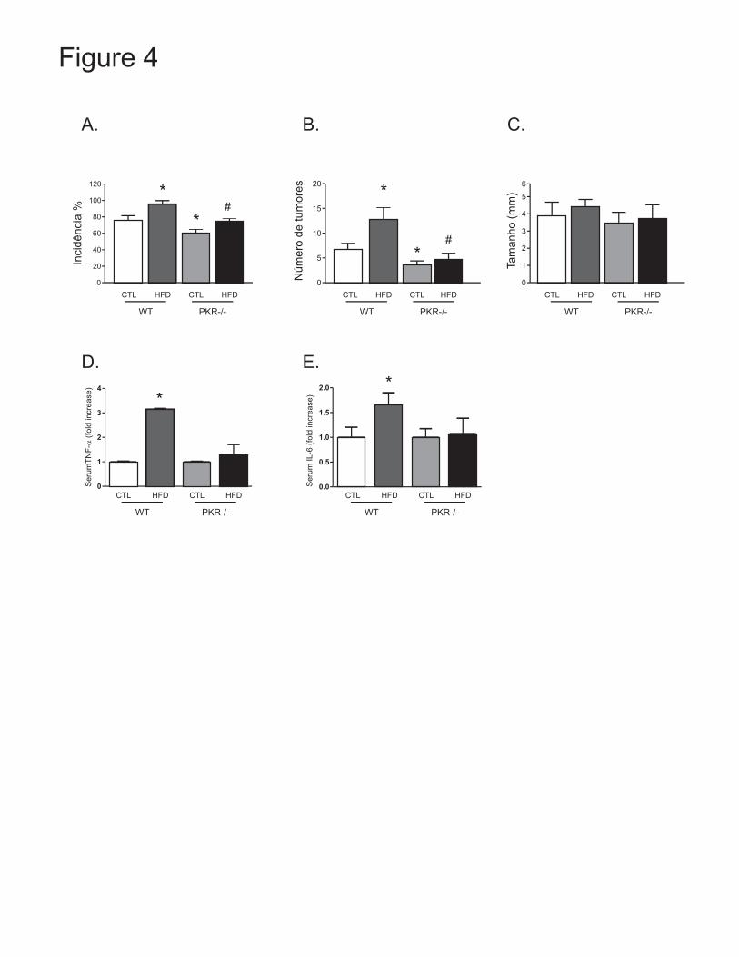


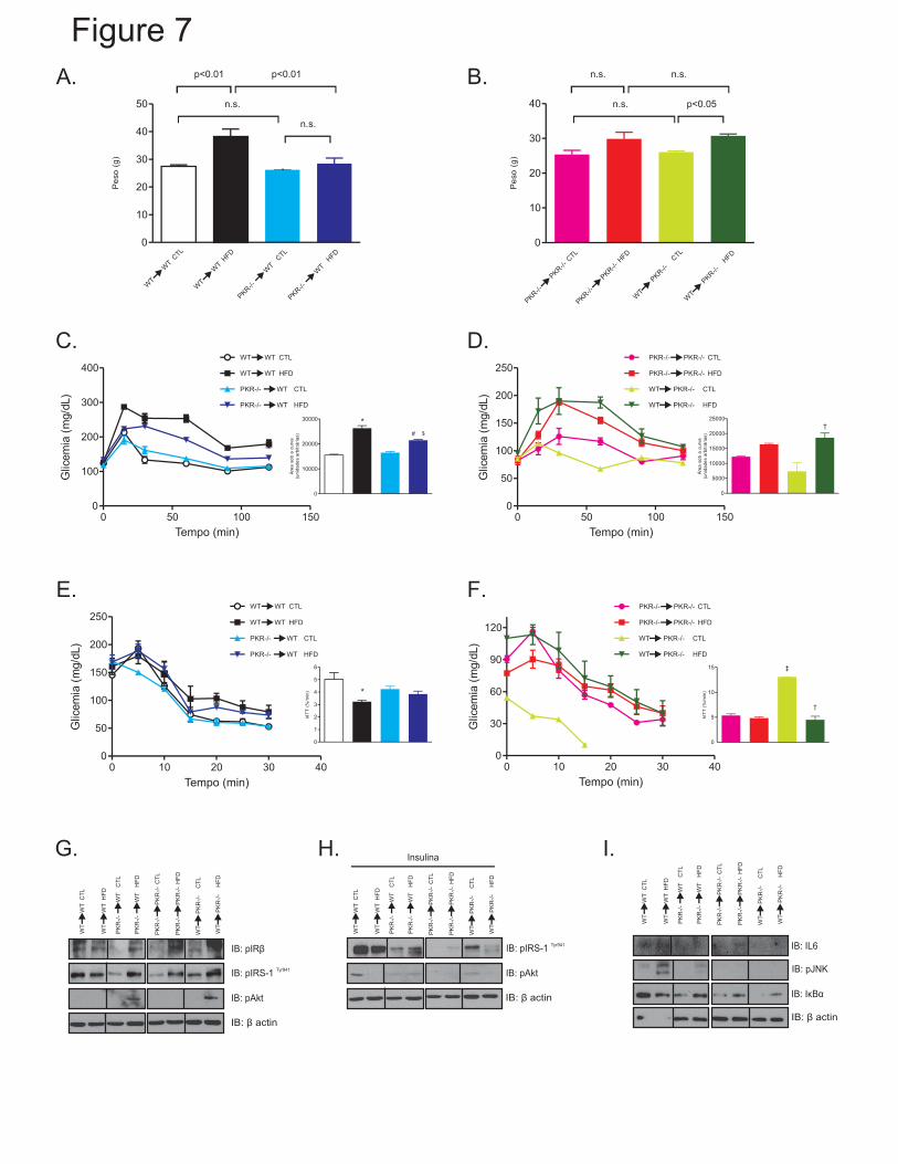

_ 93
Tese de Doutorado
DISCUSSÃO

_ 94
Tese de Doutorado
DISCUSSÃO
A relação entre inflamação e câncer é suspeita há muito tempo. O sobrepeso e a
obesidade aumentam o risco de câncer de cólon, especialmente em homens (Calle e Kaaks,
2004;. Calle et al, 2003). Apesar da magnitude do efeito e do grande número (900.000) de
indivíduos em que se baseou o estudo epidemiológico que proporcionou este conhecimento,
o mecanismo pelo qual a obesidade aumenta o risco de morte por câncer de cólon
permaneceu desconhecido. Além disso, o efeito da obesidade como indutor de tumor de
cólon depende, em grande parte, da resposta inflamatória de baixo grau por ela induzida,
que envolve a produção elevada de TNF-α e IL-6, ambas citocinas atuam como promotoras
de tumores (Lin e Karin, 2007). Notavelmente, a sinalização de IL-6 por meio de ERK e
STATt3 pode diminuir a apoptose de células epiteliais, enquanto estimula a proliferação
celular, como recentemente encontrado no câncer associado à colite (Grivennikov et al.,
2009). Além disso, a expressão de IL-6 é elevada na cirrose hepática e em carcinoma
hepatocelular (HCC) (Tilg et al, 1992;. Trikha et al, 2003). Elevadas concentrações séricas
de IL-6 também se correlaciona com um maior risco de progressão da hepatite viral crônica
para HCC (Nakagawa et al, 2009; Wong et al, 2009). Além disso, camundongos machos
sem IL-6 não mais responderam ao efeito de promoção tumoral da obesidade (Park et al,
2010). Apesar de TNF-α e IL-6 não serem os únicos mediadores responsáveis pela
promoção de tumores induzidos pela obesidade, é bem estabelecido que a sua produção é
elevada em animais obesos devido à resposta inflamatória de baixo grau causada pelo
acúmulo de lipídios (Arkan et al, 2005; Hotamisligil, 2006; Shoelson et al, 2007; Solinas et
al, 2007). Independentemente de sua origem, é reconhecido que a IL-6 pode propagar a
inflamação (Kamimura et al., 2003).

_ 95
Tese de Doutorado
Os resultados deste estudo mostram que obesidade, induzida ou genética, aumenta
significativamente o desenvolvimento do câncer colo-retal. Os nossos dados indicam que a
inflamação do cólon induzida pela obesidade aumenta a expressão e atividade de JNK e
IKKβ no cólon de camundongos tratados com AOM+DSS. Além disso, os camundongos
obesos também apresentaram um aumento significativo nos níveis de insulina e uma
deficiência na ativação da via da insulina no cólon destes animais.
Estudos epidemiológicos sugerem que a hiperinsulinemia é um importante
mecanismo pelo qual a obesidade pode conferir um risco aumentado de câncer de cólon, e
os níveis de insulina também podem influenciar o reaparecimento do câncer colo-retal
(Sandhu MS, et al., 2002; Ma J et al., 2004). No entanto, observamos uma diminuição da
sensibilidade à insulina no cólon. Consistente com estes resultados, os tratamentos de
octreotideo e pioglitazona melhoraram a sensibilidade à insulina mas não foram capazes de
diminuir a taxa de crescimento do tumor. É interessante notar que o octreotideo, além de
reduzir a secreção da insulina, também diminui a síntese de vários peptídeos intestinais, tais
como polipeptídeo insulinotrópico dependente da glicose (GIP) (Ranganath LR et al.,
1999). GIP é um hormônio que pode ativar a via PI3K/Akt/mTOR no trato gastrointestinal
(Baggio LL, et al., 2007); no entanto, não observamos diminuição do crescimento tumoral
ou fosforilação de Akt no o tratamento com octreotideo. Os nossos dados mostram que a
obesidade é acompanhada por um grande aumento do índice inflamatório do cólon, e
também por um aumento na fosforilação de JNK e IKK, bem como o tratamento com o
bloqueador de TNF-α, infliximabe, mostrou uma diminuição consistente no crescimento de
tumores em xenoenxertos de HT-29, e no desenvolvimento de câncer associado à colite.
Assim, o estado inflamatório de baixo grau observado na obesidade aumentou os níveis

_ 96
Tese de Doutorado
plasmáticos de TNF-α e criou o "solo fértil" no epitélio do cólon que aumenta a ação de
DSS e AOM.
Embora os eventos normais de células, como a proliferação e evasão de morte
controlem o desenvolvimento tumoral, o microambiente tumoral também faz uma
contribuição importante e influencia a fisiologia das células malignas (Radisky e Bissell,
2004). Quase todos os tumores contem células inflamatórias e imunes, tais como as células
dendríticas, macrófagos e linfócitos, que produzem citocinas e outros fatores que
promovem o crescimento tumoral e a sobrevivência (Balkwill et al, 2005;. Coussens e
Werb, 2002; Lin e Karin, 2007 ). O papel de promotor tumoral mais óbvio de células
imunes é manifestada em câncer associado à inflamação, em que os tumores surgem e
crescem em locais de inflamação crônica. Em trabalhos anteriores, a inibição da ativação
do NF-κB em células mielóides inibiu a proliferação de células epiteliais intestinais pré-
malignas em um modelo de câncer associado à colite (Greten et al., 2004).
Durante a indução inicial de câncer associado à colite, a IL-6 é produzida
principalmente por células mielóides (Grivennikov et al., 2009). Correspondentemente,
camundongos sem IL-6 em células derivadas de medula óssea exibiram menos tumores,
com uma redução substancial na carga total de tumores (número e tamanho) (Grivennikov
et al., 2009). Da mesma forma, nós também demonstramos que camundongos sem PKR nas
células derivadas da medula óssea também apresentaram menos tumores e de tamanhos
menores .
Tornou-se aparente que a interação entre as células transformadas e o seu ambiente,
incluindo as células imunes inatas, fibroblastos e células endoteliais pode ser um
instrumento tanto para a promoção quanto para a progressão tumoral de muitos tumores
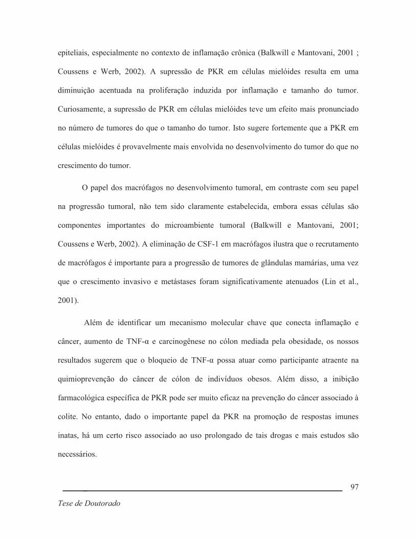
_ 97
Tese de Doutorado
epiteliais, especialmente no contexto de inflamação crônica (Balkwill e Mantovani, 2001 ;
Coussens e Werb, 2002). A supressão de PKR em células mielóides resulta em uma
diminuição acentuada na proliferação induzida por inflamação e tamanho do tumor.
Curiosamente, a supressão de PKR em células mielóides teve um efeito mais pronunciado
no número de tumores do que o tamanho do tumor. Isto sugere fortemente que a PKR em
células mielóides é provavelmente mais envolvida no desenvolvimento do tumor do que no
crescimento do tumor.
O papel dos macrófagos no desenvolvimento tumoral, em contraste com seu papel
na progressão tumoral, não tem sido claramente estabelecida, embora essas células são
componentes importantes do microambiente tumoral (Balkwill e Mantovani, 2001;
Coussens e Werb, 2002). A eliminação de CSF-1 em macrófagos ilustra que o recrutamento
de macrófagos é importante para a progressão de tumores de glândulas mamárias, uma vez
que o crescimento invasivo e metástases foram significativamente atenuados (Lin et al.,
2001).
Além de identificar um mecanismo molecular chave que conecta inflamação e
câncer, aumento de TNF-α e carcinogênese no cólon mediada pela obesidade, os nossos
resultados sugerem que o bloqueio de TNF-α possa atuar como participante atraente na
quimioprevenção do câncer de cólon de indivíduos obesos. Além disso, a inibição
farmacológica específica de PKR pode ser muito eficaz na prevenção do câncer associado à
colite. No entanto, dado o importante papel da PKR na promoção de respostas imunes
inatas, há um certo risco associado ao uso prolongado de tais drogas e mais estudos são
necessários.

_ 98
Tese de Doutorado
CONCLUSÕES

_ 99
Tese de Doutorado
CONCLUSÕES
Conclusão Geral
No presente trabalho é apresentado que em situações de obesidade, onde
encontramos um estado de baixo grau de inflamação, mas com elevação significativa dos
níveis de citocinas inflamatórias circulantes, o TNF-α contribui para a formação e
crescimento de tumores de cólon em camundongos obesos. Além disso, nossos dados
indicam que os bloqueadores de TNF-α poderiam ser usados como agentes
quimiopreventores em câncer de cólon em indivíduos obesos.
Além disso, também mostramos que a ausência da proteína PKR protege contra a
inflamação associada à obesidade e por isso também protege contra a formação de tumores
de cólon em camundongos obesos. Além de identificar um mecanismo molecular chave que
conecta inflamação e câncer, os nossos resultados sugerem que a inibição farmacológica específica
de PKR pode ser muito eficaz na prevenção de câncer associado à colite.

_ 100
Tese de Doutorado
REFERÊNCIAS BIBLIOGRÁFICAS

_ 101
Tese de Doutorado
REFERÊNCIAS BIBLIOGRÁFICAS
1. Kopelman, P.G., Nature, 2000. 404(6778): p. 635-43. 2. Kuczmarski, R.J., et al., JAMA, 1994. 272(3): p. 205-11. 3. Calle, E.E., et al., N Engl J Med, 2003. 348(17): p. 1625-38. 4. Renehan, A.G., D.L. Roberts, and C. Dive, Arch Physiol Biochem, 2008. 114(1): p. 71-83. 5. Giovannucci, E., Cancer Causes Control, 1995. 6(2): p. 164-79. 6. McKeown-Eyssen, G., Cancer Epidemiol Biomarkers Prev, 1994. 3(8): p. 687-95. 7. Rosenbloom, A.L., et al., Diabetes Care, 1999. 22(2): p. 345-54. 8. Flegal, K.M., Med Sci Sports Exerc, 1999. 31(11 Suppl): p. S509-14. 9. Kopelman, P.G., Nature, 2000. 404(6778): p. 635-43. 10. IARC, 2002. 11. Macrae, F.A., Prev Med, 1993. 22(5): p. 750-66. 12. Thorling, E.B., N.O. Jacobsen, and K. Overvad, Eur J Cancer Prev, 1993. 2(1): p. 77-82. 13. Kritchevsky, D., et al., Lipids, 1986. 21(4): p. 272-4. 14. Hu, F.B., et al., J Natl Cancer Inst, 1999. 91(6): p. 542-7. 15. Kaaks, R., et al., J Natl Cancer Inst, 2000. 92(19): p. 1592-600. 16. Schoen, R.E., et al., J Natl Cancer Inst, 1999. 91(13): p. 1147-54. 17. Will, J.C., et al., Am J Epidemiol, 1998. 147(9): p. 816-25. 18. Bird, C.L., et al., Cancer Epidemiol Biomarkers Prev, 1996. 5(8): p. 607-12. 19. Yamada, K., et al., Int J Epidemiol, 1998. 27(5): p. 794-8. 20. Calle, E.E. and R. Kaaks, Nat Rev Cancer, 2004. 4(8): p. 579-91. 21. Bruce, W.R., T.M. Wolever, and A. Giacca, Nutr Cancer, 2000. 37(1): p. 19-26. 22. Koohestani, N., et al., Nutr Cancer, 1997. 29(1): p. 69-76. 23. Komninou, D., et al., Exp Biol Med (Maywood), 2003. 228(4): p. 396-405. 24. Carvalheira, J.B., et al., Arq Bras Endocrinol Metab, 2002. 46(4): p. 419-25. 25. Park, E.J., et al., Cell, 2010. 140(2): p. 197-208. 26. Balkwill, F. and A. Mantovani, Lancet, 2001. 357(9255): p. 539-45. 27. Kuper, H., H.O. Adami, and D. Trichopoulos, J Intern Med, 2000. 248(3): p. 171-83. 28. Coussens, L.M. and Z. Werb, Nature, 2002. 420(6917): p. 860-7. 29. Warzocha, K., et al., Blood, 1998. 91(10): p. 3574-81. 30. Wang, W.H., et al., J Natl Cancer Inst, 2003. 95(23): p. 1784-91. 31. Garber, K., J Natl Cancer Inst, 2004. 96(4): p. 252-3. 32. Chang, E.T., et al., J Natl Cancer Inst, 2004. 96(4): p. 305-15. 33. Ness, R.B. and C. Cottreau, J Natl Cancer Inst, 1999. 91(17): p. 1459-67. 34. Burnet, F.M., Prog Exp Tumor Res, 1970. 13: p. 1-27. 35. Philip, M., D.A. Rowley, and H. Schreiber, Semin Cancer Biol, 2004. 14(6): p. 433-9. 36. Luo, J.L., et al., Cancer Cell, 2004. 6(3): p. 297-305. 37. Greten, F.R., et al., Cell, 2004. 118(3): p. 285-96. 38. Pikarsky, E., et al., Nature, 2004. 431(7007): p. 461-6. 39. Bonizzi, G. and M. Karin, Trends Immunol, 2004. 25(6): p. 280-8. 40. Karin, M., et al., Nat Rev Cancer, 2002. 2(4): p. 301-10. 41. Li, Z.W., et al., J Exp Med, 1999. 189(11): p. 1839-45. 42. Karin, M., T. Lawrence, and V. Nizet, Cell, 2006. 124(4): p. 823-35. 43. Schenk, S., M. Saberi, and J.M. Olefsky, J Clin Invest, 2008. 118(9): p. 2992-3002. 44. Hotamisligil, G.S., Nature, 2006. 444(7121): p. 860-7. 45. Weston, C.R. and R.J. Davis, Curr Opin Cell Biol, 2007. 19(2): p. 142-9. 46. Hacker, H. and M. Karin, Sci STKE, 2006. 2006(357): p. re13. 47. Luo, J.L., H. Kamata, and M. Karin, J Clin Invest, 2005. 115(10): p. 2625-32. 48. Williamson, R.T., British Medical Journal, 1901. 1901: p. 760-762.
_ 102
Tese de Doutorado
49. Reid, J., A.I. Macdougall, and M.M. Andrews, Br Med J, 1957. 2(5053): p. 1071-4. 50. Uysal, K.T., et al., Nature, 1997. 389(6651): p. 610-4. 51. Hotamisligil, G.S., N.S. Shargill, and B.M. Spiegelman, Science, 1993. 259(5091): p. 87-
91. 52. Jager, J., et al., Endocrinology, 2007. 148(1): p. 241-51. 53. Klover, P.J., A.H. Clementi, and R.A. Mooney, Endocrinology, 2005. 146(8): p. 3417-27. 54. Xu, H., et al., J Clin Invest, 2003. 112(12): p. 1821-30. 55. Weisberg, S.P., et al., J Clin Invest, 2003. 112(12): p. 1796-808. 56. Cinti, S., et al., J Lipid Res, 2005. 46(11): p. 2347-55. 57. Lumeng, C.N., J.L. Bodzin, and A.R. Saltiel, J Clin Invest, 2007. 117(1): p. 175-84. 58. Lumeng, C.N., et al., Diabetes, 2007. 56(1): p. 16-23. 59. Vallerie, S.N., et al., PLoS One, 2008. 3(9): p. e3151. 60. Solinas, G., et al., Cell Metab, 2007. 6(5): p. 386-97. 61. Arkan, M.C., et al., Nat Med, 2005. 11(2): p. 191-8. 62. Kanda, H., et al., J Clin Invest, 2006. 116(6): p. 1494-505. 63. Chen, C.Y., et al., Science, 1998. 280(5371): p. 1945-9. 64. Chen, C.Y., et al., Genes Dev, 2000. 14(10): p. 1236-48. 65. Senftleben, U. and M. Karin, Crit Care Med, 2002. 30(1 Suppl): p. S18-26. 66. Hirosumi, J., et al., Nature, 2002. 420(6913): p. 333-6. 67. Harding, H.P., Y. Zhang, and D. Ron, Nature, 1999. 397(6716): p. 271-4. 68. Shi, Y., et al., Mol Cell Biol, 1998. 18(12): p. 7499-509. 69. Nakamura, T., et al., Cell, 2010. 140(3): p. 338-48. 70. DuRose, J.B., et al., Mol Cell Biol, 2009. 29(15): p. 4295-307. 71. Deng, J., et al., Mol Cell Biol, 2004. 24(23): p. 10161-8. 72. Jiang, H.Y., et al., Mol Cell Biol, 2003. 23(16): p. 5651-63. 73. Polak, P. and M.N. Hall, Curr Opin Cell Biol, 2009. 21(2): p. 209-18. 74. Harrington, L.S., et al., J Cell Biol, 2004. 166(2): p. 213-23. 75. Kwiatkowski, D.J. and B.D. Manning, Hum Mol Genet, 2005. 14 Spec No. 2: p. R251-8. 76. Zhang, H.H., et al., PLoS One, 2009. 4(7): p. e6189. 77. Ozcan, U., et al., Mol Cell, 2008. 29(5): p. 541-51. 78. Khamzina, L., et al., Endocrinology, 2005. 146(3): p. 1473-81. 79. Tzatsos, A. and K.V. Kandror, Mol Cell Biol, 2006. 26(1): p. 63-76. 80. Um, S.H., et al., Nature, 2004. 431(7005): p. 200-5. 81. Lee, D.F., et al., Cell, 2007. 130(3): p. 440-55. 82. Zhang, X., et al., Cell, 2008. 135(1): p. 61-73. 83. Leaman, D.W., et al., Proc Natl Acad Sci U S A, 1998. 95(16): p. 9442-7. 84. Williams, B.R., Oncogene, 1999. 18(45): p. 6112-20. 85. Galabru, J. and A. Hovanessian, J Biol Chem, 1987. 262(32): p. 15538-44. 86. Rhoads, R.E., J Biol Chem, 1993. 268(5): p. 3017-20. 87. Bennett, R.L., et al., Blood, 2006. 108(3): p. 821-9. 88. Garcia, M.A., E.F. Meurs, and M. Esteban, Biochimie, 2007. 89(6-7): p. 799-811. 89. Williams, B.R., Sci STKE, 2001. 2001(89): p. re2. 90. Alisi, A., et al., Cell Signal, 2008. 20(3): p. 534-42. 91. Kumar, A., et al., Proc Natl Acad Sci U S A, 1994. 91(14): p. 6288-92. 92. Gil, J., J. Alcami, and M. Esteban, Oncogene, 2000. 19(11): p. 1369-78. 93. Sadler, A.J. and B.R. Williams, Curr Top Microbiol Immunol, 2007. 316: p. 253-92. 94. Clemens, M.J. and A. Elia, J Interferon Cytokine Res, 1997. 17(9): p. 503-24. 95. Koromilas, A.E., et al., Science, 1992. 257(5077): p. 1685-9. 96. Jagus, R., B. Joshi, and G.N. Barber, Int J Biochem Cell Biol, 1999. 31(1): p. 123-38. 97. Vorburger, S.A., et al., Am J Pharmacogenomics, 2004. 4(3): p. 189-98.

_ 103
Tese de Doutorado
98. Kim, S.H., et al., Oncogene, 2000. 19(27): p. 3086-94. 99. Kim, S.H., et al., Oncogene, 2002. 21(57): p. 8741-8. 100. Pataer, A., et al., Cancer Biol Ther, 2009. 8(3): p. 245-52. 101. Haines, G.K., et al., Tumour Biol, 1996. 17(1): p. 5-12. 102. Haines, G.K., 3rd, et al., Tumour Biol, 1998. 19(1): p. 52-9. 103. Shimada, A., et al., Cancer Res, 1998. 58(19): p. 4434-8. 104. Delgado Andre, N. and F.L. De Lucca, Cancer Lett, 2007. 258(1): p. 118-25. 105. Eley, H.L., S.T. Russell, and M.J. Tisdale, Mol Cell Biochem, 2008. 313(1-2): p. 63-9.

_ 104
Tese de Doutorado
APÊNDICES

_ 105
Tese de Doutorado
APÊNDICE I
_ 106
Tese de Doutorado
INSULIN RECEPTOR SUBSTRATE 1 MEDIATES IRINOTECAN-INDUCED
PI3K/AKT/mTOR PATHWAY ACTIVATION IN COLON CANCER
Running title: Function of IRS-1 in irinotecan treatment
Marília M. Dias, Kellen K. Souza, Guilherme Z. Rocha, Mario J. A. Saad, José B.
C. Carvalheira.
Department of Internal Medicine, FCM, State University of Campinas (UNICAMP),
Campinas, SP, Brazil.
Please address correspondence to: José B. C. Carvalheira, M.D., Departamento
de Clínica Médica, FCM-UNICAMP, Cidade Universitária Zeferino Vaz, Campinas,
SP, Brazil, 13081-970, Fax: +55 19 5218950, e-mail: [email protected]
Financial support: Fundação de Amparo à pesquisa do Estado de São Paulo
(FAPESP) and Conselho Nacional de desenvolvimento científico e tecnológico
(CNPq).
Conflict of interest
The authors declare no conflict of interests.
Keywords: mTOR; IRS-1; Irinotecan
_ 107
Tese de Doutorado
ABSTRACT
Aim: Resistance of tumors to treatment is a common clinical problem in human
colorectal cancer (CRC). Irinotecan, an inhibitor of topoisomerase I, potently
activates mTOR signaling, a mechanism that is involved in CRC refractoriness to
chemotherapy. Herein, we investigated the mechanisms involved in irinotecan-
induced mTOR activation.
Methods: Human CRC xenografted into severe combined immunodeficient (SCID)
mice and the CRC cell lines (HT-29 and CACO-2) were treated with irinotecan or
inhibitors of PI3K signaling or a combination of both drugs. We analyzed the effects
of irinotecan in intracellular signaling using immunoblot, immunohistochemistry, cell
viability and apoptosis assays and protein tyrosine phosphatase (PTP) 1B activity
assay.
Results: SN38, the active metabolite of irinotecan, induces multiples changes in
IRS-1 serine phosphorylation and decreased interaction of SOCS3 with IRS-1,
leading to IGFR and EGFR activation. IRS-1 knockdown as well as PI3K inhibition
abrogated the SN38 activation of IGFR, EGFR and PI3K/Akt/mTOR pathway, and
sensitized colorectal cancer cell lines to SN38 in vitro. Accordingly, treatment with
IRS-1 antisense oligonucleotide or rapamycin potentiates the anti-tumor effects of
irinotecan HT-29 xenograft tumor.
Conclusions: Irinotecan leads to IGF1-R, EGFR and PI3K/AKT/mTOR activation
through modulation of IRS-1 function in colon cancer, supporting the idea that
halting the tyrosine kinase receptors pathway is an interesting approach to
optimize irinotecan based chemotherapy as well as highlights IRS-1
phosphorylation status and SOCS3 as potential prognosis biomarker in CRC
patients treated with irinotecan.

_ 108
Tese de Doutorado
INTRODUCTION
Irinotecan, is an antitumor agent that causes cellular damage via inhibition
of topoisomerase I(1). Recently, combination of this drug with tyrosine kinase
inhibitors was approved for clinical use based on the reduced likelihood of
irinotecan resistance(2-4). Interestingly, not only blocking tyrosine kinases, but also
inhibiting PI3K/Akt/mTOR pathway enhance apoptosis induced by SN38, the active
form of irinotecan, in human hepatoma cells(5). Furthermore, rapamycin increased
irinotecan activity in colon cancer cells through a cooperative modulation of
mTOR/HIF-1a axis(6). However, the mechanism by which irinotecan modulates the
PI3K/Akt/mTOR pathway remains to be identified.
Type I insulin-like growth factor receptor (IGF1R) and epidermal growth
factor receptor (EGFR) signaling pathway are important growth-regulatory
pathways in colon cancer(7, 8). Ligand binding induces these receptor activation
and phosphorylation of tyrosine residues on substrates and adaptor proteins such
as insulin receptor substrate 1 (IRS-1)(7, 9). Tyrosine phosphorylated IRS-1 can
recruit other signaling messengers, including PI3K/Akt/mTOR pathway(7, 10),
which inhibits apoptosis and regulates proliferation and growth(11).
Several lines of evidence implicate IRS-1 as a critical target for the negative
regulation of this pathway. First, serine phosphorylation of IRS-1 by distinct
kinases, such as JNK(12), IKK(13), ERK(14), PKCζ(15), PKC q(16), Akt(17), GSK-
3(18), IRAK(19), mTOR(20) decreases tyrosine kinase induced IRS-1
phosphorylation. Second, protein tyrosine phosphatases (PTPs), such as PTP1B,

_ 109
Tese de Doutorado
dephosphorylate and inhibit IRS-1 recruitment of PI3K (21, 22). Finally, association
of SOCS3 with IRS-1 results in decreased phosphorylation of Akt (23-25).
In the current study, we evaluated if irinotecan affects IRS-1 function. We
demonstrated that irinotecan by changing IRS-1 serine phosphorylation status and
decreasing SOCS3 expression, promotes activation of EGFR and IGFR in colon
cancer. Collectively, these findings provide a molecular basis for combinatorial use
of irinotecan and tyrosine kinase inhibitors.

_ 110
Tese de Doutorado
MATERIALS AND METHODS
Antibodies and chemicals. Rapamycin was from Sigma-Aldrich, SN38 was from
Tocris (Bristol, UK). Irinotecan was from Sanofi Aventis. Anti-phospho[Ser473] Akt,
anti-Akt, anti-phospho[Thr669] pEGFR, anti-EGFR, Anti-phospho[Tyr895] IRS-1,
anti-phospho[Ser307] IRS-1, anti-phospho[Ser636/639] IRS-1, anti-
phospho[Ser1101] IRS-1, anti-IRS-1, anti-phospho[Ser2448] mTOR, anti-mTOR,
anti-phospho[Thr389] p70S6K, and anti-p70S6K were from Cell Signaling
Technology (Beverly, MA). Anti-PTP1B was purchased from Abcam (Cambridge,
MA). Tyrosine phosphopeptide (TSTEPQ-pY-QPGENL), Malachite Green solution
A, and Malachite Green were from Millipore (Billerica, MA). Routine reagents were
purchased from Sigma-Aldrich (St. Louis, MO) unless otherwise specified.
Cell culture and siRNA transfections. HT-29 and Caco-2 colon cancer cell lines
were maintained in McCoys´s and DMEM, respectively, supplemented with 10%
FBS and penicillin/streptomycin. siRNAs corresponding to human IRS-1 were
transfected into cells by using LipofectAMINEtm2000 (Invitrogen) according to the
manufacturer´s instructions.
Cell viability. Cells were seeded at a density of 20,000 cells/well and allowed to
adhere overnight and treated with the indicated drugs as described in the figures
legends. Cell viability 48h after treatment was determined by the addition of
0.3mg/ml of 3-(4,5-dimethylthiazol-2-yl)-2,5-diphenyl tetrazolium bromide. After 4h
_ 111
Tese de Doutorado
of incubation, the formazan was solubilized in ethanol and the absorbance
determined by spectrophotometer at 570nm.
Western Blot analysis. Cells were lysed in a freshly prepared ice-cold buffer(26)
and Western blot was performed. Briefly, total extracts were prepared and 50mg
total proteins were separated by SDS-PAGE and blotted onto nitrocellulose
membrane. After incubation with antibodies, immune complexes were detected
using the SuperSignal West Pico Chemiluminescent kit (Pierce Biotechnology).
PTP1B phosphatase assay. HT-29 and Caco-2 cell lines were sonicated in lysis
buffer (100mM Imidazol-HCl, 200mM EDTA, 200mM EGTA, 10mg/ml aprotinin,
10mg/ml leupeptin, 300mM Benzamidine, 100mM PMSF, 10mg/ml pepstatin). The
cell lysate was immunoprecipitated with 5µg anti-PTP1B and protein A-Sepharose
and washed three times with TBS. The measurement of PTP1B activity was
carried out in specific buffer (25mM Hepes, pH 7.2, 2.5mM EDTA, 50mM NaCl,
5mM DTT) using 250µM synthetic tyrosine phosphopeptide (TSTEPQ-pY-
QPGENL) as the substrate for 15 minutes, at 30ºC. The release of phosphate
group was measure using Malachite green detection solution at OD 660nm.
Xenografts studies. Six-week-old male SCID mice were provided by the
University of Campinas – Central Breeding Center and inoculated subcutaneously
with 1×10⁶ HT-29 cells. Once tumors became palpable, tumor volume was
calculated as previously(26). Treatments were initiated when tumors reached 50–

_ 112
Tese de Doutorado
100mm3. Mice were treated with 50mg/kg irinotecan per week, 25mg rapamycin per
day, 0.4ng/kg IRS-1 antisense oligonucleotide per day, alone or in combination. All
studies were conducted in compliance with Ethics Committee of the University of
Campinas guidelines.
Immunohistochemistry and TUNEL assay, Ki-67 and TUNEL staining were
performed as described previously(26).
Statistical analysis. All experiments were repeated two to four times after
experimental conditions were optimized. Statistical analysis was performed by
using an one-way ANOVA test with Bonferroni post hoc test, and P<0.05 was
considered to be statistically significant.
_ 113
Tese de Doutorado
RESULTS
SN38 promotes IGF1R, EGFR, and IRS-1 activation.
SN38 increased tyrosine phosphorylation of IGF1R, EGFR and IRS-1 in a
dose-dependent manner after 4 hours of SN38 treatment in HT-29 human colon
cancer cells (Figure 1A). Accordingly, p70S6K phosphorylation increased in a time-
(Figure 1B) and dose-dependent (data not shown) manner after SN38 treatment. In
contrast, IRS-1 phosphorylation at Ser-636/639 and Ser-1101 residues decreased
in HT-29 cell line after SN38 administration (Figure 1B). IRS-1 Ser-312
phosphorylation, however, increased after exposure to the drug. Consistent with
increased IRS-1 Ser-312 phosphorylation, SN38 increased phosphorylation of
JNK, IKK and p38 (Figure 1C).
In order to evaluate if the increased IRS-1 tyrosine phosphorylation after
SN38 treatment was caused by decreased expression of PTP1B (Figure 1B), we
performed a PTP1B phosphatase assay. Nonetheless, SN38 treatment did not
change PTP1B activity, as shown in figure 1D. Thus, we tested if SN38 treatment
could influence the expression SOCS3. Interestingly, SN38 treatment decreased
the association of SOCS3 with IRS-1 (Figure 1E), which might lead to the observed
increase in IRS-1 tyrosine phosphorylation.
IRS-1 knockdown sensitizes colorectal cancer cell line HT-29 and Caco-2 to
SN38 in vitro.
Since SN-38 induced activation of IRS-1, we decided to knockdown the
IRS-1 expression (Figure 2A). HT-29 and Caco-2 treatment with both IRS-1 siRNA

_ 114
Tese de Doutorado
and SN38 for 72 hours decreased cell viability by approximately 10% (Figure 2B)
and 8% (Figure 2C), respectively, when compared to SN38 group alone.
The SN38 administration increased the IGF1R phosphorylation levels, and
treatment of the cells with the IRS-1 siRNA abrogated this effect in both HT-29 and
Caco-2 cell lines (Figures 2D and 2E, respectively). The IRS-1, Akt, and mTOR
phosphorylation increased after SN38 treatment, whereas the Akt and mTOR
activation was unresponsive to SN38 administration in both cell lines lacking IRS-1.
Thus, SN38 promotes Akt/mTOR activation through IRS-1 phosphorylation, and
inhibition of this protein abrogates this effect.
PI3K and mTOR inhibitors sensitize colorectal cancer cell lines to SN38 in
vitro.
As shown in Figure 3A, the PI3K inhibitor decreased the HT-29 viability by
approximately 55%, whereas the SN38 treatment decreased the viability by
approximately 27% compared to untreated group. The combination of both drugs
dramatically decreased the viability of the HT-29 by 72%, a 16% decrease in cell
viability when compared to SN38 treatment alone. Treatment of HT-29 cell line with
10nM rapamycin reduced cell viability by about 11%. When this dose of rapamycin
was combined with SN38, viability was reduced by 44%, a 17% reduction in cell
viability versus SN38 alone.
In the Caco-2 cell lines, the viability after LY294002 treatment was also
reduced by approximately 55% of control group, whereas the SN38 treatment
decreased the viability of the cell by 40% compared to untreated group (Figure 3B).

_ 115
Tese de Doutorado
The combination of both drugs further decreased the viability to 67% of the
untreated group. Surprisingly, in the Caco-2 cell line the combination of SN38 with
rapamycin did not showed any additive effect on cell viability compared to the
SN38 treatment alone (Figure 3B).
As previously observed, SN38 treatment induced IRS-1 Tyr-895, Akt Ser-
473, mTOR Ser-2448, and p70S6K Thr-389 phosphorylation when compared to
control group (Figure 3C and D). LY294002 reduced SN38-induced Akt and mTOR
activation (Figures 3C and 3D, respectively), thereby pointing out the involvement
of PI3K in SN38-dependent AKT activation. Pretreatment of HT-29 and Caco-2
with rapamycin blunted the mTOR and p70S6K phosphorylation induced by SN38
in both cell lines, but it also increased the IRS-1 and Akt phosphorylation. mTOR
activity dampens PI3K signaling by promoting p70S6K-mediated inhibitory
phosphorylation of IRS-1, addition of the mTOR inhibitor impairs this mechanism
through the inhibition of the p70S6K activity, increasing the Akt phosphorylation
observed after SN38 administration. This feedback mechanism may be involved in
the Caco-2 resistance to the concomitant treatment with SN38 and rapamycin
(Figure 3B).
Interestingly, administration of SN-38 to HT-29 cells increased the
phosphorylation of p53, although no further increase after administration of
LY294002 or rapamycin was observed (Figure 3C). In the Caco-2 cell line,
however, p53 was not inducible by the DNA damaging agent SN38, suggesting the
absence of a functional protein (Figure 3D).

_ 116
Tese de Doutorado
Irinotecan and Rapamycin synergize to inhibit tumor growth in mice bearing
colon cancer xenografts
Tumor growth was reduced in mice treated with irinotecan (50mg/kg) or
rapamycin (25mg), while combined treatment completely arrested tumor growth
(Figure 4A). Figure 4B shows that irinotecan and rapamycin alone increased the
number of TUNEL-positive cells compared to sections from control-treated tumors.
However, the combination did not further increase the number of TUNEL-positive
cells. On the other hand, the concomitant treatment with irinotecan and rapamycin
dramatically decreased the number of Ki67-positive cells compared to either
treatment alone (Figure 4B).
As shown in Figure 4C, irinotecan significantly increase the phosphorylation
of IGF1R, IRS-1, Akt, mTOR and p70S6K protein compared to the control
xenografts. As expected, tumors from animals treated with rapamycin showed
lowered mTOR and p70S6K phosphorylation compared to control group.
Coherently, in the animals receiving irinotecan and rapamycin, the induction of
mTOR and p70S6K phosphorylation by irinotecan was blunted.
Irinotecan and IRS-1 knockdown synergize to inhibit tumor growth in mice
bearing colon cancer xenografts
Tumors were roughly half the size in mice treated with a combination of IRS-
1 ASO and irinotecan versus mice treated with either agent alone and about a third
versus mice in control group (Figure 5A). As shown in Figure 5B, irinotecan alone
significantly increase the phosphorylation of IGF1R, IRS-1, Akt, mTOR and

_ 117
Tese de Doutorado
p70S6K protein compared to the control tumors. As expected, tumors from animals
treated with IRS-1 ASO showed lowered IRS-1 content compared to control group.
We also observed a decrease phosphorylation of Akt, mTOR and p70S6K in
tumors from IRS-1 ASO treated mice. Interestingly, in the animals receiving
irinotecan and IRS-1 ASO, the induction of mTOR and p70S6K phosphorylation by
irinotecan was blunted significantly. These data demonstrate the ability of IRS-1
ASO to blunt irinotecan-dependent mTOR and p70S6K activation in vivo.

_ 118
Tese de Doutorado
DISCUSSION
The present study shows that irinotecan acts by increasing the activation of
IGF1R, EGFR and IRS-1 in HT-29 and Caco-2 colon cancer cell lines through
changes in the IRS-1 serine phosphorylation status and inhibition of SOCS3,
further activating the Akt/mTOR pathway. The administration of IRS-1 siRNA or the
PI3K inhibitor decreases this activation, and sensitizes colon cancer cell lines to
irinotecan. Moreover, in contrast to single-agent therapy, xenografted tumors
treated with a combination of irinotecan and IRS-1 ASO showed potent inhibition of
IRS-1, Akt, and mTOR, which was accompanied by a reduction in tumor volume, a
similar effect was observed with the combination of irinotecan and rapamycin.
Previous studies suggest that SOCS3 act as a negative modulator of
carcinogenesis. For instance, exogenous administration of SOCS3 inhibits
proliferation and invasion of A549 lung cancer cells(27) and loss of SOCS3
expression is associated with poor clinical outcome in breast cancer(28). In
parallel, it is observed that increased expression of SOCS3 inhibits IRS1 tyrosine
phosphorylation(29). Our results show that irinotecan in spite of not changing
PTP1B activity caused a dramatic decrease of SOCS3, and this was associated
with an increase in tyrosine phosphorylation of IRS1 and membrane tyrosine
kinases, an effect that consistently increased the PI3K/Akt/mTOR pathway. Thus,
we propose that the loss of SOCS3 expression represents a critical event,
permissive for the increased survival of HT-29 and Caco-2 cell lines.
In accordance with the hypothesis that IRS-1 is central to irinotecan-induced
activation of PI3K/Akt/mTOR pathway, SN38 treatment decreased phosphorylation
_ 119
Tese de Doutorado
of two serines residues required for inhibition of IRS-1. On the other hand, SN38
administration increased kinase activity of JNK, IKK and p38 and phosphorylation
of IRS-1 on Ser-312. Interestingly, the inhibition of NF-B, a target of IKK, sensitizes
chemo resistant tumors to the apoptotic potential of irinotecan resulting in tumor
regression(30, 31). In agreement with the observed increased activity of
PI3K/Akt/mTOR pathway, Copps et al. showed that IRS-1 Ser-307
phosphorylation, the mice analogous to human Ser-312, plays a permissive role in
insulin-stimulated IRS-1 tyrosine phosphorylation and signal transduction(32). In
aggregate, our findings suggest the existence of a short negative feedback loop,
wherein, IRS-1 serine phosphorylation status modulates its additional activation
through changes in the tyrosine kinase receptor activity(33). Another mechanism
that may co-occur to sustain the increased activity of PI3K/Akt/mTOR pathway is a
gain of interaction between irinotecan-induced IRS-1 serine phosphorylation and
PI3K(34).
Intriguingly, despite the additive effects of irinotecan and IRS-1 ASO
combination we just observed a synergistic effect of mTOR inhibition and
irinotecan in HT-29 cells. Considering that the proliferation of Caco-2 cells was
inhibited with both IRS-1 ASO and PI3K inhibitors and that it was recently reported
a gain of interaction between IRS-1 and mutated PI3K, our finding suggest that
IRS-1/PI3K interaction is critical to Caco-2 cells replication(34, 35) Since Caco-2
cell lines did not showed increased p53 phosphorylation after SN38 treatment, it is
also possible that the synergistic effect between irinotecan and rapamycin be
dependent on activation of p53. In accordance, Beuvink and colleagues(36)
_ 120
Tese de Doutorado
showed that the antitumor activity of rapamycin and cisplatin treatment in cultured
tumor cells depends on an intact p53 response.
Collectively, we suggest that the irinotecan-induced modification of IRS-1
function leads potentiation of the activity of IGFR and EGFR as well as their
downstream effectors contribute to the amplified responses observed in the
combinatorial use of tyrosine kinase inhibitors and irinotecan. Thus, the irinotecan-
induced changes in IRS-1 function may be associated with the clinically significant
activity of cetuximab or panitumumab, monoclonal antibodies that specifically block
the EGFR, combined with irinotecan, even in patients with irinotecan-refractory
colorectal cancer(2, 37, 38). In this line, we can also speculate that irinotecan-
induced changes in IRS-1 function could be involved in the increased overall
survival observed in the association of vascular endothelial growth factor (VEGF)
blockade and irinotecan in the treatment of human colon cancer (39, 40).
Therefore, the role of the commonly used chemotherapy in the activation of
intracellular signaling pathways should be focused to the discovery of biomarkers
and so IRS-1 serine phosphorylation status and its association with SOCS3 should
be explored as biomarkers in colorectal cancer patients treated with the
association of irinotecan and inhibitors of tyrosine kinases.
Taken together, our findings identify IGF1R, EGFR and PI3K/Akt/mTOR
pathway activation as a consequence of irinotecan-induced changes in IRS-1
activity and underscore, the potential of a combined therapeutic approach with
irinotecan and inhibitors of tyrosine kinase receptors and of PI3K/Akt/mTOR
pathway for the treatment of colon cancer.

_ 121
Tese de Doutorado
CONFLICT OF INTERESTS STATEMENT
The authors have declared no competing interests.
_ 122
Tese de Doutorado
REFERENCES
1. Pommier Y. Topoisomerase I inhibitors: camptothecins and beyond. Nat Rev Cancer
2006;6(10):789-802.
2. Cunningham D, Humblet Y, Siena S, Khayat D, Bleiberg H, Santoro A, et al. Cetuximab
monotherapy and cetuximab plus irinotecan in irinotecan-refractory metastatic colorectal cancer.
N Engl J Med 2004;351(4):337-45.
3. Peeters M, Price TJ, Cervantes A, Sobrero AF, Ducreux M, Hotko Y, et al. Randomized
phase III study of panitumumab with fluorouracil, leucovorin, and irinotecan (FOLFIRI) compared
with FOLFIRI alone as second-line treatment in patients with metastatic colorectal cancer. J Clin
Oncol 2010;28(31):4706-13.
4. Schultheis B, Folprecht G, Kuhlmann J, Ehrenberg R, Hacker UT, Kohne CH, et al.
Regorafenib in combination with FOLFOX or FOLFIRI as first- or second-line treatment of colorectal
cancer: results of a multicenter, phase Ib study. Ann Oncol 2013;24(6):1560-7.
5. Koizumi N, Hatano E, Nitta T, Tada M, Harada N, Taura K, et al. Blocking of PI3K/Akt
pathway enhances apoptosis induced by SN-38, an active form of CPT-11, in human hepatoma
cells. Int J Oncol 2005;26(5):1301-6.
6. Pencreach E, Guerin E, Nicolet C, Lelong-Rebel I, Voegeli AC, Oudet P, et al. Marked activity
of irinotecan and rapamycin combination toward colon cancer cells in vivo and in vitro is mediated
through cooperative modulation of the mammalian target of rapamycin/hypoxia-inducible factor-
1alpha axis. Clin Cancer Res 2009;15(4):1297-307.
7. Pollak M. The insulin and insulin-like growth factor receptor family in neoplasia: an
update. Nat Rev Cancer 2012;12(3):159-69.
8. Walther A, Johnstone E, Swanton C, Midgley R, Tomlinson I, Kerr D. Genetic prognostic and
predictive markers in colorectal cancer. Nat Rev Cancer 2009;9(7):489-99.
9. Lemmon MA, Schlessinger J. Cell signaling by receptor tyrosine kinases. Cell
2010;141(7):1117-34.
10. Taniguchi CM, Emanuelli B, Kahn CR. Critical nodes in signalling pathways: insights into
insulin action. Nat Rev Mol Cell Biol 2006;7(2):85-96.
11. Wullschleger S, Loewith R, Hall MN. TOR signaling in growth and metabolism. Cell
2006;124(3):471-84.
12. Aguirre V, Uchida T, Yenush L, Davis R, White MF. The c-Jun NH(2)-terminal kinase
promotes insulin resistance during association with insulin receptor substrate-1 and
phosphorylation of Ser(307). J Biol Chem 2000;275(12):9047-54.
13. Gao Z, Hwang D, Bataille F, Lefevre M, York D, Quon MJ, et al. Serine phosphorylation of
insulin receptor substrate 1 by inhibitor kappa B kinase complex. J Biol Chem 2002;277(50):48115-
21.
14. De Fea K, Roth RA. Modulation of insulin receptor substrate-1 tyrosine phosphorylation
and function by mitogen-activated protein kinase. J Biol Chem 1997;272(50):31400-6.
15. Ravichandran LV, Esposito DL, Chen J, Quon MJ. Protein kinase C-zeta phosphorylates
insulin receptor substrate-1 and impairs its ability to activate phosphatidylinositol 3-kinase in
response to insulin. J Biol Chem 2001;276(5):3543-9.
16. Li Y, Soos TJ, Li X, Wu J, Degennaro M, Sun X, et al. Protein kinase C Theta inhibits insulin
signaling by phosphorylating IRS1 at Ser(1101). J Biol Chem 2004;279(44):45304-7.
17. Paz K, Liu YF, Shorer H, Hemi R, LeRoith D, Quan M, et al. Phosphorylation of insulin
receptor substrate-1 (IRS-1) by protein kinase B positively regulates IRS-1 function. J Biol Chem
1999;274(40):28816-22.

_ 123
Tese de Doutorado
18. Eldar-Finkelman H, Krebs EG. Phosphorylation of insulin receptor substrate 1 by glycogen
synthase kinase 3 impairs insulin action. Proc Natl Acad Sci U S A 1997;94(18):9660-4.
19. Kim JA, Yeh DC, Ver M, Li Y, Carranza A, Conrads TP, et al. Phosphorylation of Ser24 in the
pleckstrin homology domain of insulin receptor substrate-1 by Mouse Pelle-like kinase/interleukin-
1 receptor-associated kinase: cross-talk between inflammatory signaling and insulin signaling that
may contribute to insulin resistance. J Biol Chem 2005;280(24):23173-83.
20. Ozes ON, Akca H, Mayo LD, Gustin JA, Maehama T, Dixon JE, et al. A phosphatidylinositol
3-kinase/Akt/mTOR pathway mediates and PTEN antagonizes tumor necrosis factor inhibition of
insulin signaling through insulin receptor substrate-1. Proc Natl Acad Sci U S A 2001;98(8):4640-5.
21. Goldstein BJ, Bittner-Kowalczyk A, White MF, Harbeck M. Tyrosine dephosphorylation and
deactivation of insulin receptor substrate-1 by protein-tyrosine phosphatase 1B. Possible
facilitation by the formation of a ternary complex with the Grb2 adaptor protein. J Biol Chem
2000;275(6):4283-9.
22. Egawa K, Maegawa H, Shimizu S, Morino K, Nishio Y, Bryer-Ash M, et al. Protein-tyrosine
phosphatase-1B negatively regulates insulin signaling in l6 myocytes and Fao hepatoma cells. J Biol
Chem 2001;276(13):10207-11.
23. Sachithanandan N, Fam BC, Fynch S, Dzamko N, Watt MJ, Wormald S, et al. Liver-specific
suppressor of cytokine signaling-3 deletion in mice enhances hepatic insulin sensitivity and
lipogenesis resulting in fatty liver and obesity. Hepatology 2010;52(5):1632-42.
24. Torisu T, Sato N, Yoshiga D, Kobayashi T, Yoshioka T, Mori H, et al. The dual function of
hepatic SOCS3 in insulin resistance in vivo. Genes Cells 2007;12(2):143-54.
25. Rui L, Yuan M, Frantz D, Shoelson S, White MF. SOCS-1 and SOCS-3 block insulin signaling
by ubiquitin-mediated degradation of IRS1 and IRS2. J Biol Chem 2002;277(44):42394-8.
26. Rocha GZ, Dias MM, Ropelle ER, Osorio-Costa F, Rossato FA, Vercesi AE, et al. Metformin
amplifies chemotherapy-induced AMPK activation and antitumoral growth. Clin Cancer Res
2011;17(12):3993-4005.
27. Zhang S, Wang W, Wang E, Qiu X. SOCS3 expression is inversely correlated with Pyk2 in
non-small cell lung cancer and exogenous SOCS3 inhibits proliferation and invasion of A549 cells.
Pathology 2012;44(5):434-40.
28. Ying M, Li D, Yang L, Wang M, Wang N, Chen Y, et al. Loss of SOCS3 expression is
associated with an increased risk of recurrent disease in breast carcinoma. J Cancer Res Clin Oncol
2010;136(10):1617-26.
29. Jorgensen SB, O'Neill HM, Sylow L, Honeyman J, Hewitt KA, Palanivel R, et al. Deletion of
Skeletal Muscle SOCS3 Prevents Insulin Resistance in Obesity. Diabetes 2012.
30. Piret B, Piette J. Topoisomerase poisons activate the transcription factor NF-kappaB in
ACH-2 and CEM cells. Nucleic Acids Res 1996;24(21):4242-8.
31. Wang CY, Cusack JC, Jr., Liu R, Baldwin AS, Jr. Control of inducible chemoresistance:
enhanced anti-tumor therapy through increased apoptosis by inhibition of NF-kappaB. Nat Med
1999;5(4):412-7.
32. Copps KD, Hancer NJ, Opare-Ado L, Qiu W, Walsh C, White MF. Irs1 serine 307 promotes
insulin sensitivity in mice. Cell Metab 2010;11(1):84-92.
33. Hotamisligil GS, Peraldi P, Budavari A, Ellis R, White MF, Spiegelman BM. IRS-1-mediated
inhibition of insulin receptor tyrosine kinase activity in TNF-alpha- and obesity-induced insulin
resistance. Science 1996;271(5249):665-8.
34. Hao Y, Wang C, Cao B, Hirsch BM, Song J, Markowitz SD, et al. Gain of Interaction with IRS1
by p110alpha-Helical Domain Mutants Is Crucial for Their Oncogenic Functions. Cancer Cell
2013;23(5):583-93.

_ 124
Tese de Doutorado
35. Jhawer M, Goel S, Wilson AJ, Montagna C, Ling YH, Byun DS, et al. PIK3CA mutation/PTEN
expression status predicts response of colon cancer cells to the epidermal growth factor receptor
inhibitor cetuximab. Cancer Res 2008;68(6):1953-61.
36. Beuvink I, Boulay A, Fumagalli S, Zilbermann F, Ruetz S, O'Reilly T, et al. The mTOR
inhibitor RAD001 sensitizes tumor cells to DNA-damaged induced apoptosis through inhibition of
p21 translation. Cell 2005;120(6):747-59.
37. Douillard JY, Siena S, Cassidy J, Tabernero J, Burkes R, Barugel M, et al. Randomized, phase
III trial of panitumumab with infusional fluorouracil, leucovorin, and oxaliplatin (FOLFOX4) versus
FOLFOX4 alone as first-line treatment in patients with previously untreated metastatic colorectal
cancer: the PRIME study. J Clin Oncol 2010;28(31):4697-705.
38. Van Cutsem E, Kohne CH, Hitre E, Zaluski J, Chang Chien CR, Makhson A, et al. Cetuximab
and chemotherapy as initial treatment for metastatic colorectal cancer. N Engl J Med
2009;360(14):1408-17.
39. Hurwitz H, Fehrenbacher L, Novotny W, Cartwright T, Hainsworth J, Heim W, et al.
Bevacizumab plus irinotecan, fluorouracil, and leucovorin for metastatic colorectal cancer. N Engl J
Med 2004;350(23):2335-42.
40. Van Cutsem E, Tabernero J, Lakomy R, Prenen H, Prausova J, Macarulla T, et al. Addition of
aflibercept to fluorouracil, leucovorin, and irinotecan improves survival in a phase III randomized
trial in patients with metastatic colorectal cancer previously treated with an oxaliplatin-based
regimen. J Clin Oncol 2012;30(28):3499-506.
_ 125
Tese de Doutorado
ACKNOWLEDGMENTS
We thank Silvia Gatti for providing HT-29 and Caco-2 cell lines and Luiz Janeri and
Jósimo Pinheiro for technical assistance. Funding source: Fundação de Amparo à
pesquisa do Estado de São Paulo (FAPESP) and Conselho Nacional de
desenvolvimento científico e tecnológico (CNPq). The funders had no role in study
design, data collection and analysis, decision to publish, or preparation of the
manuscript.
_ 126
Tese de Doutorado
FIGURE LEGENDS
Figure 1. SN38 increases the activity of the IRS-1/Akt/mTOR pathway. (A) HT-29 cell
were treated with increasing concentrations of SN38 for 4 hours and the phosphorylation
levels of IGF-I, EGFR, and IRS-1 analyzed by Western blot; (B) HT-29 cells were treated
with 100 nM SN38 for 30 minutes, 1, 2, 4 and 6 hours and the phosphorylation levels of
IGF-IR, IRS-1, and p70S6K analyzed by Western blot. (C) HT-29 cell were treated with
increasing concentrations of SN38 for 4 hours and the phosphorylation levels of JNK, IKK,
and p38 analyzed by Western blot. (D) PTP1B-specific phosphatase activity. (E) HT-29
cell were treated with increasing concentrations of SN38 for 4 hours and the association
between IRS1 and SOCS3 analyzed by Western blot; Representative blots of three
independent experiments.
Figure 2. IRS-1 knockdown reverses the p70S6K activation caused by SN38 treatment in human colon cancer cell lines. (A) IRS-1 was efficiently knockdown by
siRNA treatment; (B) HT-29 and (C) CACO-2 cell lines were transfected with IRS-1 siRNA
and 24 hours post-transfection treated with SN38 for 48 hours. Cell viability was measure
using the MTT assay. Data are mean values ± SD from experiment performed in triplicate
and repeated and least three times. *p<0.01; (D) HT-29 and (E) CACO-2 cell lines were
transfected with IRS-1 siRNA and after 24 hours treated with SN38 for 4 hours. IGF1R,
IRS-1, Akt, mTOR, and p70S6K phosphorylation levels were detected by Western blot.
Representative blots of three independent experiments.
Figure 3. The PI3K inhibitor, LY294002, reverses the p70S6K activation caused by SN38 treatment in human colon cancer cell lines. (A) HT-29 and (B) CACO-2 cell lines
were treated with SN38, LY294002, and rapamycin for 48 hours. Cell viability was
measure using the MTT assay. Data are mean values ± SD from experiment performed in
triplicate and repeated and least three times. *p<0.01; (C) HT-29 and (D) CACO-2 cell pre-
treated with LY294002 or rapamycin for 2 hours and then treated with SN38 for 4 hours.
IRS-1, Akt, mTOR, and p70S6K phosphorylation levels were detected by Western blot.
Representative blots of three independent experiments.
Figure 4. Irinotecan and rapamycin synergize in vivo to reduce HT-29 tumor growth. 1.0 x 10⁶ HT-29 cells were injected subcutaneously into the flank of SCID mice. Once the
tumor reached 50-100 mm3, treatments were initiated as indicated in Methods, n=8-12
mice per group mean ± SEM; (A) Tumor growth was measured daily after treatment with
Irinotecan (50 mg/kg) or Rapamycin (25 µg) alone or in combination; (B) Representative
microphotographs of TUNEL-positive and of Ki-67-positive staining cells per field, 4 field
per tumor section, mean ± SEM; (C) The HT-29 tumor lysates were detected by Western

_ 127
Tese de Doutorado
blot with the indicated antibodies. Representative blots of three independent experiments.
*, P ≤ 0.05 vs. control; #, P ≤ 0.05 vs. irinotecan; †, P ≤ 0.05 vs. rapamycin.
Figure 5. Irinotecan and IRS-1 knockdown synergize in vivo to reduce HT-29 tumor growth. 1.0 x 10⁶ HT-29 cells were injected subcutaneously into the flank of SCID mice.
Once the tumor reached 50-100 mm3, treatments were initiated as indicated in Methods,
n=6-10 mice per group mean ± SEM; (A) Tumor growth was measured daily after
treatment with Irinotecan (50 mg/kg) or IRS-1 antisense oligonucleotide (ASO IRS-1) alone
or in combination; (B) The HT-29 tumor lysates were detected by Western blot with the
indicated antibodies. Representative blots of three independent experiments.
Figure 1
SN38 (nM)
p-IRS-1 (Tyr)
p-IGF-1R
IGF-1R
β-actin
IRS-1
HT-29
A.
Vehic
le
50 100 5001000
p-EGFR
EGFR
HT-29
Hours 0 0.5 1 2 4 6
p-IGF-1R
IGF-1R
β-actin
p-IRS-1 (Tyr)
p-IRS-1 (Ser312)
p-IRS-1 (Ser636/639)
p-IRS-1 (Ser1101)
IRS-1
p-p70S6K
p70S6K
B.
PTP1B
SN38 (nM)
p-JNK
JNK
HT-29
Vehic
le
50 100 5001000
p-IKK
C.
SN38
0
20
40
60
80
100
120
Vehic
le
25
nM
50
nM
100
nM
Phosphata
se
Activity
%o
fco
ntr
ol
D.
p-p38
p38
β-actin
IKK
SN38 (nM)
IP: IRS-1Blot: SOCS3
IRS-1
Vehic
le
50 100 5001000
E.
SN38 (100 nM)
p-Akt
IRS-1
Akt
p-mTOR
mTOR
p-p70S6K
p70S6K
β-actin
p-IRS-1 (Tyr)
p-Akt
IRS-1
Akt
p-mTOR
mTOR
p-p70S6K
p70S6K
β-actin
p-IRS-1 (Tyr)
HT-29
SN38
IRS-1 siRNA- - + --- - + +
+Control siRNA- + - - -
SN38
IRS-1 siRNA- - + --- - + +
+Control siRNA- + - - -
Caco-2
Caco-2
120
0
20
40
60
80
100
IRS
-1siR
NA
SN
38
SN
83
IRS
-1siR
NA
Contr
olsiR
NA
Contr
ol
IGF-1R
p-IGF-1R
IGF-1R
p-IGF-1Rβ-actin
IRS-1
siRN
Control
A 1iR
A1
IRS-
sN
IS-
iRNA
3
R1
s
IRS-1
siRNA
2A.
C.
D. E.
0
20
40
60
80
100
120
IRS
-1siR
NA
SN
38
SN
83
IRS
-1siR
NA
Contr
olsiR
NA
Contr
ol
HT-29
*
B.
Figure 2
Cell
via
bility
%o
fco
ntr
ol
Cell
via
bility
%ofco
ntr
ol

p-IRS-1 (Tyr)
Caco-2
LY294002Rapamycin
SN38-+-
---
++ - + - +
- -- - + +
p-Akt
IRS-1
Akt
p-mTOR
mTOR
p-p70S6K
p70S6K
β-actin
HT-29
LY294002Rapamycin
SN38-+-
---
++ - + - +
- -- - + +
p-Akt
IRS-1
Akt
p-mTOR
mTOR
p-p70S6K
p70S6K
β-actin
p-IRS-1 (Tyr)
HT-29 C. D.
Caco-2S
N38
Rapam
ycin
Rapam
ycin
SN
38
LY
294002
LY
294002
*
SN
38
Contr
ol
0
20
40
60
80
100
SN
38
Rapam
ycin
0
20
40
60
80
100
Contr
ol
**
Cell
via
bility
%o
fco
ntr
ol
A.
B.
Rapam
ycin
SN
38
LY
294002
SN
38
LY
294002
p-p53p-p53
Figure 3
Cell
via
bility
%ofcon
trol

Figure 4.
A.
B. C.
1200
1000
800
600
400
200
0
Control
Rapamycin
Irinotecan
Irinotecan+Rapamycin
1 5 10 15 20 25 30
3T
um
or
vo
lum
e(m
m)
Days after treatment
HT-29
Rapamycin
p-Akt
IRS-1
Akt
p-mTOR
mTOR
p-p70S6K
p70S6K
β-actin
p-IRS-1 (Tyr)
Irinotecan-
+ -+- ++-
IGF-1R
p-IGF-1R
TUNEL
vs
%TunelPositieCell
nr
Co
to
lI
no
en
rit
ca
Ra
cin
+pa
my
In
nri
ote
ca
pR
aam
ycin
30
20
10
0
Cnt
rol
o
Irno
a
ite
cn
R
mc
apa
yin
Rap
amcin
+
yI in
oec
n
rt
a
**
40
50
Ki-67
nr
Co
to
ln
oe
nIr
it
ca
Ra
cn
+pa
my
iI
nn
riote
ca
30
20
10
0
ntr
Co
olIri
no
an
tec
R
amcn
ap
yi
aR
pam
ycin
+
Irino
teca
n
%6
sit
lKi-
7Po
iveCels
*
*
Rapam
ycin
#
#
†
*

A.
0 5 10 15 20 25 30
0
200
400
600
800
1000
1200
CTL
ASO IRS-1
Irinotecan
ASO IRS-1+Irinotecan
Days after treatment
3T
um
or
vo
lum
e(m
m)
HT-29
siRNA
p-Akt
IRS-1
Akt
p-mTOR
mTOR
p-p70S6K
p70S6K
β-actin
p-IRS-1 (Tyr)
Irinotecan- + - +
- + +-
IGF-1R
p-IGF-1R
B.
Figure 5.

_ 133
Tese de Doutorado
APÊNDICE II

_ 134
Tese de Doutorado
Lista de Publicações no período
Reis, L.O., Ferrari, K., Zamuner, M., ROCHA, G.Z., Billis, A., Fávaro, W.J. “Urothelial carcinogen resistance driven by stronger Toll-like receptor 2 (TLR2) and Uroplakin III (UP III) defense mechanisms: a new model”. World J Urol. 2014 May 29. Oliveira, A.G., Araujo, T.G., Carvalho, B.M., Guadagnini, D., ROCHA, G.Z.., Bagarolli, R.A., Carvalheira, J.B., Saad, M.J..”Acute exercise induces a phenotypic switch in adipose tissue macrophage polarization in diet-induced obese rats”.Obesity (Silver Spring). 2013 21(12):2545-56. Pimentel, G.D.; Ropelle, E.R.; ROCHA, G.Z.; Carvalheira, J.B. “The role of neuronal
AMPK as a mediator of nutritional regulation of food intake and energy homeostasis”.
Metabolism, 2013 v. 62 (2): 171-8. Flores, M.B.S.*; ROCHA, G.Z.*; Damas-Souza, D.M.; Osório-Costa, F.; Dias, M.M.; Ropelle, E.R.; Camargo, J.A.; de Carvalho, R.B.; Carvalho, H.F.; Saad, M.J.A.; Carvalheira, J.B.C. “Obesity-Induced Increase in Tumor Necrosis Factor-α Leads to Development of Colon Cancer in Mice”. Gastroenterology, 2012, v.143 (3): 741-753. *Equal contribution Tobar, N.; Oliveira, A.G.; Guadagnini, D.; Bagarolli, R.A.; ROCHA, G.Z.; Araujo, T.G.; Prada, P.O.; Saad, M.J. “Comment on: Ramos-Zavala et al. Effect of diacerein on insulin secretion and metabolic control in drug-naïve patients with type 2 diabetes: a randomized clinical trial. Diabetes Care 2011;34:1591-1594.” Diabetes Care, 2012, v. 35(2): e13. Tobar, N.; Oliveira, A.G.; Guadagnini, D.; Bagarolli, R.A.; ROCHA, G.Z.; Araujo, T.G.; Santos-Silva, J.C.; Zollner, R.L.; Boechat, L.H.B.; Carvalheira, J.B.C.; Prada, P. O.; Saad, M.J.A. “Diacerhein Improves Glucose Tolerance and Insulin Sensitivity in Mice on a High-Fat Diet”. Endocrinology (Philadelphia), 2011, v. 152, p. 4080-4093. ROCHA, G.Z.; Dias, M.M.; Ropelle, E.R.; Osório-Costa, F.; Rossato, F.A.; Vercesi, A.E.; Saad, M.J.A.; Carvalheira, J.B.C.“Metformin Amplifies Chemotherapy-Induced AMPK Activation and Antitumoral Growth”. Clinical Cancer Research, 2011, v.17 p,3993-4005, 2011.